Anatomy of Gallbladder, Liver, Pancreas, Spleen Flashcards
Describe the duct system for the passage of bile from the liver to the duodenum and for storage in the gallbladder
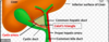
Liver produces the bile.
> Right and left hepatic ducts form the common hepatic duct
>Common hepatic duct joins the cystic duct to form the common bile duct. (At the cystic duct, bile can pass up into the gallbladder if the common bile duct is closed)
> Joins the main pancreatic duct and forms the hepatopancreatic duct/ampulla of Vater
>Hepatopancreatic duct enters the second part of the duodenum at the greater papilla.

Blood supply to the gallbladder
Cystic artery, which arises from the right hepatic artery within the triangle of calot

Venous drainage of the gallbladder
Cystic veins, which drain into the portal vein
Lymphatic drainage of the gallbladder
Cystic lymph nodes in the gallbladder neck
The liver has a dual blood supply
Portal vein (75%): nutrient-rich, but deoxygenated blood; much higher volume
Hepatic artery: higher pO2
Venous drainage of the liver
Hepatic veins drain into the IVC
Where is the gallbladder? What organs is it contact with?
On the inferior surface of the liver a fossa between the right and quadrate lobes; is also in contact with duodenum and transverse colon
Lymphatic drainage of the liver
Most drain through portal lymphatic vessels to celiac nodes?
Position, functional anatomy, and vasculature of the gallbladder and the biliary tree?

How does gallstone ileus form?
Stones that get lodged in teh fundus can ulcerate through the wall into the transverse colon or into the duodenum.
If they enter the duodenum, they produce an intestinal obstruction- the gallstone ileus
3 common sites where gallstones are impacted and the effects on bile flow
(1) Cystic duct - blocks gallbladder drainage, but bile can still flow from the liver
(2) Ampulla of vater - bile regurgitates back to pancreas
(3) Common bile duct - bile flows back into the blood, causing jaundice; choledocholithiasis
Triangle of Calot
Location of the pancreas.
What happens if you have a tumor in the head?
Mostly retroperitoneal - head that lies in the duodenum
Tumors in the head will obstruct bile flow –> jaundice

Function of gallbladder
Receive bile, concentrate it, store it, and release it during digestion in response to cholecystokinin
Cholecystitis- what is it and where would pain refer?
Inflammation of the gallbaldder caused by obstruction of the cystic duct by gall stones
–> trapped bile can cause bacterial infection and perforation.
Pain may radiate to the back or right shoulder region.
Portal triad
Hepatic artery bringing in oxygen and nutrients to liver
Hepatic veins bringing nutrient-rich and oxygen-poor blood to lvier
Hepatic ducts carrying bile out to emulsify fat in the digestive system
Blood supply of pancreas
Pancreatic branches of the splenic artery
Superior pancreaticoduodenal artery from gastroduodenal artery
Inferior pancreaticoduodenal artery from superior mesenteric arteries

Lymphatic drainage of pancreas
Head of pancreas drains into pancreaticoduodenal lymph nodes, lymph nodes in the hepatoduodenal ligament, pre-&postpyloric lymph nodes.
Venous drainage of the pancreas
Head: Superior mesenteric branches of the portal vein
Rest of pancreas: Splenic vein

Autonomic innervation of organs in the upper abdomen associated with the foregut.
Parasympathetic:
Preganglionic: Vagus nerve (CNX)
Postganglionic: Terminal gg @target organ
Sympathetic:
Pre-ganglionic: IML T5-T9, Greater splanchnic nerve
Post-ganglionic: Celiac ganglion
Referred pain among foregut organ goes where?
Epigastrium
Spleen- position, blood supply, attachments
Lies against the diaphragm against ribs 9, 10, and 11 in the left hypochondriac region.
Attachments: Supported by lienogastric/splenogastric and lienorenal/splenorenal ligaments
Supply: splenic artery
Venous drainage: splenic vein
Significance of the spleen in trauma and hematopoietic disorders?
Ruptured spleen: profuse bleeding cna cause death
Spleen is hematopoietic in early life and later destroys old RBCs to set the hemoglobin free
Collateral blood flow to the spleen
Lymphatic drainage of the foregut organs
Left hemiliver is composed of what segments?
Segments I - IV

I: caudate lobe
II-III: left lateral segment
IVa/IVb: left medial segment
Right hemiliver
Segments V - VIII

V/VIII: right anterior
VI/VII: right posterior
What duct is at the head of the pancreas?
Common bile duct
