Anatomy - Leg circulation Flashcards
What are the two different types of fascia ?
Superficial and deep

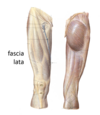
What is fascia lata ?
Deep fascia of the thigh
What does deep fascia form ?
Forms compartments separated by intermuscular speta which is deep fascia that separates two muscles

Describe the main overview of the arterial blood supply of the upper limb down to the anastomoses in the hand
Subclavian ===> Axillary ====> Brachial ===> Branches into radial (more lateral one) and ulner (more medial one) ===> deep and superficial plamar arches

What artery predominatley forms the superficial palmar arch and what artery also contributes ?
Predominatley formed by the ulner artery with a contribution from the superficial palmar branch of the radial artery.

What arteries is the deep palmar arch formed by?
Formed mainly from the terminal part of the radial artery, with the ulnar artery contributing via its deep palmar branch, by an anastomosis.

Describe the vneous draing of the upper limb
Cephalic (more lateral) and basilic (more medial) veins drain to ====> axillary vein ====>
R subclavian vein

What is the vein which runs horizontal between the cephalic and basilic veins ?
The median cubital vein

Describe the arterial supply of the lower limb
Femoral (which has a deep branch off it called the profunda femoris) =====> popliteal ====> Branches into the posterior and anterior tibial arteries
Anterior tibial ===> dorsalis pedis ===> arcuate artery (dorsum of foot)
Posterior tibial artery (has a branch come off it called the fibular artery) ===> Branches into the medial and lateral plantar arteries
Lateral plantar artery ===> plantar arch (sole of foot)

At what anatomical location does the femoral artery pass through and then become the popliteal artery ?
The adductor hiatus

Describe the venous drainage of the lower limb
Small saphenous ====> Popliteal vein ====> femoral vein
Great saphenous vein ===> femoral vein

Roughly in what region does the great saphenous vein join onto the femoral vein ?
The inguinal region
Roughly where does the popliteal vein become the femoral vein ?
As it passes through the adductor hiatus
Describe the route of the small saphenous vein
Arises from the dorsal venous arch ===> goes behind the lateral malleolus and then runs up the back of the calf drainging into the popliteal vein (posterior to the knee)

Describe the route of the great saphenous vein
Arsies from the dorsal venous arch ===> runs in front of the medal malleolus and then rises up the mdial side of the upper leg to join with femoral vein roughly at the inguinal region (around the femoral triangle)

Describe venous blood flow
- Musculovenous pump pushes blood back towards the heart
- Flow from superficial veins into deep veins through perforating veins

What is the function of valves in terms of venous blood flow and what can happen if valves become incompetent ?
Venous valves ensure unidirectional blood flow against gravity (prevent back flow)
- When valves become incompetent it results in reverse flow into superficial veins.
- These veins become weak and dilated
- Resulting in Varicose veins
Clinical note
CLINICAL NOTE
- Digital arteries are end arteries
- The ONLY blood supply to a given area of the body (no collaterals)
- Untreated occlusion of an end artery results in infarction of the area of tissue it supplies resulting in amputation
- Do not use adrenaline-containing local anaesthetic near end arteries
What are the main pulse points in the body ?
- Carotid
- In the upper limb - brachial and radial
- In the lower limb - Femoral, popliteal, posterior tibial and dorsalis pedis
Define what is meant by ischaemia
Inadequate oxygenation of cells/tissues/organ due to an interruption to blood supply (arterial or venous)
What are the two main overall causes of Ischaemia ?
- Reduced arterial perfusion pressure
- Increased venous drainage pressure
What are some of the causes of reduced arterial perfusion pressure ?
- Left ventricular failure
- Arterial bleed (injury)
- Arterial rupture (aneurysm)
- Occlusion of lumen (atherosclerosis: PVD)
- Arterial spasm
- External compression of arterial supply (e.g. tumour/compartment syndrome/crossing legs!!/inflammation/ tamponade)
What are some of the causes of increased venous drainage pressure ?
(back pressure into capillary bed then into the arteriole…preventing normal arterial in-flow)
- Right (or congestive) cardiac failure
- DVT
- External compression (e.g. tumour)
Describe the pathway of skin ulceration formation (overview not as specific as the other flashcard)
- Venous valve failure of any cause or immobility of any cause
- Resulting in (chronic) venous insufficiency (venous blood not efficiently returned to heart)
- Resulting in superficial microcirculatory deficiencies
- Then skin ulceration formation
Note after step 2 it can go either the skin ulceration pathway or DVT pathway


