Adrenal gland Physiology + Calcium Flashcards
+ clinical biochemistry of Calcium/ bone
What are the layers of the Adrenal Gland
(GFR)
- Glomerulosa
- Fasciulata
- Reticularis
- Medulla
What are the adrenal causes of hypertension?
- Primary Hyperaldosteronism (Conn’s syndrome)
- Zona glomerulosa
- adenoma
- hyperplasia
- rare genetic causes
- Zona glomerulosa
- Phaeochromocytoma (Phaeo)
- tumour of the adrenal medulla
- some forms of congenital adrenal hyperplasia
- enzyme defect (uncommon)
Who should be screened for Primary Hyperaldosteronism (Conn’s syndrome)?
those with
- hypokalaemia
- resistant hypertension (resistant to 3 drugs)
- younger people
- they have more vascular and renal pathology than people with essential hypertension &similar blood pressure
What investigations/ assessments would indicate Primary Hyperaldosteronism?
- during initial screening tests
- suppressed renin
- normal/ high aldosterone
- a confirmatory oral or IV Na+ suppression test would be done
- to get the specific aetiology the following would be done
- an adrenal CT scan
- an adrenal venous sampling
- is also secretion unilateral?
- Meeomidate PET scan
What is the treatment for Primary Hyperaldosteronism?
- unilateral vs bilateral adenoma treatment
- Unilateral Adenoma
- Laparoscopic Adrenalectomy
- Medical Treatment ( sometimes )
- Bilateral Hyperplasia
- Medical Treatment ( Aldosterone Antagonists)
- Spironolactone
- Eplerenone
- Medical Treatment ( Aldosterone Antagonists)
What are the products of the adrenal medulla?
- how is it stimulated?
- Catecholamines
- Dopamine
- Noraepinpherine (Noraadrenalin)
- Epinephrine (adrenalin)
- requires cortisol for the conversion from NE
- sympathetic neurons in the spinal cord stimulate the adrenal medulla

What are the biological effects of Catecholamines?
- Noradrenalin (Alpha 1 & 2 )
- Vasoconstriction
- Increased BP
- Pallor
- Glycogenolysis (increased blood sugar)
- Vasoconstriction
- Adrenalin ( Alpha 1, Beta 1 & 2 )
- Vasoconstriction
- Vasodilatation in Muscle
- Increased heart rate
- Sweating
What is the presentation of Phaeochromocytoma?
- “Spells” of
- Headache, Sweating
- Pallor, Palpitation
- Anxiety
- Hypertension
- Permanent
- Intermittent
- Family history
What genetic conditions are associated with Phaeo?
- Neurofibromatosis Type 1 (NF1)
- tumours under the skin that grow on nerves
- Multiple Endocrine Neoplasia type 2 (MEN 2)
- a familial disorder where one or more of the endocrine glands are overactive or form a tumour.
- Von Hippel-Lindau Syndrome
- visceral cysts and benign tumours with potential for subsequent malignant transformation
What is Neurofibromatosis Type 1 (NF1)?
- genetic condition where tumours grow under the skin or deeper
- appear at any age but predominantly during adolescence
- these tumours grow on nerves are made up of cell surrounding the nerve and other cell types called - neurofibromas
- varying number of tumours
- may or may not be painful
- Axillary freckling may be present
What is this an image of?
- in which condition does it occur?

- Cerebellar Haemangioglioblastoma in Von Hippel - Lindau
- CT + contrast. Right Enhancing cystic Mass
What biochemical investigation/ result would suggest Phaeochromocytoma?
- 24 hour urine
- Normetanephrines & Metanephrines
- 3 Methoxytyromine
- Plasma
- Noradrenalin & Adrenalin
- Metanephrines
What factors should be considered when carrying out diagnostic tests for Phaeo?
- Other things can elevate catecholamines
- obstructive sleep apnoea
- amphetamine-like drugs
- L-DOPA
- Labetalol
- Urine DA comes from the kidney 7 Nervous system, not the adrenal medulla
- so the urine methoxytyramine should be measured
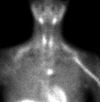
What pathology is seen in this image?

Phaeochromocytoma of the adrenal medulla
What is the management of Phaeo?
-
Alpha-blockers
- Phenoxybenzamine
- Doxazocin
- Beta-blockers
- Propranolol
- Laparoscopic adrenalectomy
Go over Post Adrenalectomy care
- Consider Genetic testing
- 30% are genetic ( 13 mutations so far)
- Annual Metanephrines (a metabolite of epinpherine)
- 24 hour urine
- Plasma
- Additional treatment if Malignant
- 10%
What should you do if someone has a fit/seizure for the first time?
- why?
- check the serum calcium levels
- hypocalcaemia can cause seizures
- decrease in extracellular Ca2+ conc. increases the neurons permeability for Na+
- allows sodium to easily depolarize the neuron’s membrane and cause an action potential
What are physical signs of hypoglycaemia?
( the same for hypoparathyroidism)
- Neuromuscular inability
- Chvostek’s sign
- when the facial nerve is tapped at the angle of the jaw, facial muscles on the same side of the face contract momentarily
- Trousseau’s sign of latent tetany
- carpal spasm when the brachial artery is occluded for 3 minutes
- less sensitive than Chvostek’s sign
- Chvostek’s sign
- Neurological sign and symptoms
- Personality disturbance
- Parkinsonism
- Irritability
- Mental status disturbed
- confusion/ disorientation
- psychosis/ psychoneurosis
- Ectodermal changes
- Dry skin
- coarse hair
- brittle nails
- Psoriasis
- alopecia
- Cardiac changes
- Ophthalmologic manifestations
- smooth muscle involvement
What are the acute and chronic consequences of hypercalcaemia
-
Acute
- Thirst & Polyuria
- Abdominal Pain
-
Chronic
- Constipation
- Musculoskeletal aches / weakness
- Neurobehavioral symptoms
- Renal calculi
- Osteoporosis
What state would serum calcium be found in?
- Protein Bound: 40%
- Albumin bound: 90%
- Globulin Bound: 10%
- Bound to Cations: 10%
- Phosphate & Citrate
- Ionised ( free ): 50%
- this should be measured directly not through the corrected total serum Ca++
- 1.1-1.35mmol/L
What is the serum Calcium level range?
2.15-2.55mmols/L
How are blood calcium levels controlled within their range?
-
Parathyroid chief cells in the parathyroid glands produce parathyroid hormones
- increased secretion of PTH –> increase in serum calcium
- Calcium-sensing receptor (CaSR) in the chief cells, sense an increase in serum Ca++ and stimulate the uptake of Ca++ by the parathyroid chief cells
How does the Calcium sensing receptor CaSR function?
- this is a type of G protein-coupled receptor that is activated by two major signal-transducing effects
-
Activation of phospholipase C
- leads to the generation of the second messenger diacylglycerol and inositol triphosphate
-
Inhibition of adenylate cyclase
- __suppresses intracellular conc. of cyclic AMP
-
Activation of phospholipase C
- the presence of this receptor in various areas in the body suggests that Calcium behaves like a hormone
How does Calcium effect PTH secretion

What are the receptors for Parathyroid Hormone
and PTHrP?
-
Type 1 parathyroid hormone receptor: Binds both parathyroid hormone and amino-terminal peptides of PTHrP
- binding activates adenylyl cyclase and phospholipase C systems
- mRNA coding for type 1 receptor is most abundant in bone and the kidneys,
- also expressed at lower levels in may other tissues
- Mutations lead to Jansen’s metaphyseal chondrosplasia ad BLomstrans chondroplasia
-
Type 2 parathyroid hormone receptor: Binds parathyroid hormone, but shows very low affinity for PTHrP
- expressed in a few tissues also coupled to adenylyl cyclase
- ligand binding induces a rise in intracellular conc. of cyclic AMP
What is the action of PTH in the bone?
- increases renal calcium reabsorption and posphate excretion
- PTH blocks reabsorption of phosphate in the proximal tubule while promoting calcium reabsorption in the ascending loop of Henle, distal tubule, and collecting tubule
- promotes the absorption of calcium from the bone
- In the rapid phase when PTH binds to receptors on these cells, the osteocytic membrane pumps calcium ions from the bone fluid into the extracellular fluid
- In the slow phase which happens over several days osteoclasts are activated to digest formed bone, and second, proliferation of osteoclasts occurs - stimulates cytokines released by by differentiation of immature osteoclast precursors that possess PTH and it. D receptors
What is Rank Ligand?
Receptor activator of nuclear factor kappa B ligand
- RANKL, produced by osteoblasts and other cells, causes osteoclast precursors to form and differentiate into active (mature) osteoclasts
- implicated in altering the adherence of osteoclasts to the bone surface and suppresses apoptosis of mature osteoclasts
- body naturally produces a protein called osteoprotegerin (OPG) that neutralizes the effects of RANKL, keeping the bone loss process in check
- RANKL has direct catabolic effects on cortical and trabecular bone including reductions in bone density, volume and strength
Renal Synthesis of Active Vitamin D

How is vitamin D production regulated in the kidney?
- PTH stimulates and
- FGF23 inhibits 1,25(OH)2D
- leads to renal phosphate excretion
- In turn 1,25(OH)2D inhibits PTH production and secretion from the parathyroid glands and stimulates FGF23 production from bone

What is Primary Hyperparathyroidism?
- what are its causes
- excess production/ secretion of Parathyroid hormone (PTH) –> increased calcium levels
Causes
- enlargement of one or more parathyroid gland
- most common in over 60’s
- women are more likely to be affected than men
- radiation to the head and neck increases risk
- rarely caused by parathyroid cancer
What are the symptoms of Primary Hyperparathyroidism?
- Fatigue
- Fractures
- Decreased height
- Upper abdominal pain
- Loss of appetite/ Nausea
- Muscular weakness/ Muscle pain
- Depression/ Personality changes
- Stupor and possibly coma
- Kidney stones/ Increased urination
Complications: Osteoporosis, bone cysts in severe cases
What investigations can be done to confirm Primary Hyperparathyroidism?
- A test called radioimmunoassay shows an increased level of PTH.
- Serum calcium is increased.
- Serum phosphorus may be decreased.
- Serum alkaline phosphatase may be increased.
- Bone x-ray may show bone reabsorption (the body breaks down the bone), or fractures.
- Imaging of the kidneys or ureters may show calcification or blockage.
- ECG may show abnormalities.
- This disease may cause changes in the results of the following tests:
- Calcium - urine
- Calcium (ionized)
- Bone density
- Markers of bone resorption (N-telopeptide, pyridinoline, and deoxypyridinoline)
What is the treatment for Primary Hyperparathyroidism?
- treatment depends on the severity of the disease
- If calcium levels are minimally raised, they may simply be monitored,
- unless renal dysfunction, renal calculi or osteoporosis are present.
- Treatment may include:
- Drinking more fluids to prevent the formation of kidney stones
- Avoiding immobilization
- Avoiding thiazide-type diuretics
- Using Bone protective treatment if osteoporosis is present
- Treating with a calcium-receptor sensitizer (cinacalcet) to decrease levels of PTH
- Surgery: if indicated by
- osteoporosis, renal calculi, <50yrs, serum Ca++ >2.8mmol/l
- dependent on patient
What type of scan is this?
- what does it show?

Sesta Mibi Parathyroid scan
Ectopic Parathyroid adenoma
What is this a scan of?
- type of scan?

Parathyroid adenoma
4D CT scan
What are the causes of Hypoparathyroidism?
- Iatrogenic
- thyroidectomy
- radical neck surgery
- Autoimmune
- Hypomagnesaemia
- Genetic mutations
What are the common causes of secondary Hyperparathyroidism?
- Low / low normal serum Calcium + HIGH PTH
- Low serum 25 OH vitamin D
- Lack of sun exposure
- Gastrointestinal problems
- Malabsorption
- Extensive surgery ( small bowel )
- Renal Failure
- Low serum 25 OH vitamin D
What is rickets?
- A softening of the bones in children potentially leading to fractures and deformity predominantly due to vitamin D deficiency
- lack of adequate calcium may also lead to rickets
- majority of cases occur in children suffering malnutrition- famine/ starvation
- Osteomalacia is used to describe a similar condition occurring in adults

What is the overall physiological effect of Parathyroid Hormone?
- Mobilisation of calcium from bone
- Enhancing absorption of calcium from the SI
- works indirectly by stimulating the production of the active form of vitamin D in the kidney.
- Vitamin D induces synthesis of a calcium-binding protein in intestinal epithelial cells that facilitates efficient absorption of calcium into the blood
- Suppression of calcium loss in urine
- loss of phosphate ions in urine in exchange for calcium
What can cause a disruption to calcium homeostasis?
- Disorders of the gut, kidney, skeleton
- Disorders of the parathyroid glands
- Abnormal vit D metabolism
- intake
- synthesis
- metabolism to 1,25-dihydroxycholecalciferol
Give an overview of Parathyroid Hormone
- it’s secreted as Intact-PTH (1-84 AA)
- this is metabolised in the periphery mainly the liver and kidney and stored in the gland and some goes into circulation
- the active form is PTH 1- 30 AA
- release stimulated in
- low plasma ionised calcium (acute stimuli)
- rise in plasma phosphate (chronic hypocalcemia)
What are the key actions of PTH?
- Increase in calcium via
- renal tubular reabsorption
- bone resorption (action of osteoclasts)
- GI tract absorption
- increased renal generation of active vit D
- acts as a Hypophosphataemic agent
- reduces proximal tubular reabsorption of phosphate by decreasing activity of type II sodium-phosphate co-transporter
How is intestinal absorption of calcium facilitated?
- biological reaction
- 1alpha-hydroxylation of 25 hydroxyvitamin D in the kidney forms calcitriol which
-
1,25 vitD (calcitriol) increases calcium and phosphate uptake,
- it also stimulates bone resorption, intestinal absorption and renal reabsorption
What is the calculation to give the corrected plasma calcium?
corrected calcium = calcium + 0.02(40-albumin)
- disturbances in albumin (the bound fraction of calcium) can cause misinterpreted values
- abnormally high albumin levels may indicate hypocalcaemia
- abnormally low albumin levels may indicate hypercalcaemia
What are the causes of Hypercalcaemia?
- Malignancy - 65%
- may be due to local bone resorption due to metastases, or the production of PTH-related protein which activates osteoclasts
- Hyperparathyroidism - most common in ambulatory patients
- Hypervitaminosis D
- Familial hypoclaciuric hypercalcaemia
- autosomal dominant - inactivation of calcium genes coding for calcium-sensing receptors on the parathyroid cells and the kidney
what does plasma PTH levels in hypercalcaemia indicated about the cause of the pathology?
- If there is suppressed PTH
- non-parathyroid cause: malignant, it D excess, sarcoidosis
- if there is Raised or detectable PTH
- PTH mediated cause: 1y or 3y hyperparathyroidism + calcium receptor defects
What are the different Hyperparathyoid states?
-
Primary
- adenoma, hyperplasia
- plasma calcium is high
-
Secondary
- a normal response to chronic hypocalcaemia: CRF malabsorption
- plasma calcium is low/normal
-
Tertiary
- developed from prolonged secondary state
- PTH secretion becomes autonomous
- plasma calcium is high
What does the plasma PTH levels in hypocalcemia indicate about the cause of the pathology?
- increased PTH
- non-parathyroid cause: vit D deficiency, renal failure
- low/normal PTH
- parathyroid cause: hyperparathyroidism, Mg deficiency
What are the markers of bone formation?
- increase osteoblast activity
- alkaline phosphatase
- bone alkaline phosphatase
- collagen peptides

What are the markers of bone resorption?
- Osteoclast activity
- collagen peptides
- pyridinolines (x-links)
- urine hydroxyproline
