ACE Fitness Essentials of Exercise Science Chapter 1 Flashcards
Anterior (ventral)
Toward the front
Posterior (dorsal)
Toward the back
Superior
Toward the head
Interior
Away from the head
Medial
Toward the midline of the body
Lateral
Away from the midline of the body
Proximal
Toward the attached end of the limb, origin of the structure, or midline of the body
Superficial
External; located close to or on the body surface
Deep
Internal; located further beneath the body surface than the superficial structures
Cervical
Regional term referring to the neck
Thoracic
Regional term referring to the portion of the body to the neck and abdomen; also known as the chest (thorax)
Lumbar
Regional term referring to the portion of the back between the abdomen and pelvis
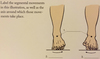
Plantar
The sole or bottom of the feet
Dorsal
The top surface of the feet and hands
Palmar
The anterior or ventral surface of the hands
sagittal plane
The longitudinal (imaginary) line that divides the body or any of its parts into right and left sections
Frontal plane
A longitudinal (imaginary) section that divides the body into anterior and posterior parts; lies at the right angle of the sagittal plane
Transverse plane
Also known as the horizontal plane; an imaginary line that divides the body or any of its parts into superior and inferior sections
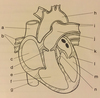
_______ and ________ carry oxygen-rich blood away from the heart
Arteries and arterioles
_______ and ________ returns oxygen-poor blood to the heart
Veins and venules
________ connects arteries and veins and provide sites for gas, nutrients, and waste exchange between the blood and tissues
Capillaries
The right two chambers (right atrium & right ventricle) push deoxygenated blood into the lungs where it releases carbon dioxide in exchange for oxygen is called _______
Pulmonary circuit
The Left two Cambers (left atrium & left ventricle) receives newly oxygenated blood from the lungs and pumps it into the various tissues of the body through the ________
Systemic circuit
Explain the route blood flow beginning when it exits the heart and ending when it re-enters the heart through the inferior and superior vena cava.
As blood leaves the heart, it is carried by the arteries. As arteries lead away from the heart, their branches to form a “tree” of smaller, microscopic vessels called arterioles. Eventually, the arterioles develop into “beds” of much smaller structures, the capillaries. Blood passes from the capillary beds to small venous vessels called venules. As venules lead back to the heart, they increase in size and become veins (eventually leading to the inferior and superior vena cava)
Explain the route blood flow beginning when it leaves the right ventricle of the heart and enters left atrium.
The right ventricle pumps the blood into lungs through the pulmonary arteries. In the lungs, the blood picks up a fresh supply of oxygen and gives off carbon dioxide. The oxygenated blood returns from the lungs to the left atrium through the pulmonary veins.
Explain the concept of “form follows function” as they relate to the skeletal system and describe the importance of exercise and maintaining bone density throughout life.
Wolff’s law indicate that changes in the bone structure coincide with the changes in bone function. That is, when a skeleton is subject to stressful forces, such as those that occur when exercise, it responds by laying down more bone tissue, thereby increasing its density. Conversely, when individuals experience prolonged period of bed rest due to illness or injury, their bone loses mineral and become less dense.
What axis? Imagine a pin that inserts through the joint from front to back (anteriorly and posteriorly). Because of the pin’s position, the only movement allowed around this axis is lateral movement (abduction or adduction) in the frontal plane.
Anteroposterior Axis (also known as the Sagittal Axis)
What axis? take our imaginary pin and insert it from a mid-point at the side of the body. The position of our pin allows only forward and backward movement (flexion and extension) in the sagittal plane around this axis.
Mediolateral Axis (also known as the Transverse Axis)
what axis? If we insert our pin through the joint from top to bottom, it will allow movement in transverse plane only (i.e., rotation)
Longitudinal Axis
what joint rotate in one axis, allowing movement in one plane? The elbow joint is a hinge joint because it only moves forward and backward (flexion and extension) in the sagittal plane
Uniaxial or uniplanar joints (also called hinge joints)
what joints rotate in two axes, allowing movement in two planes?
Biaxial or biplanar joints
what joints rotate in all three axis, allowing movement in all three planes.
Multiplanar or triaxial joints
What angular movement? The move in which the bone comprising a joint movement towards each other in the sagittal plane, decreasing the joint angle between them.
Flexion
What angular movement? Causes the angle between two adjointing bones to increase in the sagittal plane.
Extension
What angular movement? Occurs when a part of the body is moved away from the midline of the body in the frontal plane.
Abduction
What angular movement? The movement of the body part toward the midline of the body in the frontal plane.
Adduction
Move the top of the foot toward the shin in the sagittal planne (only at the angle joint)
Dorsiflexion
Moving the sole of the foot downward in the sagittal plane; “pointing the toes” (only at the angle)
Plantarflexion
Moving to the scapulae to a superior position in the frontal plane
Elevation
Moving to an inferior position in the frontal plane (only at the scapula)
Depression
ankle rolls out and your weight goes on the outside of your foot. Lifting the medial border of the foot in the frontal plane (only at the subtalar joint)
Inversion
ankle rolls in and your outside of your foot comes off the ground. Lifting the lateral border of the foot in the frontal plane (only at the subtalar joint)
Eversion
Internal (Inward) or external (outward) turning about the vertical axis of the bone in the transverse plane
Rotation
rotating palm down. Rotating the hand wrists medially from the elbow in the transverse plane
Pronation
rotating palm up. Rotating the hand and wrist laterally from the elbow in the transverse plane
Supination
Combination of eversion and abduction, rising the lateral edge of the foot in the transverse plane
Pronation
Combination of inversion and abduction, raising the medial edge of the foot in the transverse plane
Supination
From a 90° abducted shoulder or hip position, the humerus or femur, respectively, is flexed (adducted) in toward the midline of the body in the transverse plane
Horizontal flexion (adduction)
The return of the humerus or femur from horizontal flexion in the transverse plane (adduction)
Horizontal extension (abduction)
Motion describes a “cone”; combines flexion, extension, abduction, and adduction in sequence in the multiplanar plane
Circumduction
The movement unique to humans and primates in the multiplanar plane
Opposition
Define Autogenic inhibition and explain how fitness professionals can take advantage of each during a stretching routine.
Autogenic inhibition is an automatic reflex relaxation caused by stimulation of a Golgi tendon organ (GTO). This can be a advantageous during static stretching routine, in the after 7 to 10 seconds of low force stretch, a GTO response is activated, allowing further muscle stretching.
Define Reciprocal inhibition and explain how fitness professionals can take advantage of each during a stretching routine.
Reciprocal inhibition is a reflex in the inhibition of the motor neurons of antagonists when the agonists are contracted. This can be advantageous during a dynamic stretching routine, that low-grade muscle contractions of the antagonists muscle can reduce tonicity in the agonist muscle, allowing the muscle to be more easily be stretched.
what is the scapula?
shoulder blade
a. Superior vena cava
b. Pulmonary veins
c. Right atrium
d. Pulmonary valve (right atrioventricular value)
e. Tricuspid valve (Right)
f. Right ventricle
g. Inferior vena cava
h. Aorta
i. Right & left pulmonary arteries
j. Pulmonary trunk
k. Left atrium
l. Mitral (bicuspid) value (left atrioventricular value)
m. Left ventricle
n. Aortic value
- Sagittal Plane
- Frontal Plane
- Transverse Plane
- Mediolateral Axis (vertebal column)
- Extension
- Flexion
- Mediolateral Axis (shoulder joint)
- Flexion
- Extension
- Mediolateral Axis (elbow joint)
- Flexion
- Extension
- Mediolateral Axis (wrist)
- Extension
- Flexion
- Mediolateral Axis
- Flexion
- Extension
- Mediolateral Axis (Hip joint)
- Extension
- Flexion
- Mediolateral Axis (ankle)
- Dorsiflexion
- Planterflexion
- Anteroposterior Axis
- Eversion
- Inversion
- Anteroposterior Axis (shoulder joint)
- Abduction
- Adduction
- Lateral Flexion (right)
- Lateral Flexion (left)
- Anteroposterior Axis (vertebral column)
- Anteroposterior Axis (hip joint)
- Abduction
- Adduction
What is the liquid component of blood?
Plasma
How blood flows through the heart?
- Deoxygenated blood flows from vena cava
- right atrium
- right ventricle
- pulmonary arteries
- lungs
- Pulmonary veins
- left atrium
- left ventricle
- aorta
- to the rest of the body.
What is the major artery that delivers blood from the heart to the body?
Aorta
0What are the muscles that assist with breathing? (In order)
- Nasal
- Pharynx
- Larynx
- Trachea
- Primary Bronchi
- Secondary Bronchi
- Bronchioles
- Alveoli
- Lungs
The ________is the most important muscle of inspiration
diaphragm
After air enters the nose and mouth, it is warmed and passed through the ______.
pharynx (throat)
What is the order in which food travels through the gastrointestinal tract?
- Mouth
- Pharynx
- Esophagus
- Stomach
- Small intestine
- Large intestine
What are the Six basic processes of the digestive system?
- Ingestion of food into the stomach
- Movement of food along the digestive tract
- Mechanical preparation of food for digestion
- Chemical digestion of food
- Absorption of digested food into the circulatory and lymphatic systems
- Elimination of indigestible substances and waste via defecation
Common passageway for the respiratory and digestive system is the ________
Pharynx
Where does Saliva blends with food particles to enhance taste buds?
Mouth
Where is the absorption of water and salt leaves a semisolid waste that is passed through the anus?
Large intestine
Located behind the trachea, it eventually empties into the stomach is what?
Esophagus
Where in the digestive system store chime?
Stomach
The location where nutrients, vitamins, minerals, and water are digested and absorbed.
Small intestine
The Fibrous articulations/joints allows _______ movement
no
The Cartilaginous articulations/joints allows _______ movement
little to no
The Synovial articulations/joints allows what kind of movement?
to move freely
Name the muscle movement for pointing the toes toward the floor?
Plantarflexion
What nerve fiber carries impulses to the CNS?
Afferent (sensory)
What nerve fiber carries impulses from the CNS to the PNS?
Efferent (motor)
What nervous system? Your body is in a state of relaxation. It is digesting food, storing energy, and promoting growth.
Parasympathetic nervous system
What nervous system? This portion of the nervous system is under control when strength training.
Somatic nervous system
What nervous system? The receptors in your skin recognize that you are touching something cold.
Afferent (sensory) division
What nervous system is activated when there is a stressor or an emergency, such as severe pain, anger, or fear.
Sympathetic nervous system
What nervous system? Your glands function on their own, without any assistance from you.
Autonomic nervous system
In order for an impulse to move from one neuron to another, __________ must be released.
Neurotransmitter
________ conduct electrical impulses toward the cell body
Dendrites
True or falseNeurotransmitters are released to carry an impulse from one neuron to another.
True
True or false
There is only one axon in a neuron, but there can be hundreds of dendrites.
True
____________ is the kinesthetic awareness of your body.
Proprioception
What is the function of the Golgi tendon organ (GTO) ?
its located b/w the muscle belly and its tendon. Its function is to recognize when there is too much tension on the muscle. Either stretching too much or contracting too tightly. It causes an inhibition (ceased) of the contraction known as autogenic inhibition. This allows the muscles to relax and resulting in contraction of the opposite muscle group.
What is the function of muscle spindles?
Its located in the belly of the muscle and lies parallel to the fibers. When the muscles stretch, the muscle spindle stretches and causes a reflexive contraction known as the stretch reflex. This contraction causes the antagonist muscle (the opposite muscle to relax). This is known as the receptacle inhibition.
During __________ stretching, the activation of the GTOs causes the muscle spindles to relax and stretch a little further.
static
When the muscle contraction is ceased, it is known as
autogenic inhibition
Muscle Spindles sensory receptors lie ________ __ ___ ________.
parallel to the muscle fibers
Muscle spindles respond to muscle fibers being overstretched by causing a __________ ____________, known as the ________ _______.
muscular contraction , stretch reflex
When the muscle spindle’s reflex contraction occurs, it causes the antagonist muscle group to relax. This is known as __________ __________.
reciprocal inhibition.
During __________ stretching, the shortening of the agonist muscle allow the antagonist muscle to lengthen.
dynamic
What type of muscle is found in the walls of hollow organs. (Ex. Stomach) consider involuntary/not in conscious control.
Smooth
What type of muscle forms the walls of the heart, also involuntary?
Cardiac
What type of muscle is attached to the skeleton, considered voluntary b/c it’s under our conscious control?
Skeletal
What kind of connective tissue is limit in motion and resist stretch.
Collagen fibers
What connective tissue is attached bone to bone?
Ligaments
What connective tissue straightens out when fibers is stretched?
Crimp
What connective tissue transmits force from muscles to bones?
Tendons
What connective tissue surrounds the sarcomere?
Elastic fibers
What connective tissue encases muscles, bones, nerves, blood vessels, and organs?
Fascia
What muscle is involved in upward rotation and elevation of the scapula?
Upper Trapezius
What muscle is involved in upward rotation and adduction of scapula?
Middle Trapezius
What muscle is involved in depression of scapula?
Lower Trapezius
What muscle is involved in Elevation of the scapula?
Levator scapulae
What muscle is involved in Adduction, downward rotation, and elevation of the scapula?
Rhomboid major and minor
What muscle is involved in stabilization, depression, downward rotation, and abduction of the scapula?
Pectoralis minor
What muscle is involved in Stabilization, abduction, and upward rotation of the scapula?
Serratus anterior
Stabilization of the _______ muscles help prevent subluxation of the humeral head from the glenoid fossa.
rotator cuff
True or False. The muscles of the rotator cuff originate on the posterior portion of the scapula.
False b/c Subscapularis is located in the anterior portion. All other is posterior.
What is the agonist?
The muscle that cause the desired action or prime mover.
What is the antagonist?
It oppose the action of the agonist muscles
What are Synergist muscles?
It assist the agonist muscles
What are muscles work together to help stabalize our joints