7.15 Joints of the Hip Flashcards

1
Q

How many joints are involved in the hip?

A

One

A ball and socket

2
Q

Compare the hip ball and socket joint to the one of the shoulder (glenohumeral joint)

A

The socket is deeper than the glenoid fossa of the shoulder (which is designed for mobility). The hip has much more stability

3
Q

What are the three components of the hip bone?

A
  • Ileum
  • Ischium
  • Pubis
4
Q

What and where is the acetabulum?

A

It is the socket component of the hip bone

Each component of hip bone (ileum, ischium & pubis) helps form acetabulum.

5
Q

Is all the acetabulum an articular surface?

A

No

There are 2 parts to the acetabulm

  1. A lunate surface is the semicircular surface that is the weight bearing region and articular with the femur
  2. The acetabular notch & fossa are in the centre and are non- articular. This is roughened bone i with exposed trabeculae
6
Q

Describe the congruence of the three parts of the hip bone

A

The converge to each contribute (unequally) to the acetabulum.

They converge very well into a ‘Y’ shaped area of congruence and fuse (epiphysis) in late puberty

7
Q

What is the acetabular notch?

What fills this notch?

A

A depression in the margin of the acetabulum located anteroinferiorly.

It is bridged by a transverse ligament

8
Q

What is located deep in the socket and fills irregularities in the hip joint socket/acetabulum?

A

A fat pad

Intra-articular but extra-synovial fat that has both a nerve and blood supply (source of pain and bleeding) and is also important in spreading the synovial fluid.

9
Q

Where is the acetabulum ligament coming from?

What function does it serve?

A

From a depression in the head of the femur (called the fovea). The ligament that goes into the acetabulum.

  • It is an intraarticular structure in the joint
  • It doesn’t have much other function
10
Q

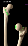
Label the major parts of the proximal femur

A
  • Head (with the fovea)
  • Anatomical neck
  • Greater and lesser trochanter
  • The intertrochanteric line
  • The shaft
11
Q

Describe the joint capsule of the hip, especially in terms of where it attaches to

A

The capsule of the hip bone attaches to the anatomical neck only in utero.

After birth…

  • Anteriorly, it migrates distally towards the intertrochanteric line
  • Posteriorly it stays near the anatomical neck (migrates slightly distally)
12
Q

What is contained in the Vascular foraminae of the head of the femur?

A

These are holes in the bone that enable the passage of blood vessels (around the neck of the femur)

13
Q

Describe what type of epiphysis the greater and lesser trochanters are

A

Both trochanters are traction epiphyses which fused and formed due to the pull of muscles/ligaments.

The greater trochanter fuses in mid-teens.

14
Q

What other kind of epiphysis is present in the head of the femur? Describe its location and formation

A

A pressure epiphyses - the weight bearing epiphyses also called the capital epiphyses

Dependent on an adequate blood supply getting to that part of the bone.

15
Q

Describe Perthe’s Disease

A

When a change (loss) in blood supply potentially affects development of the capital epiphysis of the head of the femur

It causes avascular necrosis of head of femur due to interuption of blood supply

The bone can take between two and five years to re-grow. Perthes’ disease is also known as Legg-Calve-Perthes disease or coxa plana.

16
Q

Describe the orientation of the large fossa of the head of the femur

A

It is 2/3 of a sphere

It is directed upwards, medially & forwards (the anterior most part lies outside the acetabulum)

17
Q

If the anterior most part of the head of the femur lies outside the acetabulum, how is it protected?

A

By the psoas bursa

It lies deep to the primary flexor of the hip (ileopsoas muscle before articulates to the lesser trochanter).

18
Q

The roof of the acetabulum is the region that has the thickest cartilage.

Why is this?

A

This is the primary area of articulation of the head of the femur - where most of the weight bearing is occuring

19
Q

What enables a greater range of motion of the hip joint?

A

There is a narrowing in the mid-region of the femoral neck (it is narrower than circumference of head)

This means that there is a range of motion that can be achieved without the bone making contact with the acetabulum and this increasing ROM

20
Q

How are trabecular patterns of cancellous bone formed?

A

There is a trabecular pattern of growth that follows the course of stress (weight) lines along the bone. Maximum trabeculae develop along the lines of maximum stess.

21
Q

Describe the boney architecture of the head of the femur

A

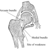
In the head of the femur there are two main intersecting trabecular systems:

  1. SUPERIOR: medial bundle & arcuate bundle – formed due to the compression through head & neck
  2. INFERIOR: medial bundle & lateral bundle – formed in response to muscle pulls on greater & lesser trochanters
22
Q

Where is the fracture risk in relation to the trabecular patterns?

A

Where the two main trabecular patterns intersect
23
Q

Describe the normal neck-shaft angle of the head of the femur

A

The angle between the neck of the femur and the head of the femur is about 125 degrees +/- 10 degrees

It is generally a smaller angle for females due to a wider pelvis

24
Q

Describe the development of the femoral head-neck angle

A

The angle is much greater in newborns (≥150°) and reduces by approx. 1° per year until the end of adolescence.

25
Q

Why does the femoral head-neck angle exist?

A

It enables the femur to swing free of pelvis in locomotion (in the anterior-posterior direction)

26
Q

Describe the pathology of the angle of inclination of the femur

A

Increase in the angle 􏰀 = ‘Coxa valga’

Decrease in the angle = ‘Coxa vara’

27
Q

What is the angle of torsion in the hip joint?

A

The relationship between the axis of the femoral head and neck and the femoral condyles (the distal end of the femur)

The normal femur has an angle of torsion between 12 and 15 degrees.

It is this angle that causes the anterior part of the head of femur to stick outside the acetabulum

28
Q

Describe the pathology of the angle of torsion for the femur

A
  • Increase in this angle is termed anteversion
  • Decrease in this angle is termed retroversion
29
Q

Stabilising the hip joint overrides any other joint/segment of the lower limb

Explain this statement

A

There is priority for joints of lower limb is to secure the hip joint.

The head of the femur needs to be brought back into the socket.

There may be resultant increased rotation of segments distal to the hip in order to achieve this

30
Q

Explain coxa vara in adolescence

A

Coxa Vara = decrease in the angle of inclination

Coxa vara increases stress on femoral neck.

Normally weight-baring forces pass along trabeculae and press on the capital epiphysis to increase stability. But if there is a decrease in the angle of inclination, forces through head of femur may result in the proximal epiphysis not pushing firmly against the head but slides off the remaining part of the head of the femur = ‘slipped capital femoral epiphysis’

31
Q

Where does the line of gravity pass through at the level of the hip?

What is the implication of this?

A

At the level of the hip it passes behind the centre of the hip joint

Thus there is a need to reinforce and stabilise the joint in front. The capsule of the hip joint is reinforced ANTERIORLY

32
Q

What is the position of greatest stability for the hip joint?

A

Called the ‘close packed’ position – is in extension, (abduction & medial rotation)

This is when all the articular surfaces are congruent, and the capsules and ligaments are all pulling tight around the joint

33
Q

Describe the loose-packed position

A

In flexion (adduction and lateral rotation)

It is the position that precipitates injury of the joint when forces are directed around it

34
Q

Why are ligamentous supports better for support of joints compared to muscles?

A

Muscles will fatigue

35
Q

What are the 3 ligaments that reinforce the hip joint capsule?

A
  1. strong iliofemoral ligament in front projecting far down to the intertrochanteric line (shaped like a V)
  2. weaker behind pubofemoral ligament
  3. weaker behind ischiofemoral ligament (this blends with the capsule not directly to femur).
36
Q

Why is the extension position the most stable for the hip joint?

A

Primarily due to the to iliofemoral ligament - which spirals & tightens with hip extension (acts like rotator cuff muscles of the shoulder)

37
Q

What is the zona obicularis?

A

An internal lining of fibres forming a ring around the capsule

38
Q

What is the labrum of the hip joint?

What are the major functions

A

A (triangular) ring of cartilage that surrounds the acetabulum of the hip.

It has multiple functions:

  • Bridges acetabular notch
  • Deepens cup & increases surface area
  • Creates a suction effect on head of femur (negative pressure)

It is often torn in hip injury

39
Q

What direction is the most common for a hip dislocation?

A

Posterior

40
Q

Describe the role of the ligament of head of femur in development

A

It directs branch of obturator artery to femoral head allowing a rich blood supply to it preventing ischemic injury (this artery regresses after puberty)

41
Q

What are the retinacular fibres of the femur?

A

Reflections of fibrous tissue from capsule to femoral neck contain ‘retinacular’ vessels. The fibres are reflected back along the neck of femur, blended with the periosteum, to the articular margin of the head

Critical blood supply to head of femur.

Across the neck of the femur they are suscptible to injury esp. when the neck is damaged (fracture)

42
Q

List the major movements of the hip joint

A
  • PRIMARY MOVEMENTS: Flexion/extension (F > E)
  • Abduction/adduction
  • Medial/lateral rotation
    • Medial rotation occurs with extension (‘screw- home’) – joint ‘locked’. Reverse in flexion
  • Medial rotation in stance, lateral rotation in swing
43
Q

What is the primary hip flexor muscle?

A

The iiopsoas muscles (inner hip muscles):

  • Psoas major
  • Iliacus muscle

They attach to the lesser trochanter of the femur

44
Q

What the extensor muscles of the hip?

A

Posteriorly:

  • The most powerful extensor is gluteus maximus
  • The hamstring muscles also contribute to extension.
45
Q

Why is the hamstring not a good extensor of the hip alone?

A

The hamstring muscles are 2 joint muscles (articulate with 2 different joints) and can be impacted by the knee (made too short by the knee)

46
Q

What is the arrangement of muscles around the neck of the femur? What do they achieve?

A

The neck is covered by a series of short muscles: act like as rotator cuff muscles (primarily lateral) they run in line with the neck of the femur - push the head of femur into socket

47
Q

What are the main actions of the gluteus muscles?

A
  • They abduct the hip but primarily maintain position of pelvis in coronal plane (i.e. act from below)
  • Glueus medius capable of both flexion & extension
48
Q

Describe the importance of the gluteus maximus and gluteus medius on posture and locomotion

A

While walking, the gluteal muscles keep the hips aligned by contracting both sides.

When walking one leg takes all the weight of the body and in order to keep the hips aligned and prevent that weight bearing hip from tilting down, the gluteus muscles contract to pull the contralateral hip down.

They prevent the hip falling away to the unsupported leg during locomotion by contracting on the weight-bearing lip to pull the hip down.

49
Q

What is ‘Trendelenberg gait’

A

If a person walks, during the stance face of the gait, the pelvis tilts, rising on the side not taking weight.

Trandellenberg gait is mostly due to a weakness of the abductor muscles of the hip. Thus in the pathologic situation the pelvis drops instead of rising on the unsupported side.

https://www.youtube.com/watch?v=ZUPQp5oxXj8

50
Q

What are the adductor muscles of the hip?

A

Made up of a big group of adductor muscles

Produce hip adduction – most powerfully from abducted position

Common source of groin pain & pathology – common at bony attachment (osteitis pubis)

51
Q

What is Hilton’s Law?

A

A joint receives pain & proprioceptive fibres from branches to muscles that move the joint – also pass to overlying skin

52
Q

What is the hip joint innervated by?

Describe referred pain

A

Hip joint innervated by articular branches of femoral, obturator & gluteal nerves

  • Referred pain from hip (via obturator nerve)
    • Anterior obturator nerve may become irritated due to arthritic changes in the joint
  • Referred pain to hip (from lumbar spine & SIJ)
    • Possible pain can be referred from lumbar spine to hip joint
53
Q

Describe the blood supply to the hip joint

A

Via anastomoses between branches of medial (primarily) & lateral femoral circumflex arteries (normally off profunda femoris)

54
Q

What is the main artery that supplies the hip joint?

A

The main supply to the thigh and hip comes from the profunda femorus artery to femur muscles

aka. deep femoral artery is a branch of the common femoral artery and is responsible for providing

Profunda gives off 2 branches: circumflex femoral arteries (medial and lateral). The medial is more important

55
Q

Any fracture within capsule and along the neck causes damage to retinacular vessels

What other sequelae can occur?

A

Muscle spasm particularly of ileopsoas muscle group causing a sterotypic posture: hip joint in external location and shortening of spasming muscles