7.12 Regions of the Neck Flashcards
The neck is composed of a series of tubes passing between head above and thorax below. These tubes have spaces in between them. What is the clinical significance of this?

These spaces between the compartments of the neck t are in direct communication with the mediastinum. Thus any collection of infection in the neck will tract down causing mediastinitus
What are the anterior borders of the neck?
From the lower border of mandible down to the manubrium
What are the posterior borders/extreminities of the neck?
From the superior nucal line down to the C7-T1 disc
The superficial fascia of the neck is unique to the body. Why is this?
It contains a thin sheet of subcutaneous muscle called platysma. It comes up and blends with the muscle of the face (and thus shares facial muscle nerve supply)
This muscle is thus considered a muscle of facial expression

There is vasculature running through the superficial fascia making up the superficial vaculature of the neck.
Describe the superficial veins [2]
- External jugular vein descending laterally down the neck from the angle of the mandible veritcally on SCM
- The anterior jugular vein descends vertically on either side of the midline

What are the layers/compartments of the neck? Label a cross sectional diagram
- Superficial fascia with platysma
- Investing fascia
- Visceral (pre-tracheal) fascia
- Carotid sheath/vascular fascia
- Pre-vertebral fascia

What structures are in each of the layers of fascia?
Superficial:
Platysma
Investing layer:
Just under skin and sup. fascia which completely surrounds the neck: splits and encloses trapezius posteriorly and SCM anteriorly.
Visceral/pre-tracheal layer:
Thyroid, parathyroid, thymus (preadolescence), trachea and oesophagus and recurrent laryngeal nerve (note: only the thyroid/parathyroid are enclosed by the fascia)
Carotid:
Contains the common carotid arteries, the internal jugular veins and the vagus nerve
Pre-vertebral:
Surrounds the cervical vertebra and postural muscles

Draw the layers of fascia onto a saggital section of the neck

The visceral compartment is surrounded by the pre-tracheal fascia. Describe the vertical borders of this fascia
It extends right up to the hyoid bone and runs down encompassing the visceral structures within it (thyroid and parathyroid glands mainly) to blend with the fascia of the arch of the aorta
What is the significance of the pre-vertebral fascia attaching to the hyoid bone?
Anything that pretracheal envelops will move up and down with swallowing
this is an important clinical sign when diagnosing lumps in the neck
Describe the buccopharyngeal fascia
The pre-tracheal fascia envelops the thyroid gland only. Surrounding the posterior part of the visceral part of the neck there is an extra layer of fascia.
Together they enclose the visceral compartment such that it contains the oesophagus, trachea and recurrent laryngeal nerve.
The BP fascia deliniates pretracheal with prevertebral

Name and Describe the regions of the neck
Anterior triangle
- In front sternocleinomastoid muscle, below the inferior border of the mandible and behind of the midline of the neck
Posterior triangle
- Behind SCM, in front of trapezius muscle and and above middle third of clavicle

How are the muscles of the anterior triangle of the neck groups?
According to their relationship to the hyoid bone:
- Above the hyoid bone: Suprahyoid muscles
- Below the hyoid bone: Infrahyoid muscles

Between what fascial layers do the muscles of the anterior triangle of the neck sit in?
Between the investing fascia & pretracheal fascia
Describe the connections of the suprahyoid muscles
What is their function?
- Connecting the hyoid bone to the scalp (floor of the mouth)
- Funtion to elevate the hyoid and the larynx (swallowing and phonation)
What is a common anatomical term to describe the infrahyoid muscles of the anterior triangle of the neck? Why is this so?
Strap muscles
They all are characteristically long and thin
What are the connections of the infrahyoid muscles?
What are their functions?
Connect hyoid bone (anchoring it) to the sternum, clavicle and scapula (essentially to the pectoral girdle).
They depress the hyoid and the larynx
Describe the path of the common carotid artery and its bifurcation site
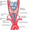
Common carotid-passes in carotid sheath, on the lateral sides of the neck. It ascends to level of C3/4 (upper border thyroid cartilage) where it bifurcates into the internal carotid artery and external carotid artery

What is an important feature of the internal carotid artery in terms of its course through the neck?
It has no branches in neck
It contains the carotid sinus (baroreceptors) & carotid body (chemoreceptors)


