7.1 Organisation of the Motor System and Spinal Reflexes Flashcards

1
Q

What components of NS control:

a) reflexes
b) posture
c) voluntary movement

A

a) LMN and spinal cord circuits
b) Brain stem and spinal cord
c) Cortex, brain stem & spinal cord

2
Q

Compare ‘open’ and closed’ loops that control voluntary and involuntary muscle control

A

1) Closed loops – reflex control
* Axial muscles (muscles of trunk and head) balance, posture, locomotion
2) Open loops – Sensory cue or desire to move
* Distal muscles, fine motor skills

3
Q

Where is the brain centre for movement control located?

A

Movement control sits just anterior to the central sulcus located in the pre-central gyrus of the frontal lobe

4
Q

State the function of each:

  1. premotor cortex
  2. primary motor cortex
  3. primary somatosensory cortex
A

premotor cortex: motor planning and sequencing for contralateral body side

primary motor cortex: controls contralateral body motor functions

primary somatosensory cortex: recieves contralateral sensory input from the body (incl taste)

5
Q

State the function of each:

  1. posterior parietal cortex
  2. primary visual cortex
  3. primary auditory cortex
A

posterior parietal cortex: integration of sensory input (sterogenosis)

primary visual cortex: recieves contralateral visual field info from both eyes

primary auditory cortex: recieves bilateral auditory sensory input

6
Q

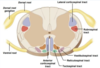
Label the following image

A
7
Q

Which regions of the spinal cord are important in control of movement?

A

Anterior and lateral funiculus

8
Q

Label the image of an axon below

A
9
Q

Which tracts are involved in conscious vs unconscious elements of the NS?

A

Conscious: Pyramidal/ Corticospinal tracts

Unconscious: Extrapyramidal tracts (4)

10
Q

What are the 4 Extrapyramidal tracts?

Label these on the image below

A

1) Rubrospinal
2) Tectospinal
3) Vestibulospinal
4) Reticulospinal

11
Q

What are the 2 components of the motor cortex?

A

1) Cerebellum
2) Basal ganglia

12
Q

Brifely explain how signals decend via the Pyramidal tracts

A

CC ➞ basal ganglion ➞ cortiocospinal tracts (pyramidal) ➞ synapses in spinal cord with peripheral motor neuron ➞ connects to muscle via NMJ

13
Q

Compare Pyramidal to Extrapyramidal tracts

A

Pyramidal tracts: originate in the cerebral cortex, carrying motor fibres to the spinal cord and brain stem. They are responsible for the voluntary control of the muscles of the body and face.

Extrapyramidal tracts: originate in the brain stem, carrying motor fibres to the spinal cord. They are responsible for the involuntary and automatic control of all muscles, such as muscle tone, balance, posture and locomotion

14
Q

What cell type comprises the Primary motor cortex?

What pathway do these travel down and where do they synaps?

A

Comprised of Betz cells which are are upper motor neurones with long axons

These travel via corticospinal tract to synapse with interneuron or alpha motor neurones.

15
Q

What tract is responsible for conscious control of skeletal muscles?

How can this be split into 3 tracts and what does each specifically control?

A

Corticospinal tract, can be split into:

1) Corticobulbar: conscious control over eye and face muscles
2) Lateral corticospinal: conscious control over limb skeletal muscles
3) Anterior corticospinal: conscious control over axial skeletal muscles

16
Q

Describe the pathway of the corticospinal tract

A

1) Originates in the primary motor cortex
2) Descends via the pyramids of the medulla
3) 75-90% decussate: mainly distal musculature
4) 10-25% don’t decussate: axial
5) Descend in anterior and lateral corticospinal tracts of the spinal cord
6) Synapse onto the ventral horn

17
Q

Which tracts are ascending? (5)

Label these on the image below

A

Dorsal column (fasiculus cuneatus and gracilis)

Dorsal spinocerebellar

Ventral spinocerebellar

Lateral spinothalamic

Ventral spinothalamic

18
Q

Which tracts are decending? (8)

Label these on the image below

A

Ventral white commissure

Lateral reticulospinal

Lateral corticospinal

Rubrospinal

Medial reticulospinal

Ventral corticospinal

Vestibulospinal

Tectospinal

19
Q

What is the role of the Extrapyramidal motor pathways?

Label the 4 impt on the image below

A

Control involuntary actions, reflexes, locomotion, complex movements and posture

20
Q

What is the specific function of the Rubrospinal tract?

A

Involuntary movements, specifically large muscle flexor movement and inhibiting extensor tone of upper limbs.

21
Q

Describe the pathway of the Rubrospinal tract

A

1) Originates in the red nuclei of the brain stem
2) Descends in the lateral part of brainstem
3) Then in the lateral funiculus adjacent to the lateral corticospinal tract

22
Q

What can be said about a lesion in the Rubrospinal tract?

A

Lesions produce minimal effects: demonstrating overlap

23
Q

What is the specific function of the Reticulospinal tract?

A

Responsible for body posture and muscle tone

24
Q

What 2 tracts comprise the Reticulospinal tract and Describe the origin and function of each

A

1) Pontine reticulospinal tract

  • Medial pathway responsible for exciting extensor muscles
  • Originates in the pontine reticular nucleus

2) Medullary reticulospinal tract

  • Lateral pathway responsible for inhibiting excitatory axial extensor muscles and automatic breathing
  • Originates in the medullary reticular nuclei
25
Q

What is the specific function of the Vestibulospinal tract?

A

Involuntary control of head balance and body posture

26
Q

Describe the pathway of the Vestibulospinal tract

A

1) Vestibular nuclei receives information from vestibulocochlear nerve about changes in the orientation of head.
2) Descends via vestibulospinal tract
3) Alters muscle tone, extension, maintaining posture

27
Q

What is the specific function of the Tectospinal tract?

A

Orientation and positioning of the head for balance in response to stimuli.

Eye-head coordination

28
Q

What is the origin of the Tectospinal tract?

A

Origin tectum of midbrain (tectum is the roof of the 4th ventricle comprising of superior and inferior colliculi)

29
Q

What is a LMN?

A
  1. Neuron w its cell body @ either Ventral horn OR Cranial Nerve nuclei of brain stem
  2. Directly innervates a muscle with its axon
30
Q

What is a Motor unit and how do these function?

What can be said about ‘number of fibres vs control of fine movement’?

A

Motor unit: the number of muscle fibres innervated by a single axon

Groups of motor units work together to coordinate the contractions of a single muscle

Smaller number = finer control

31
Q

What are the 2 types of LMNs and what does each innervate?

A

1) α-MN: innervates the muscle itself
2) γ-MN: innervatew the muscle spindle and keeps it taught

32
Q

What is a motor unit in terms of α-motor neuron?

How many are found in the eye vs the quads?

A

The α-motor neuron and the muscle fibres it innervates

  • Extra-ocularmuscles = 10
  • Quadriceps = 1000
33
Q

What are Muscle spindles?

A

Encapsulated sense organ found within the muscle that detects muscle contraction and is responsible for tendon reflex

34
Q

What is always activated alongside muscle spindles and why?

A

Coactivated with α-motor neurons so tighten as muscle contracts

35
Q

What is the function of Muscle spindles? (4 things)

A
  1. Fine motor control
  2. Proprioception
  3. Detects muscle tension (contraction / relaxation)
  4. Activates LMN in response to muscle stretch
36
Q

Describe the 3 main components of a muscle spindle

A

** Note there are extra-fusal fibres around the outside which contain the actin and myosin and contract

37
Q

Describe how Muscle spindles respond to strech

A

When we want to contract our muscles strech of the extrafusal fibres is sensed by afferent sensory neurones (1a axons) wrapped around intrafusal muscle

These send a signal to the spinal cord which stimulates the alpha motor neuron (efferent neuron) to respond causing contraction of the muscle

However, when we contract our muscles the intrafusal fibres become wobbly, and thus gamma motor neurons are activated

These gamma motor neurons are located ONLY at the ends (where actin and myosin IS present) and therefore, ensure the spindles remain taut even though the extra-fusal fibres have changed shape

38
Q

Where does the alpha motor neuron synaps?

A

Onto the muscle itself via the NMJ

39
Q

Compare the activation of a single motor neuron to multiple motor neurons at the NMJ

A

The activation of a single motor neuron leads to weak but distributed contraction

The activation of more motor neurons will activate muscle fibres, increasing the force of contraction

40
Q

What ion stimulates contraction of skeletal muscle?

A

Raised intracellular Ca2+

41
Q

How does muscle contraction occur? (10 steps)

A
  1. Motor nerve APs arrive + release ACh into Synaptic Cleft
  2. Activation of Nicotinic ACh receptors on Sarcolemma (muscle fibre membrane)
  3. Ligand gated Na channels open
  4. AP fired thru T tubule (invagination in Sarcolemma)
  5. T tubule depolarised
  6. Activation of Ryonidine Receptor on Sarcoplasmic Reticulum (SR)
  7. Stored Ca released into Cytoplasm
  8. Ca binds to Troponin subunit
  9. Myosin binding sites revealed
  10. Ca returned to SR via ATPase pumps
42
Q

Where are Golgi tendon organs located?

A

Found at the myotendinous junction between the muscle and tendon

43
Q

What do Golgi Tendon Organs sense?

What do they do when activated? (2 things)

What is the purpose of this?

A

Senses degree of muscle contraction by sensing Collagen changes when Tendon stretches @ muscle contraction

@ Golgi Tendon Organ activation:

  1. Inhibits muscle contraction
  2. Activates its antagonist

This prevents over contraction of muscles

44
Q

What is muscle tone?

A

Background continuous muscle contraction found in all skeletal muscles of the body

Allows body posture, rapid responses to environment

45
Q

What is meant by the ‘resting motor tone’ and what is it important in?

A

Resting tension in a skeletal muscle which occurs because when we are awake LMN continually activate muscle

This minimal contraction gives the muscle a small amount of force which allows body posture and rapid responses to environment

46
Q

During ‘resting motor tone’ how is minimal contraction by LMN sustained?

A

LMN are under inhibition from descending extrapyramidal pathways

47
Q

How are our muscles primed to react?

A

Because of our ‘resting muscle tone’

48
Q

In what 2 instances is our ‘resting muscle tone’ poor

A

Poor at birth and in REM sleep