7 - Knee Joint Flashcards
Where is the anterior inferior iliac spine?


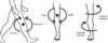
Label this saggital view of the knee joint.


Label this diagram of the femur.

- Slight medial angulation so knee closer to body’s centre of gravity

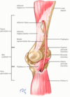
Label this diagram of the distal femur and state which leg it comes from?

- Right side
- Trochlear groove articulates with patella
- Medial condyle larger as bears more weight

What is the difference in prominences between the femoral condyles and why?
- Lateral condyle more prominent to prevent patella displacement laterally during tracking
- Those with flatter femoral condyle more prone to dislocation and instability of patella

What arises from the epicondyles of the femur?
Collateral ligaments

What arises from the intercondylar groove (femur), and where do they insert?
- Cruciate ligaments
- ACL: Attaches to medial aspect of lateral condyle
- PCL: Attaches to lateral aspect of medial condyle
PAM’s APPLES

What is the anatomy of the patella?
- Held in trochlear groove by quadriceps tendon superiorly, and patella ligament inferiorly to the tibial tuberosity

What are the three main functions of the patella?
- Stabilise and reduce friction on femoral condyles
- Protect anterior knee from physical trauma
- Extension of leg by attaching quadriceps to the tibia so acts as a fulcrum. Increase mechanical efficiency of the muscle

Label this diagram of the proximal tibia.


What is the intercondylar eminence the main attachement for and what do the intercondylar tubercles articulate with?
- Attachement of cruciate ligaments and the menisci of the tibia
- Articulate with the intercondylar fossa

What are the borders of the tibia shaft?
Prism-shaped
- Anterior: proximal aspect marked by tibial tuberosity and is palpable all the way down
- Posterior: marked by ridge of bone known as soleal line which extends inferomediall
- Lateral (Interosseous): Interosseous membrane arises here

How do the inferior portions of the leg bones articulate with the tarsals?
- Malleolus

What are the three articulations of the fibula and what is the anatomy of the fibula?
- Proximal tibiofibular joint (lateral condyle of tibia)
- Distal tibiofibular joint (fibular notch)
- Ankle joint (talus bone of foot)
Has three surfaces and lateral malleolus can be palpated at ankle joint

What is the issue with proximal fibula fractures?
Common peroneal nerve winds around posterior and lateral surfaces of the fibula neck so vulnurable to damage

What type of joint is the knee joint and what two articulations make the joint?
- Hinge synovial joint
- Tibiofemoral and patellofemoral
- All within the same joint capsule
What is the blood and nerve supply to the knee joint?
Blood: Genicular anastomoses supplies by genicular branches of femoral and popliteal arteries
Nerve: Femoral, Tibial and Common Peroneal

What is the stability of the knee joint like and how is stability improved?
- Unstable due to high motion allowed
- Deepened articular surface by menisci
- Ligaments
- Joint capsule

What is the function of the menisci of the knee?
- Deepen articular surface, increasing joint stability
- Shock absorber by increasing surface area to dissipate forces over
- Avascular by adulthood so difficult to repair

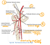
How are the menisci of the knee attached in terms of ligaments and label this diagram of them.

- Peripheral rims attached to tibia and joint capsule by coronary ligaments
- Menisci attached to each other by transverse ligament
- Posterior meniscofemoral ligament stabilises posterior horn of lateral meniscus by attaching to medial femoral condyle
- Menisci attach at both ends at the intercondylar area. Thicker at periphery than centrally

What are the ligaments involved in the knee joint?
Oblique popliteal ligament comes from semimembranosis and strengthens the back of the knee

What do the cruciate ligaments prevent?
PCL: prevents posterior tibia dislocation on the femur. main stabiliser when standing
ACL: prevents anterior translation and medial rotation of the tibia in relation the femur

What is the joint capsule of the knee joint like?
- Surrounds the sides and posteriorly but deficient anteriorly to allow supra-patellar bursa
- Strengthened at sides by vastus lateralis and medialis muscles
- Strengthened by oblique popliteal ligament posteriorly (continuation of semimembranosus tendon)

What do the collateral ligaments resist?
Medial: resists valgus angulation of tibia. Weaker and more likely to tear, taking meniscus with it. (wide flat ligament)
Lateral: resist varus angulation of tibia (thin round ligament). Reenforced by iliotibial tract