4/11 Devptal Disorder of Bone/Cartilage - Corbett Flashcards
functions of bone
- mechanical support
- transmission of force
- protection of viscera
- mineral homeostasis
- acid-base balance
- niche for production of blood cells
two processes of bone formation
1. intramembranous ossification
- bone tissue laud down directly in embryonic connective tissue or mesenchyme
- flat bones: skull, midface, jaw, clavicle
2. endochondral ossification
- bone tissue REPLACES preexisting hyaline cartilage (template for future bone)
- long bone
membranous bone devpt
bones of skull, midface, jaw, clavicle

woven bone vs lamellar bone
new bone = woven bone
remodeled into lamellar bone

long bone devpt
bones of limbs and girdle
endochondral ossification
type of collagen thats made changes (type 2 - type 10 - type 1)

zones of endochondral ossification
- proliferative zone
- hypertrophic zone
- vascular invasion zone

regulators of bone devpt
chondrocyte proliferation regulators?
- growth hormone
- thyroid hormone
- Indian hedgehog
- PTHrP
- Wnt
- Sox9
- Runx2
- FGF-receptor3 → inhibits chondrocyte prolif
chondrocyte proliferation regulators

FGFr3
- regulates ability of chondrocytes to prolif/mature
-
negative reg of bone growth via inhibition of chondrocyte prolif
- gain of fx mutation: activation of FGFr3 → short stature
- achondroplasia
- thanatrophic dysplasia

normal bone anatomy
diaphysis: in shaft
- central trabecular portion surrounded by thick cortical layer
metaphysis
- loose trabecular bone surrounded by thin cortical layer
- adjacent to growth plate
epiphysis
- end of bone, articular surface, contains subchondral regions under articular cartilage


cortical bone vs trabecular bone
cortical: outer periosteal surface + inner endosteal suface
- periosteum contains vessels/nerves/osteoblasts/osteoclasts, aids in bone formation, appositional bone growth and fracture repair
trabecular: honey comb network of rods and plates

cortical vs trabecular
summary

bone matrix
1. osteoid
- type I collagen
- GAG
- osteopontin (unique to bone)
- serum measurements indicate osteoblast activity
2. mineral component (hydroxyapatite)
- gives hardness
- repositor for 99% of body Ca, 85% of P
osteoblasts
- at surfaces of bone matrix, compose most of the flattened bone lining cells in endosteum and periosteum
-
main fx: SYNTHESIZE BONE MATRIX
- cuboidal when synthesizing matrix
functions
- formation of new bone
- regulation of osteoclastogenesis (RANKL and OPG)
- comm with osteocytes to receive mechanotrasduction signals (anabolid)
regulated via
- Runx2 (platform for hormone/cytokine action)
- osterix (interaction with NFAT2)
- Wnt-betacatenin

osteocytes
most abundant (90-95%) adult bone cells
derived from osteoblasts
fx: dendritic processes connect periosteal and endosteal surfaces
- sense stress
- load? → bone matrix synthesis
- reduced load? → bone resorption

osteoclasts
- 1-2%
- derived from circulating monocytes
- need two signals for maturation: RANKL (formation) & M-CSF (growth/survival/diff)
- large, motile, multinucleated
fx: resorbing bone
clast contact w matrix → formation of “sealing zone” for isolation of acidification of bone matrix by cathepsin K

dysostosis
localized problem in migration and/or condensation of mesenchyme
- absence of bone/digit (aplasia)
- extra bone/digit
- abnl bone fusion (syndactyly or craniosynostosis)
dysplasia
abnormal growth; global disorganization of bone and/or cartilage
- mutation in genes controlling devpt or remodeling of entire skeleton
role of tf in bone/cartilage devpt
Sox9 mutation
fx: tf regulating commitment to chondrogenic lineage (Col2) in resting zone of growth plate
mutation → camptomelic dysplasia
- auto dom
- short limbed dwarfism
- neonatal death
- bowing of long bones, small thoracic cage, failure of scapulae to form; defects in all endochondral bones
- ambiguous genitalia
role of tf in bone/cartilage devpt
Runx2 mutation
fx: tf regulating commitment to osteoblast lineage
- required for maturation of hypertrophic chondrocytes
loss of Runx2 → ossification defects - cartilage skeleton but NO BONE
mutation of Runx2 → cleidocranial dyslplasia
- auto dom
- char ft: clavicular hypoplasia
- open sutures in skull
- dental abnormalities (hyperdontia)
role of tf in bone/cartilage devpt
GDF5
growth/differentiation factor 5
fx: promotes chondrogenesis
- increases size of initial chondrocyte condensations
- incr chondrocyte prolif
diminished GDF5 or heter/homozygous GDF5 →
- shortening of appendicular skeleton
- incr severity prox to distal
- loss of some joints
achondroplasia
most common short-limb dwarfism
paternal allele is site → related to paternal age
activating mutation in FGF3R → inhibits chondrocyte proliferation
decreased ENDOCHONDRAL (not membranous) OSSIFICATION
- decr chondrocyte prolif and matrix affecting long bones
- relatively normal head size
clinical features
- shortened prox extremities
- relatively normal trunk
- enlarged head/bulging forehead (frontal bossing, flattened nasal bridge)
- spinal deformities (spinal canal stenosis, intervertebral foramen stenosis → over 50% lower ext radiculopathy

thanatorphic dysplasia
most common lethal skeletal dysplasia
mutation in FGF3R
- auto dom
- de novo activating mutations with 100% penetrance
“like an excessive form of achondroplasia”
clinical features
- severe shortening of limbs (curved long bones are worst affected)
- nl trunk length
- macrocephaly
- narrow bell-shaped thorax, shortened limbs

osteogenesis imperfecta
pathologic changes occuring in tussues in which type 1 collagen is an important constituent
- problems seen in bone, ligament, dentin, sclera
- why? type 1 collagen makes up 90% of bone
- caused by QUALITATIVE or QUANTITATIVE reduction in type 1 collagen
80% cases due to mutation in one of two genes for type 1 collagen
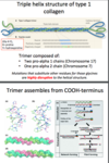
type 1 collagen
structure
triple helix
- two pro alpha1 (chr17)
- one pro alpha2 (chr7)
mutations that sub a.a.s in for Gly are v disruptive to structure
qualitative and quantitative defects of OI
mutations
defect comparison
qualitative (COL1A1, COL1A2)
- COL1A1 mutations are more severe than COL1A2
- mutations at COOH terminus are more severe
- size/polarity determine severity of mut
quantitative (from nonsense mutations, mostly COL1A)
- haploinsufficiency effect (reduced levels of normal collagen)
defect comparison?

osteogenesis imperfecta type 2
pernatal lethal (most severe form of OI)
- due to QUALITATIVE DEFECT
- auto rec/dom
skeletal deformity with numerous fractures
osteogenesis imperfecta type 1
mildest, most common form of OI
male = female
auto dom
- blue sclera (sclera is thin, so you can see the choroid! blue)
- varying bone fragility/deformity
- 20% kyphosis, scoliosis
- later fractures
- hearing loss
- normal stature
osteopetrosis
incr bone mass and mineralization
- failure of osteoclasts to resorb bone → skeletal fragility
etiologies:
- decreased numbers of osteoclasts
* genetic defects in: RANKL, RANK, osteopregenerin - defective acidification
- carbonic anhydrase 2
- CLCN7 (proton pump)
most severe osteopetrosis
severe AR infantile osteopetrosis
increased OC numbers with secondary genetic defects:
- carbonic anhydrase II → renal tubule acidosis, cerebral calcification
- CLCN7 (osteoclast Cl ion channel)

