30-09-21 - Blood Vessels and Lymphatics Flashcards
What is a brief description of systemic circulation?
- Elastic arteries
- Muscular arteries
- Arterioles
- Capillaries
- Venules
- Medium veins
- Large veins
What are the 3 layers of blood vessels starting from inside to out?
- Tunica intima
- Tunica media
- Tunica externa

What does the tunica intima create?
What is it lined with?
What is the purpose of this lining?
What does this lining sit on top of?
What is present in the tunica intima of larger arteries?
- The tunica intima creates the lumen of blood vessel (the inside of a tubular structure)
- It is lined by simple squamous epithelial cells, which forms what is known as the endothelium
- These endothelial cells release substances that control vascular constriction and relaxation, enzymes that control blood clotting, immune function, and platelet adhesion.
- These endothelial cells sit on top of a basement membrane which separates them from underlying tissue
- In larger arteries, an internal elastic membrane can also be present at this boundary of the intima and media, which provides additional support to blood vessels that stretch more.

What are large differences between the structure of veins and arteries?
Why are they different?
- The lumen of veins is significantly larger than arteries
- This is because veins carry a greater volume of blood than arteries
- Arteries (particularly muscular arteries) contain significantly more smooth muscle and veins contain more collagen and elastic fibres.
- This is due to arteries (especially muscular arteries) handling blood at higher pressure than veins

What is the Tunica Media composed of?
What might it contain?
What does it contain?
What is it responsible for?
How does it compare between other layers of arteries?
How does it compare between arteries and veins?
- Composed of concentric layers of smooth muscle cells, which surround the lumen and are used for vasoconstriction and vasodilation
- Contains a varying number of elastic fibres and an internal elastic membrane may be present between the media and externa.
- The tunica medica contains sympathetic vasomotor nerve fibres, which are used for vasoconstriction (reducing lumen size) and vasodilation (increasing lumen size)
- The tunica media is the thickest layer in the arteries, and contains the most smooth muscle
- It is much thicker in arteries than veins.

What does the Tunica externa consist of?
What does it contain?
How does is compare between other layers of veins?
- Tunica externa is a sheath of longitudinally (running length wise) oriented connective tissue composed primarily of type 1 collagen fibres and elastin.
- Contains nerve fibres and lymphatic vessels.
- Contains Vaso vasorum – the blood supply to blood vessels.
- Contains elastic fibres in larger veins

What are the Vaso vasorum?
What is their purpose?
Where do they branch?
- Network of small blood vessels which nourish the outer layers of the largest blood vessels (e.g aorta, brachiocephalic, carotid arteries)
- They branch extensively into the tunica externa and the outer areas of the tunica media

What are elastic arteries?
What do they contain and why?
How do they compare to other vessels?
How do they withstand pressure?
How do they allow for continuous blood flow?
- Elastic arteries are large, thick-walled arteries that are the closets to the heart, where pressure is very high
- They contain a larger lumen, which decreases resistance.
- They contain more elastin than other vessels (in all tunica) but tunica media contains the most.
- The elastin they contain allows them to withstand a lot of pressure
- The elastic arteries expand during systole and recoil during diastole which enable continuous flow of blood.
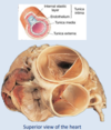
Label these arteries on a head and neck arteriogram and whereeach artery supplies

- Subclavian artery – supplies the upper limbs
- Vertebral artery – supplies blood to the neck’s vertebrae, the upper spinal column, the space around the outside of the skull, and parts of the brain
- Common carotid – main blood supply to head and neck
- Internal carotid – Main blood supply to the brain.

How do muscular arteries compare to other vessels?
How do they compare to elastic arteries?
What do they do?
What 5 things do they use vasoconstriction/vasodilation for?
- Muscular arteries have the thickest tunica media layer of all vessels
- They contain more smooth muscle and less elastic tissue in the tunica media than elastic arteries.
- Muscular arteries are more active in vasoconstriction, and use it for:
- Decreasing blood pressure to regulate blood flow
- Controlling the distribution of blood to issues
- Decreasing blood pressure before blood reaches delicate capillary beds.
- Occlusion (closure/blocking) to a principal artery or region in the case of haemorrhage (loss of blood from damaged blood vessels)
- Muscular arteries can also dilate to carry blood to ischaemic areas (deficient of blood flow.

What is the name of the artery that feeds the upper limbs?
How does its name change as it moves through the limb?
- The subclavian artery feeds the upper limb
- In the clavicle areas, it is called the subclavian artery.
- In the armpit areas, it is called the axillary artery
- In the upper arm, it is called the Brachial artery
- In the forearm, it splits into the radial and ulnar arteries.

What are anastomoses?
Where can they be found?
How are they formed in the hand?
- Anastomoses are backup routes for blood flow in case one route is blocked or compromised.
- They can be found around all joints in case blood flow is restricted during certain movements and positions.
- In the hand, superficial and deep Palmar arches are formed by both the radial and ulnar arteries of the hand.
- This arterial anastomosis ensures blood flow to the hands and fingers in any position of the upper limb.

Name some of the main abdominal arteries on this diagram


How does the femoral artery, vein and nerve lie in relation to each other in the upper leg?

Name the low limb arteries above the knee in this diagram


Name the arteries of the lower limb below the knee.


How big are arterioles?
What are the differences between large and small arterioles?
What does arteriolar diameter determine?
How do arterioles control this?
- Arterioles are the smallest of the arteries (0.3mm-10µm)
- The largest arterioles have all three tunica, however small arterioles basically consist of endothelium and a small layer of smooth muscle.
- Arteriolar diameter determines the blood flow to tissues and organs, and blood pressure
- When arterioles constrict, this results in an increase in resistance, which controls blood flow into capillary beds
- This also changes the blood pressure.

What are metarterioles?
What are pre-capillary Junctions?
What is Vasomotion?
What is vasomotion caused by?
- Metarterioles are the ends of the arterioles that go towards the capillary beds (capillary junctions)
- Pre-capillary junctions are sphincters that control the flow of blood through capillaries within the tissue or organ
- Vasomotion is when the sphincters close and blood flow is shunted (moved) through the metarterioles to maintain blood pressure to vital organs
- Vasomotion can occur in response to neural, hormonal, and chemical influences

What are capillaries?
What do they form?
What is their purpose?
How do they achieve this?
What are the 3 types of capillaries?
- Capillaries are endothelial cells supported on a basement membrane (basal lamina)
- They form networks called capillary beds
- They are the only vessels that allow the exchange of O2, CO2 nutrients, and hormones between cells/interstitial fluid (fluid around cells) and blood by:
- Active transport
- Passive transport:
- Osmosis
- Diffusion
- Filtration
- Facilitated transportation (uses transport molecule)
- There are continuous, fenestrated, and sinusoid capillaries

How common are continuous capillaries?
Where are continuous capillaries found?
What is associated with them?
How permeable are they?
- Continuous capillaries are the most common type of capillary
- They are found in the skin, muscle, lungs, and CNS.
- They have associated pericytes, which are cells that maintain structure and function
- They are the least permeable type of capillary, as the intercellular clefts are sealed by gap junctions.
Where are fenestrated capillaries found and why?
- Fenestrated capillaries are found in the kidneys, small intestine, and areas of hormone secretion.
- This is because they have large pores for active filtration
How common are sinusoid capillaries?
Where are they found?
How does blood flow in them?
What do they allow?
How permeable are they?
- The sinusoid capillaries are found in limited locations
- They are found in the liver, bone marrow, and spleen,
- Blood flows through them slowly
- They allow larger molecules and cells to pass
- They are the most permeable type of capillary.
How are venules formed?
What do they allow and how?
What do they form?
And how do these compare to corresponding arteries?
What are the layers of these like?
- Capillaries unite to form venules
- Venules enable white blood cells to extravasate (flow out of vessel that naturally contains it) to site of inflammation (diapedesis) as they are extremely porous.
- Venule come together to form veins
- Veins have a thinner wall and greater lumen diameter than corresponding arteries
- The tunica media layer of veins has little smooth muscle
- The tuna externa layers are the thickest layers of veins, and consist of collagen/elastin

What are veins considered as and why?
What do they contain and why?
How do veins prevent back flow of blood?
- Veins are considered capacitance vessels, as they contain most of the blood in the body (approximately 65%)
- Veins contain a large lumen, which allows for little resistance, and a low risk of bursting
- Veins contain venous valves, which are formed from their tunica intima
- When blood pressure proximal to the valves is greater than distal, valves close, preventing back flow, and only allowing one way blood flow.


