14-09-21 - Introduction to the Body Flashcards
Describe the anatomical position and why it is important
- Stand upright
- Face forward
- Upper limbs by each side
- Palms face forward, thumbs pointed away from body
- Feet together
- The anatomical position is important as it provides a clear consistent way of describing human anatomy and physiology.
- It creates clear points of reference which help to avoid confusion.

Give definitions for these terms:
• Superficial
• Deep (profundus)
• Peripheral
• Central
- Superficial – near the surface
- Deep (profundus) – Away from the surface
- Peripheral – Away from centre; on the outer edge of an area/object
- Central – At or close to the centre
Give definitions for these terms:
• Dexter-dextra
• Sinister-sinistra
• Ipsilateral
• Contralateral
• Unilateral
• Bilateral
- Dexter-dextra – Right side
- Sinister-sinistra – left side
- Ipsilateral - appearing on, or affecting the same side of the body
- Contralateral – appearing on, or acting in conjunction with a part on the opposite side of the body
- Unilateral – affecting or relating to one side of the subject (one-sided)
- Bilateral – Affecting or relating to the right and left side of the subject
Label the aspects of the anatomical position


Name these body parts


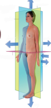
Label these planes


Name the 3 layers of skin top to bottom and what they are responsible for
- Epidermis – Protection
- Dermis – Sensory receptors (also found in epidermis) for pain, temp, pressure, touch, proprioception (sense movement, action and location). Responsible for thermoregulation.
- Subcutaneous – layer of insulation protects internal organs and muscles from shock and change in temperature

Name the 2 types of sweat glands and their features
- Merocine Sweat glands
- Acidic secretion
- Found throughout the body
- Present from birth
- Thermoregulation sweat glands
- Apocrine sweat glands
- Becomes active in puberty
- Found in armpits, groin and feet
- Alkaline secretion can be fed on by bacteria, creating odour.
What are langers lines?
- Collagen fibres give skin structure, and they are arranged in lines called langers lines

How do langers lines affect how incisions should be made?
- Incisions are much better if made parallel to the tension in the skin (langers lines)
- If incisions are made perpendicular to the langer lines, the wound is far more likely to gape, increasing healing, time and scar tissue.
What are dermatomes? And why are they in the pattern they are in?
- Dermatomes are areas of skin supplies by a spinal nerve.
- They are given this pattern due to somites, which give segmental pattern to the human body during foetal development.
- As limbs develop, these somites are stretched, giving the dermatomes this pattern

What is the tri-laminar disk? What is the name of its layers, and what does each layer develop into?
- Tri-laminar disk is a human at 3 weeks of development
- The disk has 3 layers: The ectoderm, the mesoderm and the endoderm
- The ectoderm becomes the epidermis and the nervous system
- The mesoderm gives muscles, bones, cardiovascular system and splits to form cavities
- The Endoderm contributes to the gastro-intestinal tract (lines gut tube) and reproductive systems

Describe the first way in which the tri-laminar disk folds during development
- The first way it folds is cephalon-caudal (which means head - tail)
- The bottom layer of the disk (endoderm) is pinched off to form the gastro-intestinal tube running from mouth to anus.

Describe the second way in which the tri-laminar disk folds
- Lateral folds close the body wall, and enclose body cavities.
- The cavities are potential spaces around the heart (pericardium), the lunges (pleura) and the gastro-intestinal tracts and reproductive tracts (abdomino-pelvic)

What are cavities in the body lined by and why?
What does this allow for?
- Cavities in the body are lined by a few mls of lubricating serous fluid and slippery membranes.
- This allows for potential spaces, which are spaces with surfaces that are normally pressed together.
- This potential space, serous fluid and slippery membranes allow for organs to move and slide past one another without leaving big gaps in the cavities
Why does the body not like potential spaces?
- There are loads of potential spaces present in the body cavities, bodily fluids could leak out and collect.
- This could cause inflammation and infection, which could easily spread to the rest of the organs.
Name each body cavity in this diagram and what it is for


Describe organ invagination, what the visceral and parietal layers are, and what is between the visceral layer and organ.
- Structures invaginate into balloons of serous fluid and slippery membranes.
- This creates a visceral layer touching the organ and a parietal layer against the wall of the cavity.
- The potential spaces between the organ and the visceral layer is lubricated by a few mls of serous fluid

What are fascia, describe 2 types, and why fascia can be detrimental
- Fascia is a connective tissue layer that separates one structure from another.
- Superficial fascia- runs around the outside of the body (where you find subcutaneous fat and some superficial veins and nerves)
- Muscular fascia – Allows muscles to move over one another. Can be used to separate individual or compartments of muscles from one another
- Fascia can be potential tracks for infection spread and blood loss due to the potential spaces

What causes compartment syndrome and how is it treated?
- Injury can cause swelling, which leads to pressure that can compress the neurovascular bundle in a limb.
- This is a surgical emergency that must be treated with a fasciotomy

How is the skeletal system divided?
- Axial (in the midline) – Skull, vertebrae (including sacrum), ribs and sternum
- Appendicular (off to the sides) – Bones of upper and lower limbs (including scapula and clavicle – (pectoral girdle) and hip bone (pelvic girdle)).

Describe the diagram for the divisions of the nervous system

What does somatic and visceral mean in relation to the nervous system
- Somatic – conscious movement and conscious sensation ex. Picking something up
- Visceral – Unconscious ex. The body will slow down or speed up the heart rate depending on if you are calm or anxious without you consciously doing anything.
What is the enteric nervous system and why is it unique?
- The enteric nervous system is the semi-autonomous, self-contained nervous system of the gut.
- Heavily mediated by the visceral nervous system but it can function on its own to a certain degree.


