24. Bone Tumors Flashcards
What is the source of these FCs?
Memoli’s bizarre lecture AND First Aid (Primary Bone Tumors)
What are the two main types of benign Primary Bone Tumors?
- Giant Cell Tumor (osteoclastoma)
- Osteochrondroma (exotosis)
Giant Cell Tumor: Benign or malig?
what bones does it tend to appear on? where on the bone does it tend to appear?
Distal femur, Proximal tibia (knee)
Appears on epiphysis (end of bone).
Often resembles a soap bubble.
Osteochondroma: benign or malignant? What part of the bone does it tend to appear on?
Benign
Tends to appear on mature bone with cartilaginous cap. Often originates from long metaphysis. Looks like a kickstand growth directly jutting out from bone
May rarely transform to chondrosarcoma (malignant)
Osteosarcoma: benign or malig?
What bones does it tend to appear on? What part of that bone? What are characteristic features?
Prognosis?
Malignant, poor prognosis.
Appears on metaphysis of long bones, often around distal femur/prox tibia (knee)
Characteristic Codman’s triangle or sunburst pattern on xray

Ewing’s Sarcoma: benign or malignant?
On what bones does it appear? characteristic appearance?
Behavior?
Malignant.
Appears in diaphysis of long bones, pelvis, scapula, ribs.
Aggressive with early metastases, responsive to chemo.
Onion skin appearance in bone (“going out for eWings and onion rings”)

Chondrosarcoma: benign or malig?
What bones does it usually locate to?
Characteristics?
Malignant.
Located in pelvis, spine, scapula, humerus, tibia, femur
Cartilaginous tumor.
May be primary or from osteochondroma (benign)
Expansile glistening mass within medullary cavity (in the diaphysis)
What’s this?

Picture of Ewing’s Sarcoma, published in 1921.
Also a fracture
Names for the different parts of the bone: where is the diaphysis, metaphysis, epiphysis?

What do the radiographic margins of a lesion tell you about the lesion?

Tell you about the interaction of the normal bone with the growing lesion.
A bone lesion with a sclerotic margin indicates what kind of growth pattern?

Sharp, sclerotic margin indicates slow growth of lesion. Slow growth allows for the remodeling of the bone (according to Wolff’s law)
Reason: bone has responded by increasing its density. This process takes time.
The sclerosis in this situation refers to the bone’s increased density. Can have sharp borders without sclerosis.
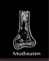
Motheaten lesion with an ill-defined margin means what about the lesion’s growth?

Don’t see the outline of the lesion: appears in a ragged pattern. Lesion is growing faster than ones with sharp margins or with more well-defined geography.
Lesions with a permeated pattern on xray indicate what growth pattern/speed?

Faster and infiltrative lesions will give a permeated (permeative) pattern on the x-ray since the normal bone does not have sufficient time to respond to the faster growing mass.
Cannot see the margins of the lesion
For the fastest-growing bone lesions, what will we see on imaging?
We’ll either see a permeated pattern, or possibly nothing at all.
Nothing at all would indicate that the bone has not had time to respond. Ex = leukemia.

What do these represent? Patterns of what?
Ivory appearance means what?
Rings/Arcs means what?

Patterns of mineralization.
tells you whether the lesion is mineralizing or not.
“Ivory” appearance = more dense than usual level of mineralization I Suggests osteosarcoma.
Rings/arcs are typical of cartilage lesions.
Generally, what do these images represent?

Radiographic appearance of the periosteum.
Fibrous membrane, responds to things both inside and outside the bone. Will respond to lesion by making new bone.
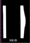
What does this periosteal growth pattern indicate about the lesion that is causing it?

Lesion is growing slowly and cortex has time to repair: will get solid thickened bump (solid).
What does this periosteal growth pattern indicate about the lesion that caused it?

Onion skin: lesion grows, then slows down and bone grows, then tumor grows – layers of bone and tumor.
What does this periosteal growth pattern indicate about the lesion that caused it?

Lesion is growing rapidly and invading into periosteum –> response is more spiculated pattern. New bone forming in response to tumor growing out.
What is Wolff’s Law?
A slow growing lesion allows the bone to respond and remodel, according to Wolff’s law (increased bone at sites of increased mechanical stress, decreased bone where stress is diminished).
Faster growing lesions do not allow for the bone to respond as effectively. In the periostium, complex types of reactions (spiculated, lamellar) occur as the lesion penetrates the periostium.

Why do a number of lesions tend to localize to the distal ends of bones (epiphysis/metaphysis)?
Bone is dynamic in these areas, lots of activity and vascularization.

Ewing’s Sarcoma tends to be where in the bone? Osteosarcoma tends to be where in the bone?
(repeated facts but maybe impt)
osteosarcoma: metaphysis.
Ewing sarcoma: diaphysis.

What elements of the clinical presentation can help you distinguish what lesion type your patient might have?
Age
Location within the bone
History (trauma, cancer)
Systemic findings (fever, WBCs, Ca)
Single or multiple lesions.
Is a primary bone neoplasm likely to be a single lesion, or multiple lesions?
What about metastatic lesions?
Primary bone neoplasm typically single lesion
Metastasis often multiple lesions (ex: multiple myeloma)


