2. Neural Tissue Flashcards
In the body superior means up and inferior means down, but in the brain what terms refer to up and down?
- dorsal = up or top like a dolphins fin on the top
- ventral = down

In the body ventral means up and dorsal means back, but in the brain what terms refer to front and back?
- front = rostral
- back = caudal

On the brain there are areas that sink in, similar to the crypts in the GI and then there are area that bulge out, as per the image below. What are the 2 terms used to describe these anatomical landmarks?

- gyrus = look like bumps
- sulcus** = brain **Sinks in (depressions) resembling the crypts

What is the the theory that describe the below:
- nervous system is made up of discrete individual cell
- All cells are individual BUT mutually rely on one another
neuronal doctrine theory
What is the cerebral cortex?
1 - outermost region of the cerebrum
2 - inner most layer of the cerebrum
3 - attached to the top of the brain stem
4 - located at the back of the brain, referred to as the mini brain
1 - outermost region of the cerebrum
- made up of neuronal cell bodies**, so appears **grey

What is a Brodmann area?
- boundaries of the brain
- determined by histological architecture and function, essentially the tissue and cell type
How many Brodmann areas are there in the brain?
- 52

Why are Brodmann areas of the brain important?
- they generally posses specific functions
Brodmann area 17 is located on either side of the calcarine sulcus, on the medial surface of the occipital lobe. What is this area also commonly referred to as and what is its function?
- primary somatosensory cortex
- primary visual cortex
- primary motor cortex
- primary auditory cortex
- primary visual cortex

Brodmann area 4 is located in the posterior portion of the frontal lobe. What is this more commonly known as and what is its function?
- primary somatosensory cortex
- primary visual cortex
- primary motor cortex
- primary auditory cortex
-
primary motor cortex
- heavily involved in motor function

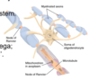
The head of a neuron is called the cell body. It has appendages that are designed to receive communications from other cells, what are these called?
- dendrites
- axon hillock
- axon terminal
- oligodendrocyte
- dendrites

The head of a neuron is called the cell body. What is the name where the cell body attaches to the axon and what is its role?
- dendrites
- axon hillock
- axon terminal
- oligodendrocyte
- axon hillock
- initiates an action potential

What is the end of the axon furthest away from the cell body called?
- dendrites
- axon hillock
- axon terminal
- oligodendrocyte
- axon terminals
- synapses with other neurons or tissues

Axon collaterals can be found along the axon, between the cell body and the axon terminals. What is the function of these?
- provide modulation of the cell firing
- wrap around the axon body ensuring good conduction
- connect the axon hillock the the axon
- provide modulation of the cell firing

How do we know if a neuronal cell is a white matter neuron?
- if it is encapsulated in myelin sheath
- myelin is a white protein
In the CNS which cell is responsible for myelinating neurons?
- microglia
- schwann cell
- oligodendrocytes
- astrocytes
- oligodendrocytes
- able to do this for multiple neurons

In the PNS which cell is responsible for myelinating neurons?
- microglia
- schwann cell
- oligodendrocytes
- astrocytes
- Schwann cells
- one neuronal cell per schwann cell
Each neuron is a separate entity with a limiting cell membrane, like all other cells in the body. Why is this important?
- confirms neuronal doctrine
- all cells are individual but dependent on one another
Morphology refers to the size and shape of neurons. How many different types of neuronal cells are there?
- 1
- 2
- 3
- 4
- 4

Morphology refers to the size and shape of neurons. There are 4 different types of neuronal cells. What are they?
- unipolar, bipolar, pseudo-unipolar, facet-polar
- membrane polar, bipolar, pseudo-unipolar, multipolar
- synaptic polar, bipolar, pseudo-unipolar, multipolar
- unipolar, bipolar, pseudo-unipolar, multipolar

What are dendrites in neuronal cells?
- part of neuron responsible for immunity
- part of neuron that facilitates conduction
- protoplasmic appendages of cell membrane
- part of nucleus of neuronal cells
- protoplasmic appendages of cell membrane
- designed to facilitate communication between cells
In some cells, what proportion of the neurones can consist of dendrites?
- 10%
- 30%
- 60%
- 95%
- up to 95%
In a neuronal cell, what is the soma?
- neuronal cell body containing nucleus
- appendages of neuronal cell
- connection between neuronal cell and axon
- connection between post synapse and pre synapse
- neuronal cell body containing nucleus
- metabolic activity and protein synthesis occur here

Neuronal transmitters are produced where in the neuronal cell, before being stored in the synaptic vesicles?
- soma (cell body)
- nucleus
- axon hillock
- axon terminal (pre synapse)
- soma
Which part of the neuronal cell is responsible for generating an action potential due to the high density of the voltage gated Na+ channels, allowing the threshold to be reached, which then triggers the action potential?
- dendrites
- axon hillock
- axon terminal
- oligodendrocyte
- axon hillock

What is saltatory conduction?
- propagation of action potential along myelinated axons
- propagation within the pre synapse
- continues propagation along non-myelinated axons
- propagation along the axon hillock
- propagation of action potential along myelinated axons
- saltatory comes from the greek to ‘leap’
- allows very fast action potentials to travel from node to node in neurons
- 120m/s

What is the difference between an afferent and efferent neuron?
- afferent = away from the body to CNS (sensory)
- efferent = effector neurons from CNS to PNS (muscles, glands etc)
What are interneurons?
- carry messages between neurons (can be inhibitory or excitatory)
- facilitate communication
Oligodendrocytes are a form of glia cells. What is the function of these cells?
- myelinate neurons in the CNS
- blood brain barrier, energy metabolism, reaction to injury
- immune function
- synthesise neurotransmitters
- myelinate neurons in the CNS

Astrocytes are a form of glia cells. What is the function of these cells?
- myelinate neurons in the CNS
- blood brain barrier, energy metabolism, reaction to injury
- immune function
- synthesise neurotransmitters
- blood brain barrier, energy metabolism, reaction to injury

Microglia are a form of glia cells that can be activate or dormant. What is the function of these cells?
- myelinate neurons in the CNS
- blood brain barrier, energy metabolism, reaction to injury
- immune function
- synthesise neurotransmitters
- immune function
- think M like macrophage

What is the main composition of oligodendrocytes?
- peptides
- proteolipid proteins (80%)
- proteins
- glycosaminoglycans
- proteolipid proteins (80%)
- myelin basic proteins

In addition to myelin basic protein and proteolipid protein that make up oligodendrocytes, myelin associated glycoprotein is also an important protein, what is its main function?
- facilitates interactions between the axon and myelin
Myelinated neurons are larger than non-myelinated neurons, >1.5um and <1.5 um in diameter, respectively. Myelination allows nerve conduction velocity, allowing for a decrease in the nerve size. Without myelinated nerves, what would this mean for the nerve diameter?
- would make no difference
- we would need huge nerve diameters
- we would need smaller nerve diameters
- we would need to slow nerve conduction
- we would require huge nerve diameters to transmit information
If a myelinated neurons loses its myelin sheath, what can this cause?
- nothing it would grow back
- nerve would become non-myelinated, but function normally
- can cause axon degeneration and function, but can repair to some degree
- linked with diseases such as multiple sclerosis
What is the difference between demylination and unmyelinated?
- demylination = was myelinated but lost due to disease/disorder
- unmyelinated = was never myelineated
Microglia cells are the immune cells of the nervous system, with lineage to macrophages. Are they able to phagocytose pathogens?
- yes
- also clear cellular debris
Microglia mature at birth and are at the greatest number just prior to birth. Why do the microglia down regulate and become ramified (resting)?
- needed to protect the brain, but then blood brain barrier forms and does this job
- not as likely to get an infection as we get older
- blood pressure increases around the brain and kills of microglia
- microglia are unable to cross the blood brain barrier
- needed to protect the brain, but then blood brain barrier forms and does this job
Astrocytes are an important glia cell type of the nervous system, what are the main functions?
- provide structural
- glucose supply
- maintain ionic enviroment
- neurotransmitter uptake
- repair
- form blood brain barrier
What does synaptogenesis refer to?
- formation of new synapses between neurons
- regulated by astrocytes
The blood brain barrier controls what does or does not enter the nervous system. It is composed of 3 key things, what are they?
- vascular endothelial cells, astrocytes, microglia
- vascular endothelial cells, microglia, pericytes
- vascular endothelial cells, astrocytes, oligodendrocytes
- vascular endothelial cells, astrocytes, pericytes
There is evidence to suggest that an important part of the nervous system becomes more permeable as we age, and has been linked with confused states and disorders. What is it that supposably becomes more permeable in the brain?
- blood brain barrier
Astrocytes are stellate cells (star like pattern) found throughout the CNS. There are 2 broad classifications, depending on if they are located in white or grey matter, what are they?
- white matter = fibrous astrocytes
- grey matter = protoplasmic astrocytes
What is glutamate?
- neurotransmitter
- hormone
- steroid
- medicaiton
- neurotransmitter
- amino acid neurotransmitter functions as a an excitatory neurotransmitter (AMPA and NMDA)
What is the glutamate-glutamine shuttle?
- how glutamate is moved around the neuronal cell
- how glutamate is synthesised in the soma of the neuron
- how glutamate is recycled from synaptic cleft by astrocytes
- how glutamate is released by vesicles into the synaptic cleft
Glutamate-glutamine shuttle:
how glutamate is recycled from synaptic cleft by astrocytes
- recycling of glutamate
- glutamate is released into synaptic space
- glutamate binds receptors on the post synapse
- recycled by pre-synapse or astrocytes
- ultimately ends up back in pre synapse
Glutamate - glutamine shuttle:
c.80% of glutamate released at the synapse taken up by astrocytes
- astrocytes covert glutamate → glutamine
- release glutamine
- taken up by presynaptic terminal
- converted back to glutamate.
Astrocytes are connected via gap junctions, and can transport Ca2+

The glutamate-glutamine shuttle describes how glutamate, a neurotransmitter is released into synapse following an action potential. It can bind with receptors on the post synapse, be recycled by pre-synapse or be absorbed by receptors on astrocytes. Once glutamate has been re-taken up by astrocytes, what happens to it?
- converted into glutamine in astrocyte, then back into glutamate in pre-synapse
- converted into dopamine in astrocyte, then back into glutamate in pre-synapse
- converted into serotonin in astrocyte, then back into glutamate in pre-synapse
- converted into glutamine in astrocyte, then back into glutamate in synaptic cleft
- converted into glutamine in astrocyte, then back into glutamate in pre-synapse
- absorbed by glutamine transports (Na+ dependent)
- glutamine synthetase acts on it leaving glutamine
- glutamine released back into pre-synaptic neuron
- re-converted back into glutamate in pre synapse

In the CNS there is an additional type of glia cell called the ependymal cells, which are ciliated epithelial glial cells. These cells line the inside of the ventricles and the spinal spinal cords. What is the main function of the ependymal calls?

- maintain fluid homeostasis
- direct contact with cerebral spinal fluid (CSF) and brain parenchyma (brain tissue)
- facilitate transport of electrolytes and solutes into brain and CSF
Schwann cells myelinate axons in the PNS. How many axons can one schwann cell myelinate?
- one axon only
- also important for providing substrate for axons to grow long
Satellite glia cells are located in the PNS, exclusively in the peripheral ganglia, sensory parasympathetic and sympathetic ganglia. How do they support the neurons?
- wrap around the neuronal cell bodies
- function similiar to astrocytes, but unknown

Do neurons and glia cells have action potentials?
- no, only neurons
Do neurons and glia cells both divide in mitosis?
- no, only glia cells
What is a glioma?
- a group of glial cells
- a glial cell found in a lymph node
- tumour cells derived from glial cells
- glial cell that has undergone apoptosis
- tumour cells derived from glial cells
Are gliomas dangerous?
- yes, highly malignant
- glial cells can proliferate and therefore increase in size
What is the most common treatment for gliomas?
- radiotherapy due to accessibility
What does intra-axial refer to?
- within the brain parenchyma

What is neuroblastoma mean?
- neuro = nerve cells
- blastoma = immature cells
- cancer that develops from immature nerve cells
- occur outside of the cranium
- common in adrenal glands, kidneys, neck, spine

In a patient with neuroblastoma, what are the outcomes?
- neuro = nerve cells
- blastoma = immature cells
- outcomes are generally good
- patients tend to develop neurological issues in later life
Primary visual cortex is found in which lobe of brain?
Occipital lobe
What type grey matter or white matter neuron is this image?
slide 12 add image
Occipital lobe
White matter neuron because myelinated
which type of glial cell
which cells represented in scaffold
Which type of glial cell is distributed in regular mosaic patterns throughout the CNS.
Microglia