196b - Breast Histology, Pathology Flashcards

1
Q

Lobular Carcinoma in situ (LCIS)

  • Type of breast mass:
  • Pathologic changes:
  • Clinical features:
  • Relative risk of carcinoma:
A
  • Type of breast mass: Carcinoma in situ
  • Pathologic changes: several possibilities
    • Uniform, monomorphic cells
    • Bland, round nuclei in loosely cohesive clusters
    • Intracellular mucin
  • Clinical features: Incidental - no calcifications
  • Relative risk of carcinoma: 8-10 fold relative risk
    • ~1/3 of women will develop invasive carcinoma in either breast
2
Q

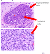
What 2 cell types line breast glands?

A
  • Cuboidal epithelial cells
    • Inner layer
    • Produce milk
  • Myoepithelial cells
    • Outer layer
    • Squeeze the milk out
    • Limiting membrane of breast ducts
3
Q

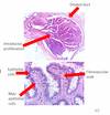
Intraductal papilloma

  • Type of breast mass:
  • Pathologic changes:
  • Clinical features:
  • Relative risk of carcinoma:
A
  • Type of breast mass: Proliferative without atypia
  • Pathologic changes:
    • Intraductal - large excretory ducts
    • Papillae - fibrovascular stalks
    • Mass-forming but benign
  • Clinical features: Mass below the nipple, bloody discharge
  • Relative risk of carcinoma: 1.5-2 fold relative risk
4
Q

What is the difference in management of ductal carcinoma in situ (DCIS) vs. lobular carcinoma in situ (LCIS)?

A
  • DCIS - Surgery and radiation
    • The DCIS lesion is the one that may become invasive - if you find the DCIS, you know what to remove
  • LCIS - Radiation and chemo
    • LCIS is basically a marker that something is going wrong in the breast - Location of LCIS does not predict the area that is likely to become invasive
5
Q

Duct Ectasia

  • Type of breast mass:
  • Pathologic changes:
  • Clinical features:
  • Relative risk of carcinoma:
A
  • Type of breast mass: Inflammatory
  • Pathologic changes:
    • Dilated ducts
    • Lymphocytes, plasma cells (one nuclei)
    • Pink squamous epithelium instead of cuboidal cells
  • Clinical features: Older, multiparous women, nipple pain, retraction, unilateral discharge
  • Relative risk of carcinoma: None
6
Q

How does pregnancy/lactation change the histology of the breast?

A

More lobules

More acini within each lobule

7
Q

What is the relative risk of breast lesions in the “nonproliferative” category?

A

Minimal or no increased risk for developing breast carcinoma

  • Acute mastitis
  • Duct ectasia
  • Fat necrosis
  • Simple cysts
  • Apocrine metaplasia
  • Fibroadenoma
8
Q

Ductal Carcinoma in situ (DCIS)

  • Type of breast mass:
  • Pathologic changes:
  • Clinical features:
  • Relative risk of carcinoma:
A
  • Type of breast mass: Carcinoma in situ
  • Pathologic changes: several possibilities
    • Architectural: solid, cribiform, papillary
    • Comedo: extensive central necrosis
    • Low grade (bland, monotonous) or high grade (pleiomorphic)
  • Clinical features: Calcifications on mammogram
  • Relative risk of carcinoma: 8-10 fold relative risk
    • At least 1/3 of women with untreated low-grade will eventually develop invasive carcinoma
9
Q

What is the relative risk of breast lesions in the “Proliferative without atypia” category?

A

1.5-2 fold relative risk of developing breast carcinoma in the future

  • Usual ductal hyperplasia
  • Sclerosing adenosis
  • Radial scar
  • Intraductal papilloma
10
Q

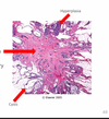
Radial scar

  • Type of breast mass:
  • Pathologic changes:
  • Relative risk of carcinoma:
A
  • Type of breast mass: Proliferative without atypia
  • Pathologic changes:
    • Stellate configuration
    • Central nidus of small entrapped glands
    • Dilated glands at the periphery with cysts or hyperplasia
  • Relative risk of carcinoma: 1.5-2 fold relative risk
11
Q

What is the relative risk of breast lesions in the “Proliferative with atypia” category?

A

4-5 fold relative risk of developing breast carcinoma in the future

  • Atypical ductal hyperplasia (ADH)
  • Atypical lobular carcinoma (ALH)
12
Q

Sclerosing adenosis

  • Type of breast mass:
  • Pathologic changes:
  • Relative risk of carcinoma:
A
  • Type of breast mass: Proliferative without atypia
  • Pathologic changes:
    • Lobulocentric proliferation (around a normal TDLU)
    • Fibrosis of stroma
    • Increased nuber of acini
  • Relative risk of carcinoma: 1.5-2 fold relative risk
13
Q

What kind of breast tissue cells stain brown with smooth muscle myosin immunohistochemical stain?

A

Outer layer myoepithelial cells

14
Q

Key differences between atypical ductal hyperplasia and usual ductal hyperplasia?

A
  • Atypical
    • 4.0-5.0 fold increased risk
    • Cells are growing in a weird pattern - monotonous, roman bridges, cribiform structures
  • Usual
    • 1.5-2.0 fold increased risk
    • More cells, but they are in a normal pattern - look like background cells in the terminal duct
15
Q

Fibroadenoma

  • Type of breast mass:
  • Pathologic changes:
  • Clinical features:
  • Relative risk of carcinoma:
A
  • Type of breast mass: Non-proliferative
  • Pathologic changes:
    • Well-circumscribed
    • Biphasic - benign glands and stroma
  • Clinical features: Most common benign neoplasm of the female breast; 20-35 y/o, firm, rubbery, mobile, rounded
  • Relative risk of carcinoma: None
16
Q

Apocrine metaplasia

  • Type of breast mass:
  • Pathologic changes:
  • Clinical features:
  • Relative risk of carcinoma:
A
  • Type of breast mass: Non-proliferative
  • Pathologic changes:
    • Apocrine gland: releases some of thier cytoplasmic secretions
    • Large, polygonal cells w/eosinophilic, granular cytoplasm
    • Small round nuclei
  • Clinical features: Pre-menopausal woman, cyclic breast pain, nodularity
  • Relative risk of carcinoma: None
17
Q

Phyllodes tumor

  • Type of breast mass:
  • Pathologic changes:
  • Clinical features:
  • Relative risk of carcinoma:
A
  • Type of breast mass: Sarcoma (cancer)
  • Pathologic changes:
    • Well-circumscribed
    • Biphasic - benign glands and stroma
  • Clinical features: Rapidly enlarging, 30-50 y/o pt
  • Relative risk of carcinoma: Is already carcinoma
18
Q

What is the functional unit of the breast?

A

Terminal ductal lobular unit

(This is where most breast lesions arise)

19
Q

Usual ductal hyperplasia

  • Type of breast mass:
  • Pathologic changes:
  • Relative risk of carcinoma:
A
  • Type of breast mass: Proliferative without atypia
  • Pathologic changes:
    • Increased number of ductal cells
  • Relative risk of carcinoma: 1.5-2 fold relative risk
20
Q

Fat necrosis

  • Type of breast mass:
  • Pathologic changes:
  • Clinical features:
  • Relative risk of carcinoma:
A
  • Type of breast mass: Inflammatory
  • Pathologic changes:
    • Hemorrhage and neutrophils early
    • Macrophages, fibrosis, calcifications later
  • Clinical features: Hx of trauma to the breast, calcification on mammogram, hard mass
  • Relative risk of carcinoma: None
21
Q

What is the relative risk of breast lesions in the “in situ carcinoma” category?

A

8-10 fold relative risk of developing breast carcinoma in the future

  • Ductal carcinoma in situ (DCIS)
  • Lobular carcinoma in situ (LCIS)
22
Q

In general, what are the differences between ductal vs. lobular breast legions?

A
  • Ductal
    • Stronger association with calcifications
    • Likely unilateral
    • Cells more densely packed, darker purple
    • Invasive ductal carcinoma = 70% of breast cancer
  • Lobular
    • No association with calcifications
    • More likely to be bilateral
    • Cells look lighter purple, may have more other stuff in between
    • Invasive lobular carcinoma = 10-20% of breast cancer
23
Q

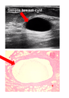
Simple cysts

  • Type of breast mass:
  • Pathologic changes:
  • Clinical features:
  • Relative risk of carcinoma:
A
  • Type of breast mass: Non-proliferative
  • Pathologic changes:
    • Dilated duct with inner luminal epithelial cells, outer myoepithelial cells
    • Involves TDLU
  • Clinical features: Pre-menopausal woman, cyclic breast pain, nodularity
  • Relative risk of carcinoma: None
24
Q

On H&E stain:

What color is breast stroma?

What color are breast ducts?

A

Fibrous stroma = pink

Breast ducts = epithelial cells = purple

25
Q

Invasive Ductal Carcinoma

  • Type of breast mass:
  • Pathologic changes:
  • Clinical features:
A
  • Type of breast mass: Cancer
  • Pathologic changes:
    • Well developed tubules with low grade nuclei
    • 2/3 express ER and PR
    • 1/3 overexpresses HER2
  • Clinical features: 70-80% of breast cancers
    • ​Hard, palpable, fixed mass
26
Q

Invasive Lobular Carcinoma

  • Type of breast mass:
  • Pathologic changes:
  • Clinical features:
A
  • Type of breast mass: Cancer
  • Pathologic changes:
    • Cells don’t stick together (due to E-cadherin mutation)
    • Invade individually, single file strands
    • Most express ER, PR
    • Rarely overexpress HER2
  • Clinical features:
    • 20% of all breast cancers
    • 10-20% are bilateral
27
Q

Atypical ductal hyperplasia

  • Type of breast mass:
  • Pathologic changes:
  • Clinical features:
  • Relative risk of carcinoma:
A
  • Type of breast mass: Proliferative with atypia
  • Pathologic changes:
    • Cytologic atypia = monotonous cells
    • Architectural atypia = arches (Roman bridges), punched out areas (Cribiform structures)
    • Ductal epithelial proliferation
  • Clinical features: Late 40’s, microcalcifications on mammography
  • Relative risk of carcinoma: 4-5 fold relative risk
28
Q

Atypical lobular hyperplasia

  • Type of breast mass:
  • Pathologic changes:
  • Clinical features:
  • Relative risk of carcinoma:
A
  • Type of breast mass: Proliferative with atypia
  • Pathologic changes:
    • Lobular - uniform, small, dyscohesive cells
    • Round nuclei that may be peripheral
    • Intracytobplasmic lumens
  • Clinical features: Multicentric, bilateral, incidental finding on core biopsy
  • Relative risk of carcinoma: 4-5 fold relative risk
29
Q

What histological changes occur in post-menopausal breast tissue?

A

More fatty stroma (less fibrous)

Acini are decreased in number, smaller

30
Q

Acute mastitis

  • Type of breast mass:
  • Pathologic changes:
  • Clinical features:
  • Relative risk of carcinoma:
A
  • Type of breast mass: Inflammatory
  • Pathologic changes: Neutrophils (multi-lobated nuclei)
  • Clinical features: Fever, enlarged, painful, red breast in a nursing mother
  • Relative risk of carcinoma: None