10. Pelvis and Gluteal Region Flashcards
Identify the following lower limb regions.
Blue: Anterior Thigh
Yellow: Medial Thigh
Green: Knee/Popliteal Fossa
Orange: Leg
Pink: Foot (dorsal and plantar)
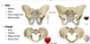
What are the bones of the pelvis?
Hip bone, sacrum, coccyx
What do the bones of the pelvis form?
the pelvic inlet=pelvic brim (hip bone, sacrum, coccyx)
Identify the following parts of the pelvis and hip.
Blue: Hip bone
Green: Sacrum
Orange: Coccyx
Purple: Femur
anterior view circle: pelvic intlet
posterior view circle: pelvic outlet
What does the false pelvis hold?
False pelvis is above the inlet (true pelvis is in between inlet and outlet)
False pelvis holds the intestines.
What articulation is found when the two parts of the hip bones meet anteriorly?
Pubic symphysis
What are the three parts of the hip bones. Identify them.
Red: ischium
blue: pubis (thinner, smaller aspect)
rest: ilium
Identify all parts of the illium of the hip bone on the posterior aspect.
What does the acetbulum hold?
the acetabulum holds the head of the femur
What articulates with the auricular surface?
the auricular surface is where the sacrum articulates with the ilium
What does the arcuate line become?
The arcuate line becomes the pectineal line
Identify the various part of the ischium of the hip bone.
Identify all the parts of the pubis of the hip bone. (of the anterior view only)
the orange part that represents the hole is the obturator foramen
Identify all the posterior parts of the pubis of the hip bone.
What inserts onto our pubic tubercle?
rectus abdominis and inguinal ligaments
When is the pelvis properly aligned?
When the ASIS (anterior superior iliac spine) is lined up with the pubis tubercle (you have anterior and posterior tilt that allows you to bear weight on your femur)
What is the ischiopubis ramus formed of?
the ramus of the ischium and the inferior pubis ramus
What does the arrow point at?
sacral promontory
What are the four joints found in the pelvis?
lumbosacral joint
sacroiliac joint
sacrococcygeal joint
pubic symphisis
What joint is this? Which ligaments reinforce this joint?
JOINT: lumbosacral joint
Blue: anterior longitudinal ligament
green: iliolumbar ligament
What joint is this? Which ligaments reinforce this joint?
JOINT: sacroiliac joint (sacrum and ilium meet at the auricular surface)
orange: anterior sacroiliac ligament
purple: posterior sacroiliac ligament
pink: interosseus sacroiliac ligament (between 2 bones)
Identify the structure in pink and green.
pink: inguinal ligament (goes from ASIS to pubis tubercle): it is the tendon extension of the external oblique
green: obturator membrane
Identify the orange and purple ligaments. Where do they insert?
purple: sacrotuberous ligament (inserts onto ischial tuberosity)
orange: sacrospinous ligament (inserts onto ischial spine)
What is the function of the sacrospinous ligament?
the sacrospinous ligament stops hinge of the pelvic and spine: maintains posture
Why are individuals more prone to shoulder dislocation rather than hip dislocation?
because your hip has the acetabulum that is uniquely for your femur to insert into. the acetabulum is deeper and there are mor ligaments attaching your femur to your hipbone
Identify the two parts of the actebulum.
blue: articular surface
green: acetabular fossa
Identify the various parts of the proximal femur.
What trick can you use to identify wether it is the right or left femur?
the tuberosity (rough line) is POSTERIOR
femur gun: if you hold the heand of your femur with your left hand and cal easily reach your lesser trochanter then it is your left femur (same with right hand)
What are the two sex differences of the angle found between your hip joint and proximal femur?
- Q ANGLE: (in blue) angle between your femur and knee
- women > men
- women’s hip articulate more laterally - Y shape
- women have a wider pelvis => require higher Q angle to hold body weight over center of mass - ANGLE OF INCLINATION: (in red) angle between head of femur and body of femur
- men > female
What is lateral minescus? Who is more prone to lateral minescus?
femur sits inside knee, all of weight is put onto lateral aspect of the tibia which puts pressure on ligaments of the knee
(females are more prone)
Identify the five ligaments found in this area. (attaches femur to hip)
- green: iliofemoral ligament
- purple: ischiofemoral ligament
- orange: pubofemoral ligament (small one)
DEEPER
- Synovial membrane
- ligament of head of femur
Describe the superficial ligaments between your femur and hip? Why are they formed that way?
3 superficial ligaments: ilifemoral, ischiofemoral and pubofemoral ligaments
they are twister: in embryo: the ligaments are straight but as fetus grow, they twist
=> it limits posterior extension of thigh but helps with flexion of thigh
Why can you flex your thigh more than extend?
due to the twisting of the three superficial ligaments between hip and femur (iliofemoral, ischiofemoral, pubofemoral)
What are the two types of hip dislocation? What action causes them?
- posterior hip dislocation: femur goes back due to internal rotation or flexion
- anterior hip dislocation: femur goes forward due to flexion or lateral rotation
What are the differences in the physical appearance of the hip and pelvis between male and female?
Who is the cutest person in town?
Little tomato
hey
Just passing by to let you know I love you :)
What is the function of fascia latae in the thigh? What does it become in the leg? Along with the fascia latae, what else do we find in the thigh and leg?
The fascia latae in the thigh becomes the crural fascia in the leg. Both fascia prevent blood from pooling into the legs (maintains blood circulation)
We find the IT band (ilitibial band)=thickening of fascia on the medial aspect of the thigh and leg (connected to fascia)
What is compartment syndrom? What is the solution?
Compartment syndrom is the swelling in 1 compartment of your thigh/leg which puts pressure on VAN: to release pressure, cut the fascia latae
Identify this muscle.
(insertion, origin, innervation, action)
GLUTEUS MAXIMUS:
ori: posterior sacroiliac ligament & sacrotuberous ligament
ins: gluteal tuberosity & iliotibial tract
inn: inferior gluteal n
action: extension @ hip & abduction
Identify the muscle indicated with the green arrow.
(insertion, origin, innervation, action)
Green:
TENSOR FASCIA LATAE
ori: ASIS (anterior superior iliac spine)
ins: from the iliotibial tract down to lateral tibial condyle
inn: superior gluteal n
action: abduction & tense fascia to keep you upright & flexion @ hip
Identify the muscle: (insertion, origin, innervation, action)
GLUTEUS MEDIUS
(deep to glute max)
ori: ilium (between ant & post gluteal line)
ins: greater trochanter
inn: superior gluteal n
act: abduction (when you stand on 1 leg, its the muscle that prevents you falling)
Identify the muscle: (insertion, origin, innervation, action) (green)
GLUTEUS MINIMIS
(deep to glute med)
ori: ilium (between ant&post lines)
ins: greater trochanter
inn: superior gluteal n
act: internal rotation & bit of abduction
Name the 6 lateral hip rotators. Name the common action they contribute to.
priformis
gemellus superior
obturator internus
gemellus inferior
quadratud femoris
obturator externus
ACTION: lateral rotation of hip, bring back to extension when flexing
Which ligaments are harmed in a posterior and anterior hip dislocation?
post: ischiofemoral lig damage
ant: iliofemoral lig damage
identify these 2 muscles: (insertion, origin, innervation, action)
blue:
PIRIFORMIS
ori: anterior sacrum
(goes thru greater sciatic foramen)
ins: greater trochanter
inn: n to piriformis
act: lateral hip rotators
OBTURATOR INTERNUS
ori: ischiopubis ramus & obturator membrane
ins: greater trochanter
(goes thru lesser sciatic foramen)
inn: n to obturator internus
act: lateral hip rotators
Identify the orange and yellow muscle: (insertion, origin, innervation, action)
OREO TRICK: gemellus superior (dark), obturator internus (light), gemellus inferior (dark)
*both gemellus are innervate by muscles below it
orange:
GEMELLUS SUPERIOR
ori: ischial spine
ins: tendon of obturator internus
inv: n to obturator internus
act: lateral hip rotators
yellow:
GEMELLUS INFERIOR
ori: ischial tuberosity
ins: tendon of obturator internus
inv: n to quadratus femoris
act: lateral hip rotators
Identify this muscle:
ori:
ins:
inv:
act:
QUADRATUS FEMORIS
ori: ischial tuberosity
ins: intertrochanteris crest
inv: n to quadratus femoris
act: lateral hip flexor
identify this muscle:
ori:
ins:
inv:
act:
OBTURATOR EXTERNUS
(very deep)
ori: ischiopubis ramus
ins: intertrochanteric crest
inv: obturator n
act: lateral hip rotators
Explain the blood flow in the hip and upper thigh (arteries).
What is the cremaster test?
light stroking of up thigh on cremaster region activates the femoral nerve (L1-L2 branch of genitofemoral n) which pulls the testes up torwards the body
What are the motor and sensory functions of the lumbar plexus?
Lumbar plexus
motor: abdominal wall, anterior thigh (femoral n), medial thigh (obturator n)
sensory: abdominal wall, anterior lateral thigh, medial thigh, anterior leg, medial , medial foot
What are the motor and sensory functions of the sacral plexus?
sacral plexus
motor: gluteal region, posterior thigh, leg, foot
sensory: buttocks, posterior thigh, posterior leg, most of foot
Draw the Lumbar Plexus.
Draw the sacral plexus.
Describe the dermatomes
hello cutie
you strong like baloo, get itttt
