10. Head and Neck - Development of the Head and Neck Flashcards
Where is the head, neck and face at by early week 4?
- The neural tube forms in week 3
- By week 4, the embryo has folded (creating primitive gut tube, including primitive pharynx)
- By early week 4 = no distinguishing features on face, but there is a head and neck
- It takes up approx 1/2 length of the embryo
Note: the image is d22

What are the pharyngeal arches and how do they form?
- Within the primitive pharynx there are ‘pharyngeal arches’
- System of mesenchymal proliferations in the neck region of the embryo
- 5 in total numbered 1-6
- Each arch has an artery, nerve and cartilage bar associated

What CN are associated with the pharyngeal arches?
- CN V, VII, IX and X
- Mixed sensory and motor functions
- CN XI and CN XII have a relationship with pharyngeal arch system

What are the muscular derivatives of the pharyngeal arches?
- Muscles of mastication are pharyngeal arch 1 derivatives.
- Muscles of facial expression are pharyngeal arch 2 derivatives
- 3rd arch = stylopharyngeus muscle (elevates the larynx, elevates the pharynx, dilates the pharynx to permit the passage of a large food bolus, thereby facilitating swallowing)
- 4th arch = cricothyroid, levator palatini, constrictors of the pharynx
- NO 5TH arch forms in humans
- 6th arch = intrinsic muscles of the larynx
What are cartilage derivates from the pharyngeal arches?
- arch1 = Meckel’s: malleus and incus plus a template for formation of the mandible
- arch2= Reichert’s: stapes plus upper part hyoid bone
- arch3= remainder of hyoid bone
- 4&6= cartilages of the larynx

What are the artery derivatives of the pharyngeal arteries?
- 1st arch artery disappears
- 2nd arch artery disappear
- 3rd arch = internal carotid
- 4th arch = arch of aorta (L) and brachiocephalic A (R)
- 6th arch = pulmonary artery

What are the pharyngeal pouches and what are the derivates of them?
- Pharyngeal pouches form on the opp side to the pharyngeal arches and clefts. It is in the pharyngeal gut tube and forms glandular derivatives
- 1st pouch = tympanic cavity + auditory tube (NOTE: other middle ear parts such as ossicles: malleus, incus, stapes are cartilage bar derivatives)
- 2nd pouch = epithelial proliferation + colonisation by lymphoid precursors = palatine tonsil
- 3rd pouch = Ventral: thymus, Dorsal: Inferior parathyroid gland
- 4th pouch = Dorsal: superior parathyroid gland
Useful pneumonic: 1A (auditory), 2P, 3TIP, 4SP

Discuss the pharyngeal clefts
- 1st cleft is all that remains - which becomes the external acoustic meatus
- 2nd arch grows down to cover all the others, obliterating the other clefts
- can be remnants = branchial cysts or fistulae

What drives the development of the face?
- Expansion of the cranial neural tube
What are the 4 components of the face embryologically?
- Frontonasal prominence (FNP)
- Stomatodeum (buccopharyngeal membrane)
- 1st Pharyngeal arch which can be divided into:
- Maxillary prominence
- Mandibular prominence

What do each of the components of the face become i.e. what are their derivatives?
How is the nose formed?
- Nasal placodes appear on the FNP (Frontal Nasal Prominence)
- Then sink to become the nasal pits
- Medial and lateral nasal prominences form on either side of the pits
- Maxillary prominences grow medially, pushing the nasal prominences closer together in the midline
- Maxillary prominences fuse with medial nasal prominences
- Medial nasal prominences then fuse in the midline (creates the intermaxillary segment)
Labial component: philtrum
Upper jaw: 4 incisors
Palate: primary palate
Main part of definitive palate is secondary palate - derived from palatal shelves derived from maxillary prominences

How are the nasal and oral cavities separated?
- Medial nasal prominences then fuse in the midline (creates the intermaxillary segment)
Labial component: philtrum
Upper jaw: 4 incisors
Palate: primary palate
Main part of definitive palate is secondary palate - derived from palatal shelves derived from maxillary prominences
How is the oral cavity formed?
- Maxillary prominence gives rise to two palatal shelves
- These grow vertcally downwards into the oral cavity on each side of developing tongue
- Mandible grows large enough to allow tongue to ‘drop’
- Palatal shelves then grow toward each other and fuse in the midline
- Nasal septum develops as a midline down growth and ultimately fuses with palatal shelves
Don’t really understand this!

What are cleft lip and palate, and how do they happen?
- Lateral cleft lip = failure of fusion of medial nasal prominence and maxillary prominence
- Cleft lip and cleft palate = failure of fusion of medial nasal prominence and maxillary prominence combined with failure of palatal shelves to meet in the midline

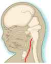
How does the ear develop?
- External auditory meatus develops from the 1st pharyngeal cleft
- Middle ear cavity and ossicles develop from 1st pharyngeal pouch and cartilages of 1st and 2nd arches respectively
- auricles develop from proliferation within the 1st and 2nd pharyngeal arches surrounding the meatus
How are the ears positioned?
- External ears develop near the neck initially
- As the mandible grows, the ears ascend to the side of the head to lie in line with the eyes
- All common chromosomal abnormalities have associated external ear abnormalities
Describe fetal alcohol syndrome
- There is no safe level of alcohol consumption during pregnancy
- Facial skeleton derived from neural crest cells populating the pharyngeal arches
- Neural crest migration as well as development of the brain are known to be extremely sensitive to alcohol
Summarise the pharyngeal arch derivatives

Summarise the trigeminal nerve in association with the pharyngeal arches
- The nerve of the first arch
- principal sensory nerve of the head: skin of the face, lining of mouth and nose
- motor innervation to: muscles of mastication, muscles derived from mandibular process

Summarise the Facial Nerve in association with the pharyngeal arches
- Nerve of the second arch
- passes through the stylomastoid foramen and parotid gland
- mostly motor: muscles of facial expression, muscles derived from 2nd pharyngeal arch
- small sensory component: taste buds in ant 2/3 of tongue

Summarise the Glossopharyngeal Nerve in association with the pharyngeal arches
- Nerve of the third arch
- Innervates stylopharyngeus muscle and provides general and special sensory to posterior 1/3 tongue

Summarise the Vagus Nerve in association with the pharyngeal arches
- Nerve of the 4th and 6th arches
- 4th arch branch is superior laryngeal nerve: innervates cricothyroid, constrictors of pharynx
- 6th arhce branch: recurrent laryngeal nerve: innervates intrinsic muscles of the larynx
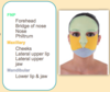
What are the fates of the facial prominences?
- FNP: forehead, bridge of nose, medial and lateral nasal prominences
- Medial nasal: philtrum, primary palate, mid upper jaw
- Lateral nasal: sides of the nose
- Maxillary: cheeks, lateral upper lip, secondary palate, lateral upper jaw
- Mandibular: lower jaw and lip
