1. Tibia & Fibula Flashcards
overall fxn of tibia
directly articulates w/ femur
weight is transferred thru condyles
overall fxn of fibula
serves as attachment site for muscles
not much weight/if at all is transferred
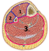
name the structure on the tibial plateau:
in green
what attaches here?

tibial tuberosity;
patellar ligament/tendon, & quadriceps tendon both attach here

name the structure on the tibial plateau:
in BLUE

intercondylar eminence/ prominences
*aka “spines” or “processes”

- name the structure(s) on the tibial plateau:
in yellow
- What attaches here?

anterior intercondylar area
ACL attaches here

- name the structure(s) on the tibial plateau:
in purple
- What attaches here?

posterior intercondylar area
PCL attaches

which color is the MEDIAL articular surface?
how do you know?

pink
•medial condyle is elongated, larger, less C-shaped,
whereas lateral is more C-shaped
which bone is this?
identify the surfaces

tibia
- lateral surface - more concave
- medial surface - less concave
- posterior surface

identify MEDIAL surface?
what attaches here?

2.
pes anserinas attaches here

on what surface is the nutrient foramen located?
what is it a branch of?

posterior surface (3)
nutrient artery is a branch of the posterior tibial artery (near it’s origin)

name the borders

- anterior border
- medial border
- lateral/interosseus border

name the muscular compartments located on the tibia
which surface does it correspond with?

(1) anterior compartment muscles (on lateral surface)
(3) posterior compartment muscles (on posterior surface)

describe the course of the anterior border of the tibia?
starts from TIBIAL TUBEROSITY proximally,
extends down anteriorly
forms anterior border of medial malleolus
describe origin and course of soleal line
starts below articulation of tibia and fibula
descends obliquely towards medial side, joining medial border

what inserts ABOVE soleal line?
what originates ON the soleal line?
popliteus inserts above soleal line (deep muscle of posterior compartment)
soleus originates ON the soleal line
the vertical line of tibia separates which 2 muscle attachments?
Tibialis posterior (laterally) and
Flexor digitorum longus (medially)
relationship of nutrient foramen and vertical line?
nutrient foramen is on posterior surface, LATERAL to the vertical line
where does the fibula attach distally?
what type of joint is this?
at the fibular notch
forms a syndesmosis
shape of articular facet on tibia for the talus?
comma shaped

from the lateral view, which COLLICULUS extends further distally?
the anterior colliculus

name the grooves, from medially –> laterally
on the posterior view of the tibia

- medial malleolus
- groove for TIBIALIS POSTERIOR
- groove for FLEXOR HALLUCIS LONGUS
- fibular notch

name the inferior surface of the tibia, which articulates w/ dome of the talus
the PLAFOND

of the plafond, which surface is longer?
(anterior/posterior)
Anterior is longer than posterior

CC: Trimalleolar fx includes…
Posterior malleolus (can incl part of plafond), medial malleolus, and malleolus of talus
significance of colliculi of tibia?
• Important attachment sites for ligaments/muscles, like deltoid ligaments of the ankle
where is apex of fibula located, relative to tibia?
approx 2 cm inferior;
lateral and posterior, on the tibial condyle

how to side a fibula?
take distal end;
facet is inverted triangle, & malleolar fossa is where you place the thumb.
You have the correct side if the thumb is in fossa and does NOT cross over the facet

identify bone:
name the key landmarks.

fiibula:
apex, head, neck, and medial flat portion to articulate w/ tibia

name key landmark on distal fibula

lateral malleolus

which muscles/ligs attach at:
head of fibula
- biceps femoris muscle
- lateral (fibular) collateral ligament
cc: clinical significance of neck of fibula
neck is weak area and is susceptible to fx
which nerve “Swoops” around the neck/superior aspect of fibula?
common fibular nerve
what are the 3 subcutaneous areas of the fibula?
- head of fibula
- triangular area immediately above lateral malleolus
- lateral malleolus

identify/name the borders

- top left: anterior
- top right: interosseus CREST
- bottomr right: medial
- bottom left: posterior

identify/name the surfaces

- top: anterior/medial surface
- right: posterior medial surface
- bottom: posterior lateral surface
- left: lateral surface

name the compartments

- anterior
- posterior
- lateral

identify the bones;
name the compartments

right and larger is TIBIA; left and smaller is FIBULA
- ANTERIOR
- N/A
- POSTERIOR
- LATERAL

COMPARTMENT 1: identify
- muscle category?
- action?
- pre or postaxial?
- anterior or posterior division?
- anterior compartment;
- dorsiflexors/extensors
- post-axial
- posterior division
COMPARTMENT 2: identify
muscle category?
action?
pre or postaxial?
anterior or posterior division of ventral rami?

- lateral compartment
- evertors/plantar flexors
- post-axial
- posterior division
COMPARTMENT 3: identify
- muscle category?
- action?
- pre or postaxial?
- anterior or posterior division of ventral rami?

- posterior compartment
- flexors/plantar flexors
- preaxial
- anterior division
name the 4 landmarks on the distal fibula

- purple: attachment of interosseus tibiofibular ligament
- green: triangular articular facet for talus
- blue: apex of lateral malleolus
- yellow: lateral malleolar fossa

what is found behind the lateral malleolar fossa?
malleolar crest
a combined tendon of the fibularis longus and fibularis brevis runs behind the lateral malleolus
gluteus maximus and tensor fasciae latae via iliotibial tract:
- name region/compartment
- list distal attachment/insertion
- gluteal region
- tubercle of IT band/Gerdy’s tubercle
quadriceps femoris:
- name region/compartment
- list distal attachment/insertion
- anterior comparmtent of thigh
- tibial tuberosity
sartorius:
- name region/compartment
- list distal attachment/insertion
- anterior compartment of thigh
- superior part of medial surface of tibia (pes anserinus)
gracilis:
- name region/compartment
- list distal attachment/insertion
- medial comparetment of thigh
- superiror part of medial surface of tibia (pes anserinus)
semitendinosus:
- name region/compartment
- list distal attachment/insertion
- posterior compartment of thigh
- superior part of medial surface of tibia (pes anserinus)
semimembranosus:
- name region/compartment
- list distal attachment/insertion
- posterior compartment of thigh
- posterior part of medial condyle of tibia
biceps femoris:
- name region/compartment
- list distal attachment/insertion
- posterior compartment of thigh
- lateral side of head of fibula
tibialis anterior
- name region/compartment
- list distal attachment/insertion
- anterior compartment of leg
- lateral condyle and superior half of lateral surface of tibia & interosseus membrane
extensor digitorum longus:
- name region/compartment
- list distal attachment/insertion
- anterior compartment of leg
- lateral condyle of tibia, and superior 3/4 of medial surface of fibula and interosseus membrane
extensor hallucis longus:
- name region/compartment
- list distal attachment/insertion
- anterior compartment of leg
- middle part of medial surface of fibula & interosseus membrane
fibularis tertius:
- name region/compartment
- list distal attachment/insertion
- anterior compartment of leg
- inferior 1/3 of medial surface of fibula and interosseus membrane
fibularis longus:
- name region/compartment
- list distal attachment/insertion
- lateral compartment of leg
- head and superior 2/3 of lateral surface of fibula
fibularis brevis:
- name region/compartment
- list distal attachment/insertion
- lateral compartment
- inferior 2/3 of lateral surface of fibula
soleus:
- name region/compartment
- list distal attachment/insertion
- SUPERFICIAL posterior compartment of leg
- multiple attachments
- posterior aspect of head & superior 1/4 of posterior surface of fibula
- soleal line & middle 1/3 of medial border of tibia
- tendinous arch extending between bone attachments
popliteus:
- name region/compartment
- list distal attachment/insertion
- DEEP posterior compartment of leg
- posterior surface of tibia; superior to soleal line
flexor hallucis longus:
- name region/compartment
- list PROXIMAL attachment/insertion
- DEEP posterior compartment of leg
- inferior 2/3 of posterior surface of fibula; inferior part of interosseus membrane
tibialis posterior:
- name region/compartment
- list PROXIMAL attachment/insertion
- deep posterior compartment of leg
- interosseus membrane; posterior surface of tibia inferior to soleal line; posterior surface of fibula
