1 - Pelvic Walls (Add Pictures) Flashcards
Pelvic Girdle:
Overview / Anatomical Function
Bony Components
- Attachment of lower extremities
- Transmits weight to lower extremities
- Protects pelvic organs
- Outlet for GI, Urinary, Reproductive systems
- Bones:
- R / L Pelvic Bones
- Sacrum
- Coccyx
Pelvic Girdle: Sacrum
Primary Body
Sacral Pomontory
Sacral Canal
Sacral Hiatus
- Primary Body
- Five rudimentary vertebrae which are fused together; articulates with L5 above, and coccyx below
- Sacral Promontory
- Anterior-Superior margin of vertebra S1 that projects forward
- Sacral Canal
- Continuation of vertebral canal; contains the cauda equina
- Sacral Hiatus
- Inferior opening of sacal canal; site of entry of needle for caudal injection/epidural

Pelvic Girdle: Coccyx
Body
Purpose
Canals
- Body:
- Formed from 4 coccygeal vertebrae fused
- Purpose:
- Site for muscle and ligament attachment
- Canals:
- None present
Pelvic Girdle: Pelvic Bones
Bone Components
Landmark of Intersection
Clinical Juvenile Structural Difference?
- Bone Components: Fusion of -
- Ilium
- Ischium
- Pubis
- Intersect at acetabulum
- Juvenile Difference:
- Three bones joined only by cartilage; thus gaps on x-ray in young patients could be confused for fractures
Landmarks of Ilium
Iliac Crest
Iliac Fossa
Anterior Superior Iliac Spine
Anterior Inferior Iliac Spine

Pelvic Girdle: Landmarks of Ischium
Ischial Tuberosity - What attaches here?
Ischial Spine - What attaches here? (2x)
Ischial Ramus
Greater Sciatic Notch
Lesser Sciatic Notch
- Ischial Tuberosity
- Roughened, posterior, inferior part of ischium
- Attachment site for sacrotuberous ligament
- Ischial Spine
- Pointed process from posterior border of ischium; lies between greater and lesser sciatic notches
- Attachment site for sacrospinous ligament and coccygeus muscle
- Ischial Ramus
- Inferior border of ischium; inferior to obturator foramen
- Greater Sciatic Notch
- Deep indention in posterior border of innominate bone superior to ischial spine
- Lesser Sciatic Notch
- Deep indentation in posterior border of ischium inferior to ischial spine

Pelvic Girdle: Landmarks of Pubis

N2K:
Pubic Symphysis
Body of Pubis
Obturator Foramen
Superior Pubic Ramus
Inferior Pubic Ramus
Pubic Arch

Pelvic Girdle: Pelvic Brim
Overview/Location
Components

- Overview:
- Bondy ridge surrounding and defining pelvic inlet
- Components:
- Promontory and Ala of Sacrum
- Right and Left Linea Terminalis
- Arcuate Line of Ilium
- Pecten Pubis
- Pubic Crest

Pelvic Girdle: Divisions of Pelvis in Relation to Pelvic Brim
False Pelvis (Greater Pelvis)
True Pelvis + Boundaries (Lesser Pelvis)
- False Pelvis
- Located above pelvic brim
- Part of posterior wall of abdominal cavity
- True Pelvis
- Located below pelvic brim
- Lower part of GI tract and urogenital organs
-
Boundaries:
- Pelvic Inlet (superior opening)
- Pelvic Outlet (inferior opening)
Pelvic Girdle: Fracture of Pelvis and Mortality
What organs must you assume damage to?
Fractures of pelvis are associated with high mortality due to pelvic organ damage and hemorrhage; one must assume damage to urinary bladder and urethra
Pelvic Girdle - Joints and Ligaments of Pelvis
Vertebral Joints (Vertebrae to Sacrum, Sacrum to Coccyx)
Sacroiliac Joint (Sacrrum to Iliac)
- Joints of Vertebral Column
- Lumbosacral / Sacrococcygeal Joints
- Sacroiliac Joint
- Transmits weight of most of body to hip bones
- Held together by strong, interosseuous ligaments and anterior and posterior sacro-iliac ligaments
Pelvic Girdle - Joints and Ligaments of Pelvis
Sacrotuberous Ligament
What is this ligaments relation to the sacrospinous ligament?
Broad attachment to sacrum and narrows inferiorly to attch to ischial tuberosity
Is located posterior to the sacrospinous ligament

Pelvic Girdle - Joints and Ligaments of Pelvis
Sacrospinous Ligament
What is its relation to the sacrotuberous ligament?
Triangular
Located anterior to sacrotuberous ligament

Pelvic Girdle - Joints and Ligaments of Pelvis
What prevents the upward tilting of sacrum due to the weight of trunk? (2x major ligaments)
What is an additional purpose of these structure?
Sacrotuberous / Sacrospinous Ligaments
Convert greater / lesser sciatic notches in Greater and Lesser Sciatic Foramina, e.g. make a “hole” from 1/2 bone + 1/2 ligament
Pelvic Girdle - Joints and Ligaments of Pelvis
Greater Sciatic Foramen - What passes through here?
Lesser Sciatic Foramen - What passes through here?
- Greater Sciatic Foramen
- Window through which neurovascular structures and piriformis pass from true pelvis into gluteal region
- Lesser Sciatic Foramen
- Window through which neurovascular structures and obturator internus pass from gluteal region (buttock) to the perineum (area around anal canal, external genitalia)
Measurements for Obstetrics:
Purpose
Methods
Purpose: Used to determine size of birth canal
Methods: Manually during vaginal exam, or with MRI
Measurements for Obstetrics:
Obstetric Conjugate
What blocks this measurement during pelvic exam?
- Minimum AP diamete of the lesser pelvis and narrowest fized distance through which baby’s head must pass in vaginal delivery
- From sacral promontory to the thickest margin of pubic symphysis
- Can NOT be measured during pelvic exam due to the bladder

Measurements for Obstetrics:
Diagonal Conjugate
What two structures are palpated? (will need both hands)
Measured by palpating the sacral promontory with tip of middle finger while using other hand to mark level of the inferior margin of the pubic symphysis on the examing hand
Measurements for Obstetrics:
Interspinous Distance
What structures does this measure between?
What does this represent?
Distance between ischial spines
Narrowest part of pelvic canal
Male vs Female Differences of Bony Pelvis
Pelvic Inlet - Shape?
Pelvic Outlet - Size?
Pelvic Cavity - Girth?
Angle of Pubic Arches - Hand mnemonic?
- Pelvic Inlet
- F - Oval
- M - Kidney (protrusion of sacral promontory)
- Pelvic Outlet
- F - Large
- M - Small
- Pelvic Cavity
- F - Wide
- M - Narrow
- Angle of Pubic Arches
- F - Wide (80-85 deg)
- M - Narrow (50-60 deg)
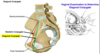
Walls of True Pelvis: What forms the following?
Lateral Wall - Bones? Muscle? Ligaments?
Anterior Wall - Bones? Ligamentous structure?
Posterior Wall - Bones? Muscle?
- Lateral Wall:
- Pelvic Bones
- Obturator Internus
- Sacrospinous and Sacrotuberous Ligaments
- Anterior Wall:
- Pubic Symphysis / bodies of pubic bones
- Posterior Wall
- Sacrum, Coccyx, Piriformis

Pelvic Diaphragm:
Anatomical Location
Passage of Urethra - Opening name?
Pierce @ midline = Anorectral Junction, what is this?
- Anatomical Location
- Floor of true pelvis
- Passage of Urethra
- Urogenital Hiatus (both sexes) and vagina
- Anorectral Junction
- Jx of anal canal and rectum
Pelvic Diaphragm:
Region Above - Major organs?
Region Below - Major organs?
- Region Above
- Main pelvic cavity of true pelvis
- Urinary bladder, rectum, uterus, etc
- Region Below
- Perineum
- External genitalia and outlet of gut and urogenital system
Pelvic Diaphragm:
What muscles make up the pelvic diaphragm? (2x)
What are they innervated by?
- Insert on midline structures; Innervated by Anterior Rami of Lower Sacral Nerves (S4, S5)
- Levator Ani
- Coccygeus

Levator Ani:
Origin
Insertion
Perinal Body & Child Birth clinical implications?
- Origin
- Ant - Body of pubis
- Mid - Tendinous Arch
- Post - Ischial Spine
- Insertion (structures located midline)
- Coccyx
- Anococcygeal Body
- Perineal Body
- Maintaining support of pelvic viscera
- If torn during childbirth, can compromise integrity of pelvic floor; could result in collapse of organs (prolapse)
- Subdivisions
- Puborectalis - Sling around anorectal jc (sphincter-like)
- Puboco

Levator Ani:
Subdivisions (3x)
Purpose of puborectalis?
Pubococcygeus
Illiococcygeus
- Puborectalis
- Sling around anorectal junction (sphincter-like)
- Pubococcygeus
- Inserts on anococcygeal body and coccyx
- Illiococcygeus
- Extends from tendinous arch and ischial spine to insert on coccyx and anococcygeal body
Urogenital Gap difference in male / female?
(vagina/urethra/perineal body orientation)
Male - Urethra in Urogenital Gap
Females - Vagina is anterior to perineal body; Urethra is anterior to vagina
Endopelvic Fascia
Anatomical Description - What is it continuous with?
Parietal Pelvic Fascia
Visceral Pelvic Fascia - Continuous with?
- Anatomical Description
- Lines pelvic cavity and covers pelvic organs
- Continuous with endoabdominal fascia
- Parietal Pelvic Fascia
- Invests muscles of pelvic walls
- Condensations of pelvic fascia, holds organs in place
- Visceral Pelvic Fascia
- Fascia on surface of viscera
- Continuous with parietal pelvic fascia
Sacral Plexus (Posterior Pelvis Wall)
Anatomical Purpose
Clinical Correlate and Sciatica?
Provides motor nerve supply to most lower limb muscles, pelvic diaphragm, muscles of perineum, pelvic viscera, and cutaneous sensory fibers to lower buttock and back of thigh
Clinical: Can be compressed by pelvic tumor (rectal tumor) or by head of fetus during childbirth resulting in sciatica
Sacral Plexus
Componsition
Location (in reference to major muscle)
Anterior Rami of L4, L5 (lumbosacral trunk) and S1 - S4
Located anterior to piriformis muscle
Sacral Plexus branches which pass through Greater Sciatic Foramen: (location in ref. to piriformis?)
Superior Gluteal Nerves
Inferior Gluteal Nerves
Sciatic Nerve (composed of?)
Posterior Cutaneous Nerve of Thigh
- Superior Gluteal Nerves
- Leaves pelvis through greater sciatic foramen above piriformis muscle
- Supply gluteal muscles of buttock
- Inferior Gluteal Nerves
- Leaves pelvis through greater sciatic foramen below piriformis muscle
- Supply gluteal muscles of buttock
- Sciatic Nerve
- Largest nerve in body
- Composed of L4-L5, S1-S3
- Leaves pelvis through greater sciatic foramen below piriformis muscle
- Posterior Cutaneous Nerve of Thigh
- Innervates back of thigh ; has perineal branch to posterior scrotum/labium majus
Sacral Plexus
Branches which pass through greater and lesser sciatic foramina:
Pudendal nerve - Somatic and Sensory?
Nerve to Obturator Internus Muscle
-
**Pudendal Nerve**
- Branches of Anterior Rami of S2-S4
- Passes through greater and lesser sciatic foramina to innervate somatic, skeletal muscles of perineum
- Provides sensory fibers to lower half of anal canal and _skin of external genitali_a
- Nerve to Obturator internus Muscle
Sacral Plexus
Branches which do not leave the true pelvis:
Branches to Pelvic Diaphragm
Pelvis Splanchnic Nerves - Nerve type?
- Branches to Pelvic Diaphragm
- Pelvis Splanchnic Nerves
- Composed of parasympathetic preganglionic fibers
- Innervates terminal ganglia, which innervate all hindgut derivatives
- Upper half anal canal, urinary bladder
