03-07 Non-Melanoma Skin Cancer Flashcards
Most common form of skin cancer? Second most common?
1 - Basal Cell Carcinoma #2 - Squamous Cell Carcinoma
What percent of skin cancers are associated w/ UV damage?
90%
6 Subtypes of BCC?
- Superficial
- Nodular
- Morpheaform
- Infiltrative
- Micronodular
- Pinkus Tumor
3 Subtypes of SCC?
- Actinic keratosis
- Squamous cell carcinoma in situ (SCCis)
- Aka Bowens Disease
- Squamous cell carcinoma
Dx?
- Typical Presentation
- Histo ∆s
- How malignant/concerning/Prognosis

Actinic Keratosis
- Presentation: Red, scaly plaques usu in sun-exposed areas
- Sometimes easier to feel (like sandpaper) than see
- Rarely: horn
- Pt may report pinprick pain or “doesn’t feel right”
- Tender to palp is good clue
- Histo (see image here)
- atypical keratinocytes only in the lower epidermis
- vs. full thickness in SCC
- Concern: Not alarming but 10% can progress to SCC

Dx?
- A.k.a.?
- Typical Presentation
- Histo ∆s
- How malignant/concerning/Prognosis

SCCis
- A.k.a. Bowen’s Dz
- Typical presentation: Well defined pink/brown scaly plaques
- Usu. on sun exposed skin
- Histo: Full thickness epidermal keratinocyte atypia
- vs. A.K. where atypia is only in lower epidermis
- Concern: ~26% go onto SCC

Name this subtype of SCCis?
- Etiology
- Other subtypes of SCCis

This is Erythroplasia of Queyrat, an erythroplakia of the glans of the penis in uncirc’d men.
- These sub-types are viral not UV-induced
- Erythroplakia can occur elsewhere as can:
- Leukoplakia (oral)
- Bowenoid papulosis (single or multiple small, red, brown or flesh-coloured spots or patches on the genitals) [seen on vulva here]

Dx?
- Typical Presentation
- Causes what % of skin cancer?
- Etiology
- Histo ∆s
- How malignant/concerning/Prognosis

SCC
- Presentation: red, scaly plaque or nodule in sun-exposed area
- scale usually central
- painful
- older folks
- maybe w/ horn
- may be erosive
- Causes 20% of skin cancer
- Etiology: Usually due to UV
- 90% have TP53 mutation
- Immunosuppression
- HPV
- Other causes: chronic inflammation, xrays, arsenic, BRAF inhibs, tobacco, EtOH
- Histo:
- hyperproliferative, eosinophilic keratinocytes
- varying degrees of atypia
- keratin pearls
- varying degrees
- can cause peri-neural invasion —> runs along nerves —> bad news
- hyperproliferative, eosinophilic keratinocytes
- Concern/Prognosis worse with:
- Location (face + ears - inner cheeks)
- Size
- 6mm in hi risk (grey) areas
- 10mm med areas, white–>
- >20mm every where else
- Recurrent
- Immunosuppressed
- Prior XRT
- Peri-neural involv
- Neuro sx
- Rapid Growth
- Breslow depth > 2mm

Dx?
- Presentation
- List Subtypes
- Causes what % of skin cancer?
- Etiology
- How malignant/concerning/Prognosis
BCC
- Presentation (see indiv cards)
- 60% nodular (see on opposite side)
- 15% superficial
- ~15 % Micronodular
- 5% Infiltrating
- 3% Sclerosing or morpheaform
- Most common cause of any kind of cancer in humans
- Etiology
- Histo ∆s
- How malignant/concerning/Prognosis
- Very low mortality, but significant morbidity if allowed to grow too big
- High risk factors:
- Location (see H here)
- Size >20mm (grey), 10mm (white here), 6mm (elsewhere
- Ill defined
- Recurrent
- Immunosuppressed patient
- Prior XRT
- Peri-neural involvement
- Subtype (?)

Dx?
- How common?
- Etio?
- Typical Presentation
- Histo ∆s
- How malignant/concerning/Prognosis

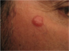
Nodular BCC
- 60% of BCC – most common
- Etio: Sun exposed skin – but not always
- Presentation: Pearly papule/plaque that can ulcerate
- “I shaved and nicked myself and have been bleeding since.
- Prognosis: Rarely metastasizes
- Histology: larger tumor nests, retraction artifact, peripheral pallisading (see here)

Dx?
- How common?
- Etio?
- Typical Presentation
- Histo ∆s

Superficial BCC
- 15% of all BCCs
- Etio: Usu sun-exposed areas
- Present w/ red, scaly plaque
- BCC vs eczema
- Histo: See here
- clefting (from washed out mucin deposits) deep to the lesion
- Pallasading around the edge

You have a patient w/ a red scaly rash recalcitrant to steroid tx. You send for biopsy and see this:
Dx?
- Typical Presentation
- Histo ∆s

Micronodular BCC
- Presentation is exactly the same as superficial
- Histo: nodules.
- ddx: vs. nodular
- this has smaller tumor rests
- tricky dx

You have a patient w/ a red scaly rash recalcitrant to steroid tx. You send for biopsy and see this:
Dx?
- Typical Presentation
- Histo ∆s

Infiltrative BCC
- Variable presentation:
- can mimic any of the other presentations of BCC
- Head and neck of old folk
- Histo
- normal epidermis
- epidermis: vertically-oriented strings of “basaloid” cells

Dx?
- Typical Presentation
- Histo ∆s

Morpheaform/Sclerosing BCC
- Scar-like presentation; very difficult to dx visually
- Hist
- wispy areas
- infiltrative strands of basaloid cells
- sclerotic stroma

Liquid Nitrogen Tx
- Appropriate For?
- MoA?
- Technique?
- Adverse effects?
- Indicated for:
- Tx of choice for A.K.
- Also for: SCCis, superficial BCC
- MoA: “Selective” Necrosis
- Damage caused directly to cell membranes and surrounding vasculature Mostly during the “freezing” portion of the cycle
- Technique: 15-60 freeze thaw cycles
- Hyperpigmentation or depigmentation
- melanocytes very reactive/sensitive
- Die at -4°C and liquid N is -196°C !
Electrodessication and Curettage
- Appropriate For?
- MoA?
- Technique?
- Adverse effects?
- Indication: hypertrophic AK, some SCCs, some BCCs
- MoA: high volt, low amp current
- gets superficial to mid-dermis lesions
- Techique: shock then scrape w/ curette
- Adverse effects:
Radiation Therapy
- Advantages
- Drawbacks
Radiation Therapy
Benefits
- No cutting
- Good cosmetic outcome on highly contoured areas
Drawbacks
- Many long tx’s
- Missing work, transportation issues
- Atrophy, scarring, dyspigmentation
- Can only treat each area one time
- May come back more aggressive.
- Can grow under scars (sneaky)
- Have to use surgery w/ bigger scar than would have had initially to remove that
Cetuximab
- MoA
chimeric (mouse/human) antibody
EGFR inhibitor
Vismodigeb
- Indication?
- MoA?
- Efficacy?
- For advanced/metastatic BCC
- Competitively inhibits “smoothened” in the hedgehog signaling pathway
- Response rates are relatively low <50% and duration only ~7months
Topical options for NMSC
- Indications
- Draw-backs?
For: AK, whole face for low-grade actinic ∆s
- 5-fluorouracil
- Imiquimod (Aldara)
- Ingenol mebutate
- Trichloroacetic Acid
- Diclofenac (NSAID)
Drawbacks
- only for superficial
- messy, hard to tolerat
- 5-FU causes this skin rash seen here (sign that it is working)

Photodynamic Therapy
- Appropriate For?
- MoA?
- Technique?
- Adverse effects?
Efficacy ~= creams
- Use for AK, some SBCC,
-
Technique Draw on patients’ lesion/whole face Use w/ 5-Ala to cause photo sensitive
- Photosensitizer applied to the skin for several hours
- Sensitized skin exposed to a given band of light to “activate” the photosensitizer (protoporphyrin IX)
- Just blue visible spectrum
- Low risk
- Very effective
- Long tx time + many txs
Surgical Resection
- Mohs vs. standard approach
- Mohs is more expensive, takes longer, requires specially trained Mohs-surgeon
- Std surgery has 90-95% cure rate, Mohs even higher
- Mohs has smaller scar b/c you removing less tissue
