Xray findings Flashcards

1
Q

What line is this?

A
  • McGregor’s Line
  • running from posteriosuperior hard palate to inferior occipital bone
2
Q

What measurement is shown here?

A
  • Atlantodental Interspace (ADI
  • adult normal ADI = <3mm
  • child normal ADI = <5mm
3
Q

What line does this image show?

A
  • George’s Line
  • alignment of the posterior vertebral body
4
Q

What alignment is shown here?

A
  • Atlantoaxial alignment
  • malalignment:
  • jefferson’s fracture
  • odontoid fracture
  • alar ligament instability
  • rotatory atlantoaxial subluxation
  • overhang of lateral mass and tilted dens
  • odontoid fracture
5
Q

What does an altered cervical curve signify?

A
  • not usually a correlation between altered curvature and symptomology
  • reduced or reversed curve could signify:
  • trauma
  • muscle spasm
    • degenerative spondylosis
6
Q

What is the normal Retropharyngeal Interspace (RPI)?

A
  • 5 to 7mm
7
Q

What is normal Retrotracheal interspace (RTI)?

A
  • Children: <14mm
  • Adults: <22mm
8
Q

What does this image show?

A
  • increased retropharyngeal interspace
  • increased retrotracheal interspace
9
Q

What are the ways of measuring a scoliosis?

A
  • Cobb’s Method
  • Risser-Ferguson Method
10
Q

What is a normarl Thoracic Kyphosis?

A
  • 20 - 30 degrees
11
Q

What is the significance of increased Kyphosis?

A
  • old age
  • osteoporosis
  • scheuermann’s disease
  • congenital abnoramlities
  • muscular paralysis
  • cystic fibrosis
12
Q

When would vertebral disc space be decreased?

A
  • degeneative disc disease
  • post surgery
  • postchemoneucleolysis
  • infection
  • congenital hypoplasia

*poor correlation between loss of disc space and LBP

13
Q

What does an increased lumbar lordosis do?

A
  • moves th nucleus pulposus anteriorly
14
Q

What angle does this image show?

A
  • The Lumbosacral Lordosis angle
15
Q

What does an increased lumbosacral angle signify?

A
  • produces low back pain by increasing the shearing and compressive forces on the lumbosacral posterior joints
16
Q

What angle does this image show?

A
  • A Lumbosacral disc angle
17
Q

What is the significane of an increased lumbosacral disc angle?

A
  • greater than 15 degrees is related to LBP casued by facet impaction
18
Q

What is the significance of a decreased lumbosacral disc angle?

A
  • acute disc herniation
19
Q

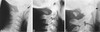
Describe the vertebral malpositions shown.

A
  • A: Overextension of the L4 vertebrae
  • B: Lateral flexion at specific segment b/c agle can be seen between the superior and inferior endplate
  • C: Laterolisthesis (lateral deviation) of L4 and Rotation of L3
  • D: Anterolisthesis shown by white arrow, retrolisthesis shown by black arrow
20
Q

What line is this an image of?

A
  • Macnab’s line
  • line drawn through the inferior enplate of a vertebrae through the superior articular facet
  • if the line is inferior to the superior articulating facet then there may be facet subluxation/dislocation
21
Q

What does anterior or posterior displacement on a flexion or extension view show?

A
  • this could mean instability usualy due to trauma
22
Q

What does this image show?

A
  • the 4 grades of spondylolisthesis
  • better shown in picture
  • the posterior inferior endplate of L5 aligns with the grade of slipage listed on the sacrum
23
Q

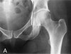
What is the normal value for the Teardrop sign?

A
  • <11mm
24
Q

How is the teardrop sign measured?

A
  • the distance between the medial margin of the femoral head and the outer part of the pelvic tear drop
25
What does a difference of \>1mm between legs mean?
- this is present in 90% of hip effusion
26
What is Waldenstrom's sign?
- lateral shift of the femur or medial widening of the joint space - sign of hip effusion
27
What line is this?
- Kohler's Line - line from the pelvic inlet to the outer border of the obturator foramen
28
What is shown in this image?
- Protrusio Acetabuli
29
What line is this?
- Shenton's line - this is the curved line from the femoral neck to the inferior margin of the superior pubic ramus
30
What is this an image of?
- this is an example of a hip dislocation - a sign of hip dislocatio is when shenton's line is disrupted
31
What is this line?
- Iliofemoral line
32
What does it mean if the Iliofemoral line is similar symetrically in bot hips?
- congenital hip dysplasia - slipped femoral capital epiphysis - dislocation - fracture
33
What is the range of normal for the femoral angle?
- from 120 to 130
34
What is the femoral angle called when \<120 degrees?
- coxa vara
35
What is the femoral angle called when \>130?
- coxa valga
36
What is this angle an example of?
- coxa vara
37
What is shown in these images?
- A: this is Klein's line, a line across the outer margin of the femoral neck, this should overlap the femoral head - C and D: Slipped femoral Capital Epiphysis can be seen as the Kelin's line does not intersect through the femoral head
38
What is Patella position?
- the relation between the patella length and the patellar tendon length - the patellar tendon lenth is within 20% of patella length
39
What could the patient be suffering from if the patella tendon length 20% mroe than the patella length?
- chondromalacia patellae - polio - achondroplasia - RA - tibial transposition
40
What joint space is shown here?
- Acromiohumeral Joint Space - this is the distance between the inferior acromiom and articular cortex of the humeral head
41
What is the Noraml Acromiohumeral Joint Space?
- 7 to 11 mm
42
What could a patient present with if the acromiohumeral joint space is \<7 mm?
- rotator cuff tear - degenerative tendonitis
43
What could the patient present with if Acromiohumeral Joint space is \>11 mm?
- dislocation - joint effusion - stroke - brachial plexus lesion
44
What joint space is shown in the image?
- Acromioclavicular joint space
45
What is the normal Acromioclavicular joint space?
- on average 3 mm - there also should not be a difference of 3 mm between the 2 AC joints
46
What can cause decreased acromioclavicular space?
- DJD
47
What can cause increased acromioclavicular space?
- traumatic separation - hyperparathyroidism - RA
48
What line is shown in the picture?
- Radiocapitellar line - drawn from the center of the radius through the capitellum - this line should always pass through the elbow
49
What may the patient be suffering from if the radiocapitellar line does not pass through the elbow?
- elbow dislocation
50
What sign is shown here?
- metacarpal sign - the line must pass through the articular cortex of the 4th and 5th metacarpals and must remain distal to head of the 3rd metacarpal
51
What doe sit mean if in the metacarpal sign the line passes through or proximal to the 3rd emtacarpal head?
- turner's syndrome - fracture deformity
52
What can be seen here?
- basilar impression as the odontoid process passes the McGregor's line
53
What does basilar impression usually accompnay?
- occipitalisation
54
What are the radiographical features of an arnold chiari malformation?
- MRI must be used to obtain a proper diagnosis in which the cerebellar tonsils lie lower then noraml - kinking or elongation of the fourth ventricle
55
Radiographic features of the occipitalisation of the atlas?
- anterior arch and posterior arch of the atlas is fused to the occiput - often see basillar impression as well - contrindication to adjustment b/c increases pressure on the vertebral arteries
56
What is this an image of?
- paracondylar process
57
What is this an image of?
- paracondylar process
58
What is this an image of?
- epitransverse process
59
Clinical presentation of a paracondylar process?
- jaw/facial muscle pain
60
What is shown on this radiograph?
- vertebralization of the atlas
61
What can be seen with vertebralization of the atlas?
- fusion of the anterior arch of the atlas with the basion - fusion of the posterior arch of the atlas with the axis (C2)
62
What does this radograph show?
- Agenesis of the Posterior Arch
63
What can be seen in the image?
- A: posterior tubercle of the posterior arch of the atlas is present but there is stress atrophy of the anterior tubercle (arrow points to this - B: partial agenesis of posterior atch (missing the middle connecting portion) - C: partial agenesis of the posterior arch (only a chunk of it is present) \*\* It is important to be able to differentiate bewteen the presence of a partial agenesis and a fracture, this can be differetiated by the hypertrophy of the anterior tubercle being present with agenesis
64
How to differentiate between the agenesis of the posterior tubercle and the fracture of ther posterior tubercle?
- if congenital agenesis the patient will also ahve hypertrophy of the anterior tubercle - if there ahs been a fracture there will not be hypertrophy of the anterior tubercle
65
What can be seen in this radiograph?
- Accessory Atlantoaxial Joint (Cervical Baarstrups diseas - this is the enlargment of the posterior arch of C1 which has made a psuedo joint witht he spinous process of C2
66
What is spina bifida occulta?
- congenital failure in the ossification of the midline of the vertebral neural arches
67
What is the following a radiograph of?
- C1 Spina Bifida Occulta
68
What are the radiological features of a spina bifida occulta?
- on pa non union of sp - on the lateral there is an absence of the spinolaminar junction line - on the lateral the anterior tubercle is usually enlarged
69
What are these radiographs of?
- posterior ponticles
70
Is there any clinical significance to the posterior ponticle?
- NO
71
When does agenesis of the Atlas Anterior Arch occur?
- congenital - seondary to: * RA * Tumor * infextion -
72
What is Ossiculum Terminale Persistens?
- non- fusion of the odontoid process - looks like a triangle on the odontoid
73
What does this radiograph show?
- Ossiculum Terminale - note the triangle corticie
74