White Blood Cells and Lymph nodes, Part 2: Acute Leukemias Flashcards
what is this an example of ?

neoplastic proliferations of white cells
what is this an example of?

neoplastic proliferation of white cells
what are the different types of Neoplastic proliferations of white cells?
- leukemia
- lymphoma
- plasma cell neoplasms
what is leukemia?
- neoplastic disease because of uncontrolled proliferation and incomplete maturation of hematopoietic precursors in bone marrow; displacing the remaining “normal” cells
- cells then enter into circulation and may damage other reticuloendothelial organs
neoplastic proliferation seen in leukemia is usually refered to as what?
presence of leukemia cells in peripheral blood leads to what?
“blast”
increase in total white cell counts
what is this?

lymphoma
what is this?

lymphoma
what are lymphomas?
what is an example of the tissues where it arises?
what are the 2 main categories of lymphomas?
proliferations arising as discrete tissue masses
example: lymph nodes
- Hodgkins lymphoma
- non-hodgkins lymphoma
what is this?
plasma cell neoplasm
what are plasma cell neoplasms?
where do they arise?
plasma cell tumors or terminally diff. B cell
arise mostly from bone marrow,
what are the Etiological and pathogenetic factors in white cell neoplasia?
what causes some of these?
1) Chromosomal translocation and other acquired mutations
2) Inherited genetic factors: Down syndrome, Bloom syndrome, Fanconi anemia, Ataxia telangiectasia
3) Viruses: HTLV-1, EBV, Kaposi sarcoma herpes virus/ human herpesvirus – 8 (KSHV/HHV-8)
4) Chronic immune stimulation: Helicobacter pylori association with gastric B- cell lymphoma (MALToma)
- gluten sensitivity enteropathy with intestinal T- cell lymphoma
Immune deficiency: Wiskott-Aldrich syndrome, HIV associated with B - cell lymphoma
5) Iatrogenic agents: radiation, chemotherapy, cigarette smoking
what are the types of acute leukemias?
1) Acute Lymphoblastic Leukemia (ALL)
2) Acute Myelogenous Leukemia (AML)
In what age group do we see ALL mostly?
In what age group do we see AML mostly?
what cells are seen in acute leukemias?
pediatric
40-60
“blasts”
what are these?

blasts seen in acute leukemia
what type of cell is ALL composed of?
the 85% of ALL cells are?
what cells does ALL affect in adolescents? What organ gets affected usually?
what are the risk factors for ALL?
What is the risk factor for T-cell ALL?
What is the risk factor for B-cell ALL?
what is the prognosis of B-cell ALL with t(9; 22) in adults?
what is the prognosis of B-cell ALL with t(9; 22) in children?
precursor B cells or T cells
pre-B cells
pre-T cells, thymus
chromosomal changes: especially Down syndrome (very likely to develop ALL)
NOTCH1 mutation
PAX5 mutation or translocation (12;21) involving ETV6/RUNX1, which promote maturation arrest
poor prognosis, good prognosis
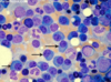
what is found in this peripheral blood smear?

leukemic blast cells of lymphoid origin
what can we expect from these lab values from ALL?
WBC
Hb
Platelet
Peripheral smear
Bone Marrow
anemia, neutropenia and thrombocytopenia will be seen
WBC = elevated
Hb = decreased
Platelets = decreased
Peripheral blood smear = circulating leukemic blast cells
Bone Marrow = presence of leukemic blast cells of lymphoid origin (usually more than 20%)
what is the most important organ to look at in a lab report for ALL?
how do you differentiate ALL from AML?
to make the distinction, what tool is used?
Bone Marrow
presence of lymphoblast will be seen and absence of Myeloblasts
use of histochemical staining for differentiation
what is this?

lymphoblast
what is this?

myeloblasts
what is being used here? what is it determining?
what is it negative for?

myeloperoxidase positive test
determines AML
ALL
what is being used here?
what is it determining?
what is it negative for?

sudan black positive
determines AML
ALL
what is being used here?
what is it determining?
what is it negative for?

PAS
ALL
AML
What will immunophenotyping reveal for ALL?
what is the only thing that wont be expressed by pre-B cell that will be by pre-T cell?
- terminal deoxynucleotidyltransferase (TdT)
- its expressed by both pre-B/T cell
- positive tdt indicates that the cells are blasts, as mature cells would be negative for tdt
- pre-B cell will show: CD10, CD19, PAX5
- pre-T cell will show: CD2, CD3, CD4, CD7 positive
CD10
what can a biochemical test reveal about ALL?
what other non-specific testing can reveal about ALL?
- serum lactate dehydrogenase (LDH) = increased
- serum uric acid = increased
- chest X-ray:
- thymic enlargement (with T- cell type ALL)
- mediastinal lymph node (with T- cell type ALL)
How important is CSF testing in ALL?
What other area is tested in ALL, considered the same as CSF?
essential
testes
what is this?

lymphoblasts in a peripheral blood smear
what are the clinical features of ALL?
What is a characteristic symptom of T-ALL?
- anemia, neutropenia and thrombocytopenia\
- abrupt stormy onset, bone marrow depression, bone pain and tenderness, generalized lymphadenopathy, CNS manifestations
T-ALL = anterior mediastinal lymphadenopathy
What is the treatment for ALL?
traditionally, the four components of ALL treatment are induction, consolidation (daunorubicin and cytosine arabinoside (Ara-C)), maintenance, and central nervous system (CNS) prophylaxis
what is Acute Myelogenous Leukemia?
In what age group does it happen the most?
what causes it?
tumor of hematopoietic progenitors caused by acquired oncogenic mutations that impede differentiation leading to accumulation of immature myeloid blasts in bone marrow
- adults 80%
- radiation exposure, chronic benzene exposure, chemotherapeutic drugs, smoking, Down synd, Bloom synd, Fanconi anemia. AML following MDS.
what is the pathophysiology of Acute Myeloid Leukemia?
- associated with acquired genetic alteration that inhibit terminal myeloid differentiation
- marrow is then replaced by undifferentiated ‘blasts’ with features of early myeloid differentiation
what are the most common chromosomal translocations leading to AML?
t(15;17) with M3,
inv 16 with M4,
t(8;21) with M2
AML following MDS
what environmental factors may lead to AML?
rubber, paint, embalming fluid, ethylene oxide, petroleum products, ionizing radiation
AML: M3 occurs on what population age?
what can be seen characteristically in myeloid cells?
what symptoms can be expected?
what risk is there?
what common feature can be seen?
with what translocation is it associated?
young males mostly
numerous Auer rods present
bleeding tendencies
DIC
Bone Marrow failure
t(15;17)
what is this?

Predominantly promyelocytes
Note: numerous granules
also, numerous auer rods

what is this?

M3 (Acute promyelocytic leukemia): majority of cells are heavily granulated leukemic promyelocytes
what is the clinical sign of Acute Monocytic leukemia M5?
for what test are people positive with AML M5?
what is the prognosis for this?
bleeding tendency + CNS involvement and extramedullary features (include: cutaneous and gingival infiltration)
positive for non-specific esterase (NSE)
poor prognosis
what can be expected in the lab values for Acute Myeloid Leukemia?
Total white count
Anemia?
Thrombocytopenia?
peripheral blood smear?
Bone Marrow?
Cytogenic studies?
Total white count = increased (could be decreased)
Anemia = yes
Thrombocytopenia = yes
peripheral blood smear = presence of atypical myeloid cells and circulating “blasts”
Bone Marrow = more than 20% blasts
what can be noted in this peripheral blood smear?

Auer rods
what are the clinical features of Acute Myeloid Leukemia?
Clinical Features:
- fever, splenomegaly, hepatomegaly, lymphadenopathy
- sternal tenderness, evidence of infection, hemorrhage
- easy bruising, petechiae, epistaxis, gingival bleeding, conjunctival hemorrhages, and prolonged bleeding from skin injuries…
- reflect thrombocytopenia and are frequent early manifestations of the disease
what are the clinical signs for Acute Myeloid Leukemia for M3?
what are the clinical signs for Acute Myeloid Leukemia for M5?
what are the clinical signs for Acute Myeloid Leukemia for M7?
gastrointestinal, intrapulmonary or intracranial hemorrhages, DIC
gingival infiltration, meningeal involvement
increased association with Down syndrome
what is a Myeloid (Granylocytic) Sarcoma?
how does it occur?
Myeloid sarcoma (also known as granulocytic sarcoma, chloroma, myeloblastoma, monocytoma) is a tumor composed of myeloblasts, monoblasts, or megakaryocytes
occur as extramedullary masses (including the skin; orbit; paranasal sinuses; chest wall; breast; heart; gastrointestinal, respiratory, or genitourinary tract; central or peripheral nervous system; and spleen)
what is the prognosis of AML?
what is the only means to induce a sustained remission in patients with AML who do not enter remission with cytotoxic drug therapy or who relapse after a first remission?
15-30% remain free of disease for 5 years
using allogeneic stem cell transplantation
