Week 3: Dermatology Flashcards
What are the eight characteristics that the FNP should describe of every skin lesion?
Number
Size
Color
Shape
Texture
Primary lesion
Location (specific landmarks)
Configuration (describing patterns)
What are examples of primary lesions?
Erosions Ulcers Nodules Ecchymosis Petechiae Palpable purpura
Flat lesion
can’t palpate with eyes closed
Macule
flat and < 1 cm
Patch
flat and > 1 cm
Raised lesion
palpate with eyes closed
Papule
raised, < 1 cm, not fluid filled
Plaque
raised, > 1cm, not fluid filled
What are some terms used to refer to the shape of a lesion?
Circular
Oval
Annular: ring-like with central clearing
Nummular: coin like, no central clearing
Polygonal

What are some terms used to refer to the texture of a lesion?
Smooth Fleshy Verrucous / warty Scaly Fine and dry (Tinea pedis) Hard and keratotic (Actinic keratoses) Greasy (Seborrheic dermatitis)
What characteristics would you expect of actinic keratoses? What conditions mimic actinic keratoses?
Hard and keratotic
- mimics superficial xerosis or seborrheic dermatitis

What characteristics would you expect of basal cell carcinoma (BCC)? What conditions mimic BCC?
pinky patch that doesn’t heal, possible focal scaling
mimics actinic keratosis and SCC in situ

What exam findings are concerning for malignant melanoma?
A - assymmetry
B - irregular border, esp notching
C- color, esp colors black, blue and red
D- diameter >6mm
E- evolving
E - elevated
F- firm
Growing
What are the characteristics of a spider angioma?
Fiery red, very small to 2cm
Central body, sometimes raised, surrounded by erythema and radiating legs
Pressure on body causes blanching of the spider
Face, neck, arms, upper trunk
Almost never below waist
normal/common on face and chest
Also seen in pregnancy and liver disease

What are the characteristics of a spider vein?
Blueish
Size variable; small to several inches
Shape: variable, spider, linear, irregular, cascading
pulsatility and effect: absent; pressure over center DOESN’T blanch but diffuse pressure blanches the veins

What are the characteristics of a cherry angioma?
Bright ruby read, purplish with age
1-3mm
Shape: round, flat, sometimes raised, may be surrounded by pale halo
Pulsatility and effect of pressure:
Absent, partial blanching, esp if pressure applied with edge of pinpoint
Location: trunk, extremities
Sig: none, increases in size and #’s w/ aging

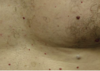
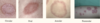
What are some examples of sun damage? How do they present?
Solar lentigo: bilateral symmetric brown macules located on sun exposed skin, face, shoulders, arms, hands
Solar elastosis: Yellowish white macules or papule in sun exposed areas, foreheads
Actinic purpura: ecchymoses only on dorsal forearms and hands but NOT extending above “shirt sleeve” line on upper arm
Poikiloderma: red patches in sun damaged areas, esp V neck, with fine telangiectasis and both hyper and hypopigmentation
Wrinkles: increased sun damage and tanning lead to deeper wrinkles at earlier age
Cutis rhomboidalis nuchae: deeper wrinkles on posterior neck that “criss cross)

What are some abnormal nail findings?
- paronychia: staph aures/strep; superficial infection of lateral nail folds; red swollen, tender
- clubbing: vasodilation w/ inc blood flow, hypoxia, CHD, interstitial lung disease, lung cancer, IBD, malignancies
- Habit tic deformity: depression on central nail; “christmas tree” ; repetitive trauma from rubbing index finger over thumb
- melanonychia: increased pigmentation in nail matrix; streaks; normal ethnic variation
- onycholysis: painless separation of opaque nail bed from pink nail bed; trauma, psoriasis, fungal infection, allergic reactions to nail cosmetics
- onychomycosis: nail thickening and debris; from tinea pedis, fungal
- terry nails: white nail plate with ground glass; liver disease, cirrhosis, HF, diabetes
- transverse linear depressions (beau lines): temp disruption of proximal nail growth from systemic illness; severe illness, trauma, raynaud’s disease
- pitting: punctuate depressions of nail plate by defective layering of superficial nail plate; psoriasis

Skin findings of someone with chronic renal disease?
pallor
xerosis
uremic frost (crystallized urea)
pruritus
half & half nails
calciphylaxis (Ca accumulates in small blood vessels of fat and skin tissues)
blood clots, painful skin ulcers, infxns
skin findings of somone with crohn’s disease?
- Erythema nodosum
- Pyoderma gangrenosum
- Enterocutaneous fistulas
- Aphthous ulcers
skin findings of somone with Cushing disease?
Striae
Atrophy
Purpura
Ecchymoses
Telangiectasis
Acne
Moon facies
Buffalo hump
Hypertrichosis
skin findings of someone with diabetes
Pruritus
Diabetic dermopathy
Acanthosis nigricans
Candidiasis
Neuropathic ulcers
Necrobiosis lipoidica
Eruptive xanthomas
skin findings of Dyslipidemias
Xanthomas (tendon, eruptive, tuberous; Lesions on skin with cholesterol and fat)
Xanthelasma

skin findings of Hypothyroidism
Dry, rough, pale skin
Coarse and brittle hair
Myxedema (think coma!!!)
Alopecia (lateral 3rd of eyebrows to diffuse)
Skin cool to touch
Thin and brittle nails
skin findings of hyperthyroidism
Warm, moist, soft, velvety skin
Thin and fine hair
Alopecia
Vitiligo
Pretibial myxedema (in graves disease)
Hyperpigmentation (local or generalized)
skin findings of Kawasaki disease
Mucosa erythema
Lips, tongue, pharynx
Strawberry tongue
Cherry red lips
Polymorphous rash (trunk)
Erythema of palms and soles
Desquamation of fingertips
skin findings of liver disease
Jaundice
Spider angiomas
Telangiectasias
Palmar erythema
Terry nails
Pruritus
Purpura
Caput medusae
skin findings of leukemia/lymphoma
Pallor
Exfoliative erythroderma
Nodules
Petechiae
Ecchymoses
Pruritus
Vasculitis
Pyoderma gangrenosum
Bullous disease
skin findings of meningococcemia
Angular stellate purpuric patches
Plaques with gunmetal gray center
Ecchymoses
Bullae
Necrosis

skin findings of Neurofibromatosis
Neurofibromas
Cafe-au-lait spots
Freckling in axillae (Crowe sign)
Plexiform neurofibroma
skin findings of Systemic lupus erythematosus
palmar erythema (mid cheeks, spans bridge nose)
Relative sparing of nasolabial folds
Periungual erythema
Interphalangeal erythema
In considering the APGAR, what does the score tell use about the newborn’s skin?
skin: 0 (pale, blue) 1 (pink body, blue extremities), 2 (pink all over)

How would you assess an infant’s skin for hydration?
Check turgor for hydration
Roll a fold of loosely adherent skin on Abdomen wall between thumb and forefinger
Tenting = NO NORMAL
Where would you assess an infant’s skin for cyanosis? What would you consider on the DDx for an infant with cyanosis?
Acrocyanosis (blue cast on hands and feet when exposed to cold) very common first few days
Central cyanosis at any age = congenital heart disease
Best area to look for central cyanosis: tongue and oral mucosa (NOT nail beds, lips, or extremities)
What are the four common dermatologic conditions in the newborn? How do they present?
Miliaria rubra: scattered vesicles on erythematous base; face/trunk; From obstructed sweat gland ducts; Disappears w/in few weeks
Erythema toxicum: erythematous macules with central pinpoint vesicles scattered diffusely over entire body; “flea bites”, disappear w/in 1st few weeks of birth
Pustular melanosis: small vesiculopustular over brown macular base; lasts several months; common in black infants
Milia: pinhead-sized smooth, white raised areas without surrounding erythema on nose, chin, forehead; retention of sebum in openings of sebaceous glands

What would be considering abnormal findings in the newborn skin assessment? What would be the DDx for these?
- jaundice: abnormal if present within 24 hours (hemolytic disease of newborn); normal on 2nd or 3rd day, peaks day 5, gone 1 week
- midline hair tuft: over lumbosacral spinal region; Spinal cord defect
What are some benign birthmarks found in infants? What do they look like?
Eyelid patch - fades w/in 1st year
Salmon patch / stork bite / angel kiss
Cafe au lait spots: Isolated lesions have no significance but multiple lesions with sharp vorders suggest neurfibromatosis; light brown pigmented lesions have borders and uniform
monogolian spots/Slate blue patches: common dark skinned babies
Harlequin dyschromia: Transient cyanosis of ½ of body or 1 extremity, from vascular instability
In the pediatric population, what are some wart-like lesions and how do they appear on exam?
Verruca vulgaris (Dry, rough warts on hands)
Verruca plana (small, flat warts)
Plantar warts
Molluscum contagiosum
Adolescent acne
What are some commons skin lesions during childhood and how do they appear on exam?
Bacterial (impetigo, furuncle or boil, folliculitis, carbuncle)
fungal (cadidiasis, tinea (capitis, corporis, veriscolor, pedis, cruris, unguium)
viral (verrucace [common, plantar, flat], molluscum contagiosum, herpes simplex type 1 & 2)
infestations (pediculosis, pediculosis capitis, scabies)
inflammatory disorders (atopic dermatitis, diaper dermatitis, seborrheic dermatitis, contact dermatitis, acne vulgaris)

cutis mamorata
Premature infants or first few months of life, trunk/legs
Infants with conventional hypothyroidism or Down syndrome
If acrocyanosis doesn’t disappear within 8 hrs or with warming, consider congenital heart disease
in cold environment

erythema toxicum
Very very common in NB
Day 2-3 days of life
Erythematous macules with central pinpoint macule
Diffuse, flea bite looking

Pustular Melanosis
More common in black infants
Vesicle pustules that form over brown macular base
Last several months
Resolves w/o tx

Miliaria Rubra
Scattered vesicles on erythematous base on face
Caused by obstruction of sweat glands
Disappear spontaneously
No intervention needed, just reassurance

Diaper Dermatitis
Skin is raw, excoriated, sloughed off
Caused by irritant, stool, diaper
Use barrier cream, letting air get to the area
Common

Candida / yeast infection of diaper area
Erythematous papules with some satellite lesions
Any rash > 72 hrs in diaper area has to do with yeast and will be treated as yeast

impetigo
Bacterial infection of skin caused by strep or staph
Very contagious
Moist vesicles on erythematous base
When vessels rupture, produce honey colored cresting
Bullous impetigo present as larger lesions
When erupt = see red, shiny rough skin under
By staph

Molluscum Contagiosum
Very very common in peds
Caused by pox virus
Pearly, colored dome shaped papules with central umbilication
Form in clusters , even linear
If scratch = spread

head lice
Difficult to dx
Significant pruritis
Overnight worsens
Occipital or postauricular areas
Lice is oval shaped, firmly attached to hair shaft
(½ inch up from scalp)
Whitish gray nit attached to shaft

Fifth disease (erythema infectiosum)
caused by parvovirus B19
Starts with mild fever, rhinitis, h/a
Slapped cheap appearance 3-5 days into the illness
Once rash shows, no longer contagious
5th disease bc 5th disease we used to see in childhood illness

Coxackie Virus (Hand, foot, mouth disease)
Sometimes pruritic
Mouth lesions very uncomfortable
a/s with anorexia, sore throat, LOA, very very fussy kids/infants
Supportive care for tx

Roseola
By herpes virus
Most kindergartens exposed by 5
Very common under 2 yo
2-3 days of high fever and irritability
Break out in rash over trunk and extremities
Pink maculopapular rash

varicella/chicken pox
“Dew drop on a rose petal”
Vesicle on erythematous base
Pruritis, fever, unwell sx’s
Lesions become infected with staph → sepsis → death

measles
High fever, cough, runny nose, diffuse pinkish
Red macupapulses forming together
What does blanching indicate? nonblanching?
blanchable: erythematous/inflamed; erythematous wheel suggest inflammation
nonblanching (petechiae, purpura, vascular structures like cherry angioamas, vascular malformations): NOT erythematous but rather red, purple, violaceous/purple
petechia/pupura
deep red or reddish purple fading over time
petechia, 1-3mm; pupura larger
shape: rounded, sometimes irregular; flat
NON-blanchable
significance: blood outside vessels suggest bleeding disorder; palpable purpura in vasculitis

ecchymosis
purple or purplish blue, fading to green yellow and brown with time
variable in size, larger than petechiae (>3mm)
shape: rounded, oval, irregular; may have central cubcutaneous fat nodule (hematoma)
NON-blanchable
significance: trauma, bleeding disorders
