Week 1 - Introduction Flashcards
Body Regions - Anterior

1 - Head
2 - Neck
3 - Thorax
4 - Abdomen
5 - Pelvis
Limb Regions - Anterior

1 - Shoulder
2 - Arm (brachium)
3 - Cubital Fossa
4 - Forearm (antebrachium)
5 - Wrist
6 - Hand
7 - Groin
Body Regions - Posterior

6 - Back
7 - Gluteal Region
Limb Regions - Posterior

8 - Hip
9 - Thigh
10 - Popliteal fossa
11 - Leg (crus)
12 - Ankle
13 - Foot
Supine
Prone
Lying face Up
Lying face Down
Superficial
Deep
Closer to body surface
Farther from surface
Superior
Inferior
Closer to head
Closer to Feet
Proximal
Distal
Closer to heart
Farther from heart
Medial
Lateral
Closer to midline
Farther from midline
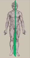
Sagittal Plane
divides body into left/right sections

Coronal Plane
divides body into anterior/posterior sections

Axial Plane
divides body into superior/inferior sections; by convention, viewed from inferiorly (used in CT/MRI)

Leg Connective Tissue

1 - Dermis (inner skin layer)
2 - Subcutaneous fat
3 - Bone
4 - Deep fascia
5 - Tendons
6 - Adventitia (outer layer of blood vessels)
7 - connective tissue surrounding nervous
Leg Muscle Groups

1 - Deep fascia
2 - Fascial Septa
3 - Periosteum
4 - Tendons
Axial Bones - Anterior

1 - Skull
2 - Mandible
3 - Teeth
4 - Sternum
5 - Ribs (12)
Axial Bones - Posterior

6 - Cervical Vertebrae (C1-C7)
7 - Thoracic Vertebrae (T1-T12)
8 - Lumbar Vertebrae (L1-L5)
9 - Sacrum
10 - Coccyx
Appendicular Bones - Posterior

8 - Ilium
9 - Pubis
10 - Ischium
11 - Femur
12 - Tibia
13 - Fibula
14 - Tarsals
15 - Metatarsals, Phalanges
Appendicular Bones - Anterior

1 - Clavicle
2 - Scapula
3 - Humerus
4 - Radius
5 - Ulna
6 - Carpals
7 - Metacarpals, Phalanges
Axial Joints - Anterior

1 - Temporomandibular
2 - Laryngeal
3 - Chondrosternal
4- Costochondral
Axial Joints - Posterior

6 - Atlanto-occipital
7 - Costovertebral, Costotransverse
8 - Lumbosacral
Appendicular Joints - Anterior

1 - Pubic symphysis
2 - Sternoclavicular
3 - Acromioclavicular
4 - Glenohumeral
5 - Elbow
6 - Radioulnar
7 - Radiocarpal
Appendicular Joints - Posterior

8 - Sacroiliac
9 - Hip
10 - Knee
11 - Talocrural
12 - Pubic symphysis (cartilaginous)
Dorsiflexion
Dorsal/superior surface moves closer to anterior surface of leg

Plantarflexion
plantar (volar, inferior) surface moves closer to posterior surface of leg

Solid Organ Components

1 - Parenchyma - Functional Epitthelial Cell
2 - Tissue Stroma - Connective tissue that supports, organizes parenchyma
Hollow Organ Components

1 - Lumen - space
2 - Inner epithelial layer
3 - Outer connective tissue layer
4 - Alternating layers of connective tissue, muscle
Respiratory System

1 - Larynx
2 - Trachea
3 - Bronchi
4 - Lungs
Digestive System

1 - Oral cavity
2 - Salivary gland
3 - Esophagus
4 - Stomach
5 - Duodenum
6 - Liver
7 - Pancreas
8 - Jejunum
9 - Ileum
10 - Colon
11 - Rectum
Male Urogenital System

1 - Kidneys
2 - Ureters
3 - Bladder
4 - Urethra
5 - Testes
6 - Vasa deferentias
7 - Seminal vesicles
Female Reproductive System

1 - Ovaries
2 - Uterine tubes
3 - Uterus
4 - Vagina
Endocrinine System

1 - Pituitary gland
2 - Thyroid gland
3 - Parathyroid gland
4 - Adrenal (suprarenal) glands
5 - Pancrias
6 - Kidney
7 - Testes
Lungs

1 - Lungs
2 - Mediastinum
2a - Parietal pleura
2b - Visceral pleura
Central Nervous System Components

- Brain
- Spinal Cord
Bones of the CNS

- Cranial Cavity surrounded by the SKULL
- Vertebrae and intervetebral disks surrounded by VERTEBRAL CANAL
Meningeal Layers of the CNS

- PIA MATER- adheres to brain and spinal cord
- ARACHNOID MATER- surrounds the pia mater
- CEREBROSPINAL FLUID (CSF)- fills the space between pia mater and arachnoid mater
- DURA MATER- adheres to the inside of the skull/lies within the vertebral canal
Peripheral Nervous System
1- Components (and # of nerves per type)
- Where each type of nerve exits the cranial cavity/spinal column
- The Cranial Nerves (1-12)
The Spinal Nerves (C1-C8, T1-T12, L1-L5, S1-S5, and coccygeal 1)
- Cranial nerves enter and leave the cranial cavity via FORAMINA (small passages) in the skull
Spinal nervers enter and leave the spinal column via INTERVERTEBRAL FORAMINA
Somatic Motor Fibers (GSE)
Convey motor impulses from the CNS to skeletal muscle directly.
The nerve cell body lies in the CNS
Visceral Motor Fibers (GVE)
Convey sensation from CNS to smooth muscle, cardiac muscle, glands, or chromaffin cells VIA the SYMPATHETIC GANGLIA (sympathetic impusles) or the PARASYMPATHETIC GANGLIA (parasympathetic impusles)
The nerve cell body lies in the CNS
Somatic Sensory Fibers (GSA)
Convey sensation from skin, musculoskeletal structures, and the parietal linings of the body back to the CNS.
The nerve cell body lies in the SENSORY GANGLIA- note that this does not mean the sensation must pass through a synapse
Visceral Sensory Fibers (GVA)
Convey sensation from the respiratory, digestive, urogenital, and circulatory systems back to the CNS
The nerve body lies in the SENSORY GANGLIA- note that this does not mean that there are more than one nerve fibers (and no synapses)
Special Sensory Fibers (SSA)
Convey sensations from the eye, olfactory mucosa, taste buds, and inner ear.
NOTE- only in cranial nerves
Spinal Nerves - Cross Section

1 - Anterior (Motor) Root
2 - Posterior (Sensory) Root
3 - Spinal Nerve
4 - Anterior Ramus
5 - Posterior Ramus
Components of Blood
Plasma (55%)
Buffy Coat (leukocytes and platelets; <1%)
Hematocrit (erethryocytes; 45%)
Heart

1 - Right Atrium
2 - Right atrioventricular (tricuspid) valve
3 - Right ventricle
4 - Pulmonary valve
5 - Pulmonary trunk
6 - Left atrium
7 - Left atrioventricular (mitral) valve
8 - Left ventricle
9 - Aortic valve
10 - Aorta
Anastomosis
Natural communication (direct or indirect) between 2 blood vessels (or other tubular structures); can provide alternative pathway for blood
Shunt
Bypass or diversion
Fistula
Abnormal communication between tubular structures (e.g. arteriovenous fistula)
Arteries

1 - Pulmonary arteries
2 - Lungs
3 - Aorta
4 - Arterial network
5 - Capillary beds
Veins

1 - Pulmonary veins
2 - Superior vena cava
3 - Inferior vena cava
4 - Capillary beds
5 - Portal vein
Abdomen

1 - Intestines
2a - Parietal peritoneum
2b - Visceral peritoneum
3 - Mesentery
4 - Retroperitoneum