Vestibular and Auditory System Flashcards
(32 cards)
What are the two types of hearing loss?
- Conductive: blockage of sound conducting path from source to cochlea–treatable and reversible
- Sensorineural: damage to inner ear or central auditory pathway (hair cell loss, auditory nerve loss, vascular damage)–incurable, permanent hearing loss
What causes pain in the ear?
Pain from noise is from activation of nerve endings which occurs over about 150 dB depending on the frequency of the sound

Describe the flow of sound in the outer and middle ear.
Sound is air pressure/movement of molecules (condensation and rarefaction of air molecules). Sound flows from the environment to the outer ear. The pinna funnels sound into the ear and sound hits the tympanic membrane. The middle ear consists of the malleus, incus, and stapes which converts and amplifies the air pressure waves into fluid pressure waves in the inner ear.
Inward movement of the tympanic membrane occurs during the compression phase of a sound wave; outward movement of the tympanic membrane occurs during the rarefaction phase of the sound wave.

Where does vibrational input to the inner ear get transmitted?
The vibrational input from the stapes footplate is into the scala vestibuli and the pressure release from these vibrations is via the round window and the end of the scala tympani. For sound to get from the upper to lower chamber, the vibrations must pass through the scala media where the hair cell of the Organ of Corti are located. This causes vibration of the basilar membrane.
What are the endolymph and perilymph? What is the relevance of their ionic concentrations?
Endolymph is located in the scala media while perilymph is located in the scala tympani and vestibuli. The ionic gradients between the endolymph and perilymph generate a positive voltage of +80 mV in the scala media called the endolymphatic potential, which is fundamental to cochlear function.
What do the outer and inner hair cells do? How do they work?
Inner hair cells are in contact with the tectorial membrane, and movement of the organ of Corti causes deflection of hair cells. When the hair cells move toward the largest hair, filaments stretch and the channels open, depolarize, and release neurotransmitter. This causes action potentials in the nerve. When the channels close, the hair cells hyperpolarize.
Basilar membrane movement causes stereocilia to bend which is translated into opening and closing of channels which becomes synaptic release and causes an action potential.
What do high frequency versus low frequency sounds cause? How are these signals transmitted?
High frequency sounds elicit vibrations at the base (thin) of the basilar membrane while low frequency sounds elicit vibrations at the apex (thick) of the basilar membrane.
Changes in hair cell potential follow low frequency sounds because there is rhythmic opening and closing of channels (and therefore vesicle release). Temporal information is maintained by spacing between action potentials.
Hair cell potential does not return to normal during high frequency sounds because membrane depolarization is constant.
What is range of hearing determined by?
The frequencies at which the basilar membrane vibrates–first third 20-200 Hz, second third 200-2000 Hz, last third 2000-20,000 Hz.
Speech is made of multiple frequencies so different parts of the membrane vibrate at the same time which stimulates different axons which carry this information to the brain.
What types of fibers does the cochlear nerve contain?
- Type I afferents: 8-12 per inner hair cell (myelinated)
- Type II afferents: 1 afferent per 14 outer hair cells (unmyelinated)
- Both types of cells receive efferent feeback
Describe the properties of the cochlear nerve–i.e. what is it about them that permits the brain to process sound?
Different fibers in the nerve will be activated depending on what region of the basilar membrane vibrates. When a particular neuron is firing, it represents the frequency (so the temporal pattern of activity is not the only thing that dictates frequency).
Describe the function of the outer hair cells.
Outer hair cells exhibit mechanical contractility–they are motor effectors. Outer hair cells feed back energy into the basilar membrane which boots the stimulus to the inner hair cell. If outer hair cells die, the sharpness of type I afferent tuning curves from the inner hair cells are blunted (poorer frequency selectivity)
What are the six major synaptic steps?
- Hair cells
- Cochlear nuclei
- Superior olivery nuclei
- Inferior colliculus
- Medial geniculate nucleus
- Auditory cortex
What is the interaural time difference? How is it computed?
The difference in the time of arrival of a sound at two ears. The eighth nerve synapses in the cochlear nucleus which projects bilaterally to many cells in the medial superior olive which act as coincidence dectectors. When a cell is stimulated by signals from both cochlear nuclei simultaneously it fires stronly and projects up through the lateral lemniscus to the inferior colliculus. The inferior colliculus relays this information to the medial geniculate complex of the thalamus which sends it to the primary auditory cortex.
What is the interaural level difference?
Sounds that are not directly ahead reach the ears with different intensities. This information can also be used to localize sound. Exons from the cochlear nucleus projects bilaterally to the lateral superior olive, however, ipsilateral input is excitatory and contralateral input is inhibitory. If the exicitory input reaching the LSO exceeds the contralateral inhibitory input, there is a net excitation which is transmitted to higher cortical centers.
What are monoaural cues used to determine?
Sound bounces around the pinna so it helps you determine if it is coming from the front/back or above/below.
Describe the main levels of processing in the auditory cortex. How is it organized?
The auditory cortex is called Heshel’s gyrus. Primary auditory cortices talk to each other as well as adjacent belt areas. Frequency information is represented in a tonographic fastion–progression of frequency from low to high.
Core regions: tonotopic, responds to simple sounds
- Includes AI (primary auditory cortex), R (rostral area) and RT (rostrotemporal area
Belt regions and parabelt regions: not tonotopically organized, respond to complex sounds
- Selectivity for certain types of sounds increases between the primary and non-primary auditory cortices
How does the cortex sort different sound sources? What clinical relevance does this have?
The cortex detects regularities in an auditory stimulus and link’s them together to form a perceptual representation called auditory objects. These representations are based on abstract, higher-order information (still recognize something even if a variable is changed) Binding keys are:
- Location
- Similarity: timbre, pitch
- Proximity: tones close in frequency
- Good continuation
Older adults and those with cochlear implants have impaired sorting ability.
Describe the ventral and dorsal stream with respect to perceptual representations of a sound.
Dorsal processing stream is used to determine where a sound is
Ventral processing stream is used to determine stimulus-object identity (what a sound is)
What types of eye movements exist?
Voluntary
- Conjugate eye movements: smooth pursuit, optokinetic, vestibular, saccades
- Disjunctive: vergence
Involuntary
- Fixation movements
Each type of eye movement has its own neural control system.
How do extraocular motor neurons have to fire in order to generate a saccadic eye movement in its direction of action?
A large high frequency pulse (burst to overcome the inertia/viscous resistance) followed by a much smaller step (to overcome the elastic restoring forces that hold the eye in its new postion). The step is the result of the neural integration of the output of the pulse generator. Both are required to move the eye and hold it in place. Conjugate eye movements are maintained during horizontal gaze.
What brainstem structures are involved in the initiation of horizontal saccades?
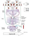
The paramedian pontine reticular formation is the pulse generator for horizontal eye movements and is under the control of the frontal eye fields and the superior colliculus. The PPRF signals to the abducens to contract the lateral rectus (pulse) and simultaneously signals to the neural integrator (nucleus prepositus hypoglossi) to produce a step in firing rate which is also sent to the abducens to maintain the position.
The superior colliculus is responsible for initiating saccadic eye movements in response to sudden appearance of visual or auditory stimuli. The FEF initiates voluntary saccades.
How is conjugate gaze mediated?
Fibers from the abducens nucleus project to the contralateral oculomotor nucleus (medial rectus portion) via the MLF to stimulate the conjugate eye movements.
What is the purpose of the vestibular system? Describe its anatomy.
The vestibular system senses motions of the head (linear and angular acceleration) and reports these to the brain. The sensory functions are mediated by hair cells in the utricle, sacculus, and in swellings of each of the three semicircular canals. Their outputs are carried by axons of the vestibular nerve (cell body lies in Scarpa’s ganglion).
Describe how vestibular receptors respond to linear acceleration.
In the utricle and saccule, the cilia protrude through a gelatinous layer that contains calcium carbonate crystals (otoconia) which cause the cilia to bend and depolarize when accelerated. Signals are tonic in the case of head tilt and transient in the case of linear acceleration.


