Thalamus and Cortex Flashcards
(44 cards)
What two major structures make up the diencephalon?
Thalamus and hypothalamus
What structures limit the thalamus laterally and medially? What structure is ventral to it?
Laterally: internal capsule
Medially: third ventricle
Ventral to the thalamus is the hypothalamus.
How is the thalamus divided?
The internal medullary lamina divides the medial and lateral nuclei of the thalamus and forks at the anterior end to divide the anterior nuclei. There are intralaminar nuclei as well. A sheet of cells forms a nucleus that surround the thalamus in the lateral and dorsal portions called the nucleus reticularis.
What three types of circuits does the thalamus have?
- Relay circuits
- Association circuits
- Non-specific circuits
What are the three types of relay circuits? Where do they project?
Sensory:
- Lateral geniculate nucleus receives visual information and projects to the visual cortex
- Medial geniculate nucleus relays auditory information to the auditory cortex
- Ventral posterolateral nucleus receives somatosensory information and projects it to the somatosensory cortex
Motor:
- VA/VL receives inputs from the cerebellum and basal ganglia and projects it to the cerebral cortex
Limbic:
- Mammillary bodies send projections to the anterior nucleus of the thalamus which relays it to the cingulate gyrus (Papez circuit)
What are the two types of association circuits? Where do they project?
The association circuits receives cortical inputs and projects back to the same areas
- Pulvinar: serves the parietal-occipital-temporal association cortex (receives and projects back to these areas of the cortex)
- Mediodorsal nucleus: serves the prefrontal cortex
Which nuclei are most involved in non-specific circuits? Where do they project and what do they do?
Involves mainly intralaminar nuclei whic send non specific projections to widespread areas of the cerebral cortex to produce changes in cortical function and brain state.
Intralaminar nuclei also receive inputs from the basal ganglia and project strongly in return.
How do thalamic nuclei interact with each other? Which nucleus does not project outside the thalamus? What does it do?
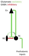
Thalamic nuclei do not project to each other at all, except for the reticular nucleus which receives collaterals from projections to and from the thalamus and cortex. Their projections are exclusively GABAergic and project only to other thalamic nuclei and do not project to the cortex at all.
What kinds of projections go to and from the cortex?
Projections to and from the cortex are reciprocal excitatory (glutamatergic) connections. Prethalamic inputs to the thalamus are also excitatory.
Describe the general structure of the cortex in terms of layers, columns, and cytoarchitecture.
The cerebral cortex is organized into six layers that differ in depth below the pia, packing densiry, and the presence of fiber bundles. These differences are aligned horizontally and differ between cortical regions (basis of cytoarchitecture).
Columnar organization is also evident in that cells within a column show the same orientation or respond to the same type of stimulus. This probably reflects massive parallel processing–applying similar operations locally to many aspects of the same information.
Cytocarchitecture involves defining cortical areas in terms of parameters revealed by stains of RNA of cell bodies–often corresponds well to functional subdivisons of teh cortex.
What is the difference between agranular and granular cortex?
Agranular cortex includes areas that give rise to many long axons and, therefore, have many large pyramidal cells in all layers (especially 5). It lacks stellate (granular) cells.
Granular cortex does not have many pyramidal cells and does not give rise to many long axons, and is instead dominated by small cells.
Where does thalamic input to the cortex go? Describe the cortical circuit that follows.
Thalamic inputs go to layer IV of the cortex. This information then travels up to supragranular layers (II/III) and then down to infragranular layers (V/VI) while activating inhibitory cells in the process.

Where do the different layers of the cortex send outputs to?Where do they receive inputs from?
Layer II sends projections to other cortical layers, layer III sends projections to the opposite hemisphere, layer V projects to subcortical structures (striatum, superior colliculus, brainstem, spinal cord), layer VI projects to the thalamus.
All layers receive input from brainstem modulatory systems.
Layers I, II, IV, and V receive inputs from other cortical areas.
Only layer IV receives thalamic input.

What is the gate function of the thalamus?
Control of the functional state of the forebrain–transition between waking and sleep states. This is mediated by changes in neuromodulation.
What are the functions of the cortex?
- To generate sensory and motor representations of external and internal worlds using a comination of sensory driven activity, internally generated activity, and memory storage
- To generate consciousness
- These functions depends on the recurrent loops between the cortex and thalamus
When and where are alpha waves seen?
Alpha waves are seen in occipital leads only when eyes are closed.
How does brain activity change during wake and sleep?
When awake, waves are low amplitude and high frequency. During deep sleep, high amplitude and low frequency (delta waves).
Thalamic firing patterns change during the transition from waking to sleeping as well–during waking, irregular single thalamic spikes represent the inputs from sensory stimuli while it fires in bursts separated by silences during sleep (rhythmic). The thalamus is responsible for the change in brain firing mode.
Explain how calcium mediates the two thalamic firing states.
When the cell is hyperpolarized, the calcium channels exist in three states–they can go from inactivated to closed so depolarization can cause them to open and fire. They can then inactivate and switch to the closed state when the cell hyperpolarizes. The calcium influx causes a slow depolarizing wave that initiates a burst of action potentials.
At normal resting potential, the cell is too depolarized to allow the calcium channels to close which prevents the burst state. Instead, depolarization initiates a train of action potentials (tonic mode).
What are the three main consequences of burst mode firing?
- Unreliable responses: thalamus cannot respond faithfully to stimuli due to inactivation of channels. The bursts also do not accurates reflect the amplitude of the input.
- Oscillations: thalamic cells generate rhythmicity
- Large scale synchrony in thalamocortical networks engages the cortex (delta waves) so that all cells oscillate together
What controls the membrane potential of thalamic cells?
The membrane potential of thalamic cells is controlled by brainstem and basal forebrain neuromodulatory systems with diffuse projections to thalamus and cortex. During waking, neuromodulatory systems are active and maintain thalamic neurons depolarized close to the threshold of sodium spikes. During sleep, neuromodulatory systems are less active and the membrane potential hyperpolarizes.
What are the three types of movements?
- Reflexes: involuntary coordinated patterns of muscle contraction and relaxation elicited by peripheral stimulation–can be modulated by descending inputs
- Rhythmic: circuits in the spinal cord and brainstem can take on a repetitive task (central pattern generators)
- Voluntary: goal directed, internally generated, improve with practice, exhibits feedback and feedforward control
Briefly describe the hierarchical control of the motor system.
- Lowest level: motor nuclei in the anterior horn of the spinal cord and cranial nerves of the brainstem
- Second level: brainstem motor control centers–medial brainstem pathways for posture/somatosensory and lateral brainstem pathways for goal directed movement
- Highest level: cerebral cortex projects the corticospinal tract for voluntary muscle control
How is the motor cortex organized?
Organized in maps such that activation of specific parts of the motor cortex activates different muscles
What are the major inputs to the motor cortex? Prefrontal cortex?
Motor cortex inputs: ventral premotor area, dorsal premotor area, supplementary motor area, primary somatosensory area.
Indirect motor inputs: basal ganglia and cerebellum via thalamic motor nuclei
Prefrontal cortex inputs: working memory, location of objects in space while guiding a movement
