Urinary tract anatomy Flashcards
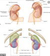
Describe the anatomical location of the kidneys
- Lie retroperitoneally
- They typically extend from T12 to L3- 3 vertebrae in length.
- Right kidney is situated slightly lower due to the presence of the liver.
The adrenal glands sit immediately superior to the kidneys within a separate envelope of the renal fascia.
Describe the external coverings kidney from superficial to deep
Renal capsule – tough fibrous capsule.
Perirenal fat – collection of extraperitoneal fat.
Renal fascia (also known as Gerota’s fascia or perirenal fascia) – encloses the kidneys and the suprarenal glands.
Pararenal fat – mainly located on the posterolateral aspect of the kidney.

Describe the anatomical structure of the kidney going inwards towards the ureter
- The cortex extends into the medulla, dividing it into pyramids
- Apex of pyramid = renal papilla.
- Each renal papilla is associated with the minor calyx, which collects urine from the pyramids.
- Several minor calices merge to form a major calyx.
- Urine passes through the major calices into the renal pelvis
- The renal pelvis drains into the ureter

Name the structure into which the renal vessels and ureter enter the kidney
The renal hilum

What are the anatomical relations of the left kidney?
Anterior
- Suprarenal gland
- Spleen
- Stomach
- Pancreas
- Left colic (splenic) flexure
- Jejunum
Posterior
- Diaphragm
- 11th and 12th ribs
- Psoas major, quadratus lumborum and transversus abdominis
- Subcostal, iliohypogastric and ilioinguinal nerves

What are the anatomical relations of the right kidney?
Anterior
- Suprarenal gland
- Liver
- Duodenum
- Right colic flexure
Posterior
- Diaphragm
- 12th rib
- Psoas major, quadratus lumborum and transversus abdominis
- Subcostal, iliohypogastric and ilioinguinal nerves

Describe the arterial supply of the kidneys
Renal arteries
Arise directly from the abdominal aorta, immediately distal to the origin of the superior mesenteric artery.
Due to the anatomical position of the AA (slightly to the left of the midline), the right renal artery is longer, and crosses the vena cava posteriorly.
The renal artery enters the kidney via the renal hilum and forms an anterior (75%) and a posterior (25%) division supplying the kidney

What is the name and location of the avascular plane of the kidney and what is its clinical signficance?
Line of Brodel
Imaginary line along the lateral and slightly posterior border of the kidney
Delineates the segments of the kidney supplied by the anterior and posterior divisions.
Important access route for surgical access of the kidney, as it minimises the risk of damage to major arterial branches.
What is the importance of the renal arteries being anatomical end arteries?
There is no communication between vessels.
This is of crucial importance; as trauma or obstruction in one arterial branch will eventually lead to ischaemia and necrosis of the renal parenchyma supplied by this vessel.
Describe the venous drainage of the kidneys and the anaotmical relation of the veins
The kidneys are drained by left and right renal veins.
They leave the renal hilum anteriorly to the renal arteries, and empty directly into the inferior vena cava.
As the vena cava lies slightly to the right, the LRV is longer, and travels anteriorly to the AA below the origin of the SMA
The right renal artery lies posterior to the IVC

Lymph drainage of kidneys?
paraortic lymph nodes
What is the clinical relevence of the kidney’s proximity to the AA?
an AAA can block the renal arteries
What are the 3 main functions of the kidneys?
- Filters blood plasma to produce urine
- Homeostasis
- Endocrine function – secretes renin, 1a-hydroxylase and erythropoietin.
Briefly explain the physiology of blood filtration in the nephron
ULTRAFILTRATION
- Filtering of blood through fenestrated capillaries of the glomerulus
- All plasma proteins and cells remain in blood – everything else passes into Bowman’s capsule of the nephron.
REABSORPTION
- Material is retained from the ultrafiltrate – includes amino acids, ions, glucose, small proteins, water when necessary
- PCT is responsible for 70% reabsorption
LOOP OF HENLE COUNTER CURRENT MECHANISM
- This creates a hyper-osmotic extracellular fluid
CONCENTRATION OF URINE
- Occurs in the collecting tubule
- Controlled by vasopressin


