Acute Kidney Injury Flashcards
Define AKI
An acute decline in renal function leading to a rise in serum creatinine and/or a fall in urine output.
- Over a period of hours-days, with accumulation of waste products
- Spectrum of injury which may progress to organ failure

State the criteria for the KDIGO Classification of AKI
- Increase in serum creatinine > 26umol/L within 48 hrs
- Increase in serum creatinine to > 1.5x baseline within the preceding 7 days
- Urine volume < 0.5 ml/kg/hr for >6 hours

State the commonest pre-renal causes of AKI
(40-70%) – INADEQUATE PERFUSION
Anything causing renal hypoperfusion:
- Hypotension (e.g. shock, sepsis, anaphylaxis)
- Hypovolaemia (e.g. haemorrhage, severe vomiting)
- Renal artery stenosis, ACEi, NSAIDs, ARBs
- Heart failure – cardiorenal syndrome
- Cirrhosis – hepatorenal syndrome

State the commonest intrinsic-renal causes of AKI
Intrinsic Renal (10-15%) – CELLULAR DAMAGE/INTRINSIC
- Glomerular - glomerulonephritis, haemolytic uraemic syndrome, autoimmune such as SLE, drugs
- Tubular - acute tubular necrosis (ATN) is commonest intrinsic renal cause; often occurs as a result of pre-renal damage or nephrotoxins (e.g. aminoglycosides)
- Interstitial - acute interstitial nephritis (e.g. NSAIDs, autoimmune), drugs, infiltration with lymphoma/infection/tumour lysis syndrome following chemotherapy
- Vascular - vasculitides (e.g. Wegener’s granulomatosis), large vessel occlusion
- Eclampsia
State the commonest post-renal causes of AKI
Post-Renal (10-25%) – URINARY TRACT OBSTRUCTION
- Luminal: stones, clots, sloughed papillae
- Mural: malignancy (ureteric, bladder, prostate), BPH, urethral strictures
- Extrinsic compression: malignancy (esp pelvic), retroperitoneal fibrosis, prostatic hypertrophy
State some risk factors for AKI
- Age > 75- associated with CKD, underlying RVD, and other comorbidities
- CKD
- Comorbidities (heart failure, peripheral vascular disease, chronic liver disease, diabetes mellitus, connectve tissue disease eg SLE)
- Malignant hypotension
- Sepsis
- Drug overdose- due to direct nephrotoxicity, rhabdomyolysis, and volume depletion.
-
Impaired renal perfusion (ischaemia) due to:
- Cardiac arrest
- Haemorrhage-
- Hypovolaemia- from hemorrhage, vomiting, diarrhea, dehydration
- Pancreatitis- severe third spacing of fluid leading to intravascular volume depletion
Summarise the epidemiology of AKI
18% of adults admitted to hospital will develop an AKI
Most common in the ELDERLY
Acute tubular necrosis (ATN) accounts for 45% of cases of AKI.
ATN is caused by sepsis in 19% of ICU patients.

What are the presenting symptoms of AKI? Give a brief explanation of each symptom
Depends on underlying CAUSE. Often may be asymptomatic.
- Oliguria/anuria
- Nausea/vomiting
- Dehydration
- Confusion, dizziness
- Orthopnea → pulmonary oedema - symptoms of volume overload may result from impaired salt and volume regulation and decreased urine production.
What are the presenting signs of AKI? Give a brief explanation of each sign
- Hypertension
- Dehydration
-
Fluid overload signs:
- raised JVP
- pulmonary and peripheral oedema
- (in heart failure, cirrhosis, nephrotic syndrome)
- Distended bladder – palpable
- Palpable kidneys (suggests polycystic disease)
- Renal bruit (sign of renovascular disease)
- Pallor, rash, bruising (vascular disease)
State some drugs which increase the risk of AKI
- aminoglycosides
- vancomycin + piperacillin-tazobactam
- cancer therapies
- nonsteroidal anti-inflammatory drugs
- ACE inhibitors
State primary investigations on suspicion of AKI
NOTE: must start with ABCDE approach, and check for urgent K+ on venous blood specimen and ECG to check for life-threatening hyperkalaemia.
- Urinalysis and culture if infection suspected
-
Bloods:
- BUN and creatinine
- FBCs
- U+Es
- FBC
- CRP
- Urine osmolality and sodium concentration
- Venous blood gasses
- Renal ultrasound
- CXR- check for pulmonary oedema and cardiomegaly
- AXR- if stones suspected

What may you see in the urinalysis of someone with AKI?
- RBC- suggest nephritic cause
- WBCs
- cellular casts
- proteinuria- suggest glomerular disease
- bacteria
- positive nitrite and leukocyte esterase (in cases of infection (UTI))
What immunology findings may be relevent in AKI?
-
Serum immunoglobulins and protein electrophoresis - for multiple myeloma
- Also check for Bence-Jones proteins in the urine
-
ANA - associated with SLE
- Also check anti-dsDNA antibodies (high in active lupus)
-
Complement levels
- low in active lupus
- Anti-GBM antibodies - Goodpasture’s syndrome
- Antistreptolysin-O antibodies - high after Streptococcal infection
- Virology - check for hepatitis and HIV

What can be seen on RUSS in AKI?
- Check for post-renal cause
- Can help distinguish obstruction and hydronephrosis
- Shows cysts, small kidneys, masses
differentials to AKI?
-
CKD
- here are no causes of chronically elevated serum creatinine other than reduced glomerular filtration (except for minor elevations in subjects with:
-
Increased muscle mass-
- elevation of creatinine is minor and typically nonacute.
-
Drug side effect
- medications such as cimetidine or trimethoprim may lead to a minor, non-acute elevation of creatinine
FOR ALL 3: twenty-four-hour urine study for creatinine clearance should demonstrate normal/reduced function.
Identify possible complications of AKI
- Volume overload → Pulmonary and peripheral oedema- impaired volume regulation is common in AKI not occurring from prerenal azotemia.
- Metabolic acidosis- from accumulation of nonvolatile acids
- Uraemia- uremic toxins accumulate with severe and untreated AKI
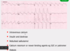
- Hyperkalaemia- impaired excretion of potassium, cell lysis, or tissue breakdown.
- Hyperphosphataemia- late complication usually arising several days after glomerular filtration falls.
- Chronic progressive/end-stage renal disease-
AKI may leave the patient with prolonged renal damage, and functional recovery may not return to the baseline.or even at all in end-stage
Outline the general management of AKI
Treatment approaches for AKI vary according to the type of insult (pre, post, intrinsic renal). The underlying illness requires treatment. Fluid and electrolyte status shold be monitored.
-
ABCDE approach + check for hyperkalaemia- treat if needed
- (10 ml 10% calcium gluconate, then 10 units actrapid in 50ml 20% glucose)
-
Intervention in electrolyte and acid/base abnormalities and optimization of volume status- euvolaemia
- replace volume in the volume-contracted patient
- fluid removal (either diuresis or renal replacement therapy) in patients with volume overload.
- Sodium, potassium and phosphorus and volume restriction are generally required
- Dose adjustment of medications is likely required in all cases and should not be overlooked- stop any nephrotoxic drugs

How should you treat pre-renal AKI?
Prerenal azotemia is managed with techniques to improve the hemodynamic status of the patient.
- The volume-contracted patient requires volume expansion and/or transfusion if there is significant anemia
- Vasopressors are recommended if hypotension is severe, to keep MAP >60 mmHg.
- Diuretics if uremia, severe metabolic acidosis, hyperkalemia refractory to medical management, or volume overload unresponsive to diuretics
- Renal replacement therapy may be needed if severe acid/base, electrolyte, or uremic complications are present while the underlying cardiac or volume issues are treated

How should you treat intrinsic renal AKI?
- Treatment of underlying disease- cease nephrotoxic agents, refer to nephrologist if specific treatment, such as dialysis, management of fluids/acid-base status, severe hyperkalemia, or immunosuppression is required
- Diuretic- furosemide if in volume overload
- Volume expansion- if pre-existing prerenal azotemia
- Renal replacement therapy- with uremia, severe metabolic acidosis, hyperkalemia refractory to medical management, or volume overload unresponsive to diuretics

How would you treat post-renal (obstructive) AKI?
-
Bladder catheterisation- mechanical decompression at the level of obstruction.
- NOTE: bladder catheter placement should be done in all cases of AKI if bladder outlet obstruction cannot be quickly ruled out by ultrasound.
-
Relief of obstruction above the bladder neck
- ureteral stenting: if there is a ureteral stricture, stone or extrinsically obstructing mass
- lithotripsy: stones present at UPJ
- exploratory laparotomy: compressing tumors may require surgical removal
- percutaneous nephrostomy
- Diuretic- if in fluid overload
- Renal replacement therapy- with uremia, severe metabolic acidosis, or hyperkalemia refractory to medical management, or volume overload unresponsive to diuretics
What is percutaneous nephrostomy?
Placement of a catheter into the renal pelvis percutaneously for drainage of urine from a distal obstruction
May be done by a urologist, surgeon or interventional radiologist

When is renal replacement therapy considered?
- Hyperkalaemia refractory to medical management
- Volume overload unresponsive to diuretics → pulmonary oedema refractory to medical management
- Severe metabolic acidaemia
- Uraemic complications
What are indicators of a poor prognosis for AKI?
- Age
- Multiple organ failure
- Oliguria
- Hypotension
- CKD
Summarise the prognosis of patients with AKI
Recovery for AKI is variable and depends on cause of injury and the severity and duration of AKI
Five-year survival rates in patients with AKI requiring renal replacement therapy range from 15%-35% (less than 10% of those patients are dialysis-dependent)
AKI is irreversible 5%-7% of adults and as many as 16% of older adult patients