Upper Limb - Part 1 Flashcards

1
Q

The body can be divided into which 2 major parts?

So the skeleton can be divided into which 2 major parts?

A
  1. The main body (head, neck and trunk)
  2. The appendages (upper and lower limbs)
  1. The axial skeleton (main body)
  2. The appendicular skeleton (appendages)
2
Q

What is the function of the upper limb?

What are the functions of the lower limbs?

A

Position hand for manipulation and grip activities

Support the body weight, locomotion (walking, running, etc.), maintain balance

3
Q

What are the 2 joints by which the upper limb connects to the trunk?

A
  1. True joints - the left and right sternoclavicular joints (
  2. Virtual joints - the left and right scapulothoracic joints (the contact between the scapula and it’s associated muscle with the thoracic wall i.e. ribs)
4
Q

What is the joint by which the lower limbs are connected to the trunk of the body?

Why is this joint particularly useful?

A

Sacroiliac joint - the joint between the pelvis and sacrum, this is a synovial joint (i.e. filled with synovial fluid)

Carries the weight of the head, neck and trunk

5
Q

Into which 4 regions, anatomically, is the upper limb divided into?

A
  1. The pectoral girdle (shoulder)
  2. Arm
  3. Forearm
  4. Hand
6
Q

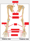
Fill in these labels onto the skeletal diagram:

Clavicle, scapula, humerus, radius, ulna, carpal bones, metacarpals, phalanges
A

Clavicle and scapula - pectoral girdle

Humerus - arm bone

Radius and ulna - parallel bones of the forearm

Carpal bones - two rows of small bones on the wrist

Metacarpals - bones of the main part of the hand

Phalanges - bones of the digits of the hands, including the thumb

7
Q

What is the clavicle commonly referred to as?

How is it positioned and what other bones does it connect to (medially and laterally)?

Why does the clavicle act as a strut?

A

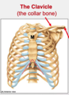
The collar bone

Medial end of the clavicle forms a joint (articulates) with the manubrium (top of the sternum), and the lateral end articulates with the acromion process of the scapula

To hold the upper limb away from the trunk to allow for a wider range of movement
8
Q

Fill in the covered labels on this skeletal diagram of the clavicle:

What are the names of the 2 ends? And then length of the whole clavicle?

A

Sternal (medial) and acromial (lateral) ends

Shaft

9
Q

What is the scapula commonly referred to as?

What is the scapula?

A

Shoulder blades

Triangular shaped bone found at the back with various bony features on which muscles and ligaments attach

10
Q

Fill in the labels of this skeletal diagram of the scapula and what they are important for:

Hints: clavicle joint, small hook, socket, ridge of bone, dividing scapula one bigger, and one smaller division, and the anterior surface of the scapula

A

Acromion - articulates with the clavicle

Coracoid process - small hook in the bone of the superior scapular, important for the attachment of muscles

Glenoid fossa - depression / shallow cup in the widened region of the lateral scapula, forming the socket for the ball and socket shoulder joint

Scapular spine - ridge of bone dividing the posterior scapula into the infraspinatus and supraspinatus fossae

Infraspinatus fossa - bigger division

Supraspinatus fossa - smaller division

Subscapular fossa - anterior surface of scapula closest to the chest wall

11
Q

What is the glenohumeral joint made of?

What is the elbow joint made of?

What do the distal ends of the radius and ulna attach to?

How are the radius and ulna connected to each other?

A

Shoulder joint - proximal humerus head forms the ball, and glenoid fossa forms the socket

Elbow joint - distal, condyles of the humerus has two articulations with the proximal heads of the radius and ulna

The proximal row of the carpal bones

Via a sheet of fibrous connective tissue called the interosseus membrane

12
Q

Why is the interosseus membrane important?

Which part of the radius is an important attachment site for the bicep?

A

Contributes to the stability of the arrangement and acts as a site for muscle attachment

Radial tuberosity (bony feature)

13
Q

What is a fancy name for the wrist?

How many bones are part of the carpal bones and how are they arranged?

What does the distal row articulate with?

Why are there many bones in the wrist?

A

The carpus

8 - 4 in the proximal row and 4 in the distal row

The bases of the metacarpals (main bones of the hand), and the proximal row

To allow for the flexibility of the wrist region

14
Q

What are the metacarpal bones and which part of the hand do they form?

What is the name given to the bones that make up the digits (including the thumb)?

A

Small, long bones of the hand, forming the palmar region, their heads forming the knuckles

Phalanges - thumb = 2 phalanges: proximal and distal; other fingers = 3 phalanges: proximal base, shaft and distal head

15
Q

Fibrous, Cartilaginous, Synovial

What are the names of the 3 structural classifications of joints and what are their properties?

What are some examples of where these joints are found?

A
  1. Fibrous:- bones connected by fibrous connective tissue, e.g. sutures of skull, syndesmosis of the arm
  2. Cartilaginous:- bones connected with cartilage, e.g. pubic symphysis
    a) Primary (synchondrosis, connected by hyaline cartilage)
    b) Secondary (symphysis, connected by fibrocartilage – mainly in the midline of the body)
  3. Synovial joints:- the articulation is surrounded by an enclosing synovial capsule; bones not directly connected at the joint surfaces but strengthened by surrounding structures e.g. interphalangeal joints
16
Q

What are epiphyseal plates?

A

Temporary cartilaginous joints observed in babies, children and young adults that allow bone growth - when bone growth ceases, they ossify

17
Q

What are the 3 types of synovial joints?

A
  1. Uniaxial - movement in only one direction, e.g. hinge joint
  2. Biaxial - movement in two different planes, e.g. saddle joint
  3. Multiaxial - movement on several axes, e.g. ball and socket joint
18
Q

What are the 3 classifications of joint mobility?

A
  1. Synarthosis - little or no mobility (mostly fibrous joints like skull sutures)
  2. Amphiarthosis - limited mobility (often fibrocartilaginous such as pubic symphysis)
  3. Diarthosis - freely mobile (many joints, mostly synovial)
19
Q

Generally, the more mobile the joint, the less stable it is.

What structures provide stability for mobile joints?

A

Ligaments (collagenous connective tissue linking bones) and tendons (collagenous connective tissue between bones and muscles)

These significatly restrict movement to prevent unwanted movement that may destabilise a joint

20
Q

What is a retinaculum?

Give and example of where it can be found?

A

A fascial feature - thickened band of deep fascia found close to a joint to hold tendons down during muscle contraction to prevent bow-stringing, which might compromise function

e.g. found on the wrist and fingers

21
Q

What is aponeurosis?

What do they provide?

An example of where they can be found?

A

Another fascial feature - flat, sheet-like structure formed from a tendon or ligament

They can provide a broad attachment for a muscle which will distribute mechanical load over a larger area than a more typical tendon

They also can provide protection for underlying structures, e.g. bicipital aponeurosis (near the elbow), in the palm of the hand and the sole of the foot

22
Q

What is a bursa?

What do they provide?

What is the name given to inflammed bursae?

An example of where they can be found?

A

A bursa is a closed sac of a serous membrane, whose interior is similar to that of synovial joints

The membranes of bursae secrete a lubricating fluid; found at body sites that are subject to friction, where they act as a “bearing” that allows free movement

Bursitis - can be extremely painful

e.g. subcutaneous bursa at the posterior of the elbow normally prevents friction between the skin and the olecranon of the ulnar bone

23
Q

What are the names of the main joints of the upper limb and where are the found briefly?

A

Sternoclavicular joint (SCJ) - where the top of the sternum and clavicle meet

Acromioclavicular joint (AVJ) - where the acromion (bony process on the scapula) and clavicle meet

Glenohumeral joint (GHJ) - shoulder joint, where the humerus and scapula meet

Scapulothoracic joint (STJ) - virtual joint, between the scapula and ribcage

Elbow joint - where the humerus, radius and ulna meet

Wrist joint - mainly where the radius and proximal row of the carpal bones meet

Numerous joints in the hand

24
Q

The sternoclavicular joint connects the upper limbs to the trunk and is one of the joints of the pectoral girdle

Which of the 3 types of joint is it?

What is it’s joint divided by?

How can this joint move?

How is this joint stabilised and why?

A

Saddle type synovial joint

Divided by a fibrous, articular disc

Stabilised a number of ligaments to limit and prevent unwanted movement

The joint can accommodate the elevation of the clavicle and the protraction and retraction of the scapula

25
Q

The acromioclavicular joint connects together which bones?

Why type of joint is it?

What type of articular disc does it have?

What movements can the joint do?

What features limit the movement / stabilise the scapula?

A

The acromioclavicular joint connects the lateral end of the clavicle with the acromion end of the scapula

Small, synovial, plain joint

An incomplete one

Limited movements - the rotation, vertical and anterior / posterior movements are more so associated with the scapula

Ligaments and muscles

26
Q

What is the scapulothoracic joint? Which bones does it involve and why is it not a true joint?

A

Not a true joint - it is how the movements of the deep surface of the scapula and its covering muscles move on the chest wall

27
Q

What is the glenohumeral joint? Which bones does it involve?

What type of joint is it?

What deepens the glenoid fossa?

How well can this joint movement?

How is this joint stabilised?

A

The shoulder joint - involves the hyaline-covered, rounded head of the humerus (ball) and the glenois fossa of the scapula (which forms a shallow hyaline cartilage line socket)

A ball and socket synovial joint

Cartilaginous labrum

Highly mobile

By numerous muscles that hold the head of the humerus against the glenoid fossa

28
Q

What is the elbow joint? Which bones are involved?

What type of joint is it?

What are the names of the pair of condyles (rounded heads) at the end of the humerus?

What movements can be made at this joint?

A

Involves 2 articulations - distal end of the humerus against the proximal ends of the radius and ulna

Synovial

Capitulum and trochlea

Flexion and extension only

29
Q

What may be a 3rd articulation in the elbow joint?

How does this part of the joint move? What movements does this result to?

A

Between the proximal ends of the radius and ulna, referred to as the proximal radioulna joint

Rotation of the head of the radius allows for the shaft to pass over the ulna - results in the movement of the palm facing anterior or posterior (pronation and supination)

30
Q

What are some adaptations of the bones at the elbow joint to allow for flexion and extension?

A

Anterior - radial and coronoid fossae sit above the condyles at the distal end of the humerus to accomodate the heads of the radius and coronoid process of the ulna during flexion

Posterior - olecranon fossa in the humerus accomodates the olecranon process of the ulna during extension (limiting further extension)

31
Q

What is the wrist joint? Which 3 of the 4 bones in the proximal row of the carpal bones are involved?

Why does the distal end of the ulna not strictly form a part of the wrist joint?

A

Articulations found between the radius and ulna, and the scaphoid, lunate, triquetrum carpal bones (the pisiform carpal bone is not involved)

Because it articulates with the articular disc, which articulates with the carpal bones

32
Q

What is the name of the joint formed between the distal ends of the radius and ulna? How do they move / when do they interact?

What type of joint is it?

The bones are connected by what type of disc?

A

Distal radioulna joint - they articulate during pronation and supination

Synovial pivot joint

Fibrous articular disc which slides over the distal ulna during the pronation and supination movements

33
Q

What are the 4 main hand joints of the hand?

A

Intercarpal joints - articulation between and within the proximal and distal rows of the carpal bones

Carpal metacarpal joints - articulations between the distal row of carpal bones and the basis of the metacarpals

Metacarpophalangeal joints - articulations between the metacarpals and phalanges (the joints visible as knuckles)

Interpharyngeal joints - articulations between the phalanges

34
Q

What type of joints are the carpal metacarpal joints?

Why type of joints are the metacarpopharyngeal joints? What are the movements limited by?

What type of joints are the interpharyngeal joints? What are the movements limited by?

A

Mainly plane, synovial joints, except the first one, which is a saddle joint - allows for the greater mobility of the thumb

Synovial condylar joints - limited by ligaments

Synovial hinge joints, capable of flexion and extension only - limited by collateral ligaments

35
Q

Why are the limb muscles divided into compartments?

What are the 7 muscle compartments of the upper limbs?

A

Compartments tend to be separated by connective tissue septae (walls) - they tend to have distinct functions, and the same nerve and blood supplies (making it easier to learn)

  1. Pectoral girdle muscles
  2. Intrinsic shoulder muscles
  3. Anterior arm muscles - flexors
  4. Posterior arm muscles - extensors
  5. Anterior forearm muscles - flexors
  6. Posterior forearm muscles - extensors
  7. Intrinsic hand muscles
36
Q

Which 2 muscle compartments are involved in the movement of the shoulder region?

What muscles do these compartments include?

A

The pectoral girdle muscles - muscles of the chest and back (the anterior and posterior axio-appendicular muscles)

and the intrinsic shoulder muscles - the muscles acting between the humerus and scapula to move the arm and shoulder (scapulohumeral)

37
Q

What are some examples of the pectoral girdle muscles and where do they act?

A

e. g. The trapezius and serratus anterior muscles - act directly on the scapula
e. g. pectoralis major and latissimus dorsi - act directly on the arm

38
Q

Which major bone do the intrinsic shoulder muscles attach to?

What are some of the names of the intrinsic shoulder muscles?

Why are these muscles important (functionally)?

A

Scapula

Deltoid - attachment to the scapula and clavicle for movement of the arm; teres major; the rotator cuff muscles (made up of the: supraspinatus, infraspinatus, teres minor, subscapularis)

As well as acting to move the arm at the shoulder joint, they stabilise the joint by acting as a grip to hold the head of the humerus in the glenoid fossa

39
Q

What are the names of the 3 main anterior arm muscles (flexors) of the arm? Where do they act?

What movements do these muscles cause?

What functions of the biceps are especially important?

A

Biceps brachii - acts across both, shoulder and elbow joints; coracobrachialis - acts only across the shoulder joint; brachialis - acts only across the elbow joint

Flexion of the shoulder and elbow joints; biceps contribute to the stability of the shoulder joint

Biceps tendon attaches to the radius - allows to act as a supinator of the forearm

40
Q

What are the 2 muscles of the posterior compartment of the arm (extensors)? Where do they attach and act?

A

Main extensor = triceps brachii, it has 3 heads, 2 of which are attached to the posterior shaft of the humerus, and the long head spans the shoulder joint attaching to the infraglenoid tubercle, a small lump of bone just inferior to the glenoid fossa. The fibres from all 3 heads converge distally to form the triceps tendon, which attaches to the olecranon of the ulna

Aconeus = small muscle, may contribute to extension of elbow, but more believed to stabilise elbow

41
Q

Why are many of the muscles that move the wrist and digits found in the forearm?

Onto which structure do these muscles attach to?

A

Wrist and digits often responsible for activities that require strength or delicacy, and bulky muscles found in the wrist or digits can impede those movements (reduce efficacy / ability)

Across the elbow joint on the distal humerus

42
Q

There are many MANY muscles of the forearm, how are these muscles named logically according to funciton / where they are found? Fill in the table:

A

Flexors and extensors are found on the anterior andposterior forearm respectively

Muscles acting on the wrist are carpi muscles, and they do this by attaching onto the ulna (ulnaris) or radius (radialis)

Movers of the digits can act on groups of digits (digitorium) or individual digits

Pollicus = acts only on thumb

digiti minimi = only act on little finger

indicis = acts only on index finger

Longus / brevis and superficial / profundus = only applied if this descriptor is required, so if there is a longus, there must be a brevis, and if there is a superficial, there must be a profundus

43
Q

Using the terminology from the last flashcard, fill in the names of these muscles:

  1. Do they cause flexion or extension
  2. What are they moving? (wrist or digits)
  3. Descriptor
A
44
Q

Using terminology, fill in the names of these muscles:

  1. Do they cause flexion or extension
  2. What are they moving? (wrist or digits)
  3. Where is the muscle found?
  4. Descriptor
A
45
Q

What are all the names of the muscles of the forearm? Name as many as possible:

A
46
Q

How are the digits of the hand numbered?

A

1 = thumb

2 = index finger

3 = middle finger

4 = ring finger

5 = little finger

47
Q

What are the names of the 2 muscle bulges that can be seen in the palmar aspects of the hand?

Fill in the labels on the diagram:

A

Thenar eminence - formed on the lateral side of the palm by a number of small muscles that act on the thumb

Hypothenar eminence - found on the medial aspect of the palm, group of muscles that act on the little finger

48
Q

What are the names of 3 more groups of muscles found in the hand?

Fill in the labels on the diagram:

A