Transplantation Flashcards
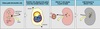
Autograft
(Autologous)
Self-tissue transferred from one site of the body to another on the same individual.
Histocompatible.
Isograft
(Syngeneic)
Tissue transferred between genetically identical individuals.
Histocompatible.
Allograft
Tissue transferred between genetically different members of the same species.
Histoincompatible.
Xenograft
Tissue transferred between members of different species.
Histoincompatible.
Histocompatible
A tissue that is antigenically similar to the recipient’s tissue and does NOT induce an immunological response that leads to tissue rejection.
Histoincompatible
A tissue that is antigenically dissimilar to the recipient’s tissue and induces an immunological response that leads to tissue rejection.
Transfusion
Involves the transfer of blood from one individual to another.
Transplantation
Involves the transfer of any organ or tissue from one individual to another.
- Whole organs: kidney, liver, lung, heart, pancreas etc.
- Tissues: bond, skin, cornea etc.
- Cellular: bone marrow, pancreatic islet cells etc.
Genes Determining Histocompatibility
- ABO antigens
- most important parameter in solid organ grafts
- blood group type can change with bone marrow transplantation
- MHC/HLA
- Matching class II MHC important in solid organ transplant
- Must match both class I and II for bone marrow transplantation
- Minor histocompatibility antigens
- > 40 different genes important in preventing rejection
Host-versus-Graft
(HvG)
- Alloreactive host lymphocytes damages the graft
- Follows transplantation of a histoincompatible tissue organ
- May lead to destruction of the organ

Graft-versus-Host
(GvH)
- Follows transfer of immunologically competent alloreactive lymphocytes into an immunocompromised host
- bone marrow transplant
- passenger lymphocytes in an organ
- Graft mounts an immunological attack on the host.
- CD4 T-cells promote damaging immune function
- CD8 T-cells destroy tissue
- Host cells can aid donor cells in tissue destruction
- Removal of T cells using T-cell reactive mAb and complement decreases incidence and severity of GvH
- If bone marrow completely purged of T cells using anti-CD3+ complement treatment engraftment failure dramatically increases
- Occurs in HLA matched siblings and during autologous transplants
- Acute GvH:
- epithelial cell necrosis of skin, liver, and GI tract
- rash
- jaundice
- diarrhea
- Chronic GvH:
- fibrosis of skin, liver, and/or GI tract without necrosis
- can lead to complete organ dysfunction
Hyperacute Rejection
HvG following allograft solid organ transplant.
Occurs within minutes to ~12-24 hours post reperfusion of the organ.
Type II hypersensitivity.
Preformed Ab binds to tissues → complement activation → recruitment of phagocytic cells, platelet activation and deposition → thrombosis, swelling, hemorrhage, and necrosis.
Cell-mediated immunity is generally NOT involved.
Characterized by thrmobotic occlusions with endothelial injury, neutrophil influx, and fibrinoid necrosis.
No treatment, only prevention through ABO matching and PRA screening for pre-existing Ab.
Explanations for Pre-existing Antibodies
- ABO incompatible organ.
- Multiple pregnancies.
- Prior incompatible transplants.
- Prior blood tranfusions.
Acute Rejection
HvG following allograft solid organ transplant.
Occurs within 10-14 days in non-immunosuppressed patient and within several months with suppresion.
Due primarily to T-cell mediated immunity.
Transplant desctruction by CTLs → phagocytosis → presentation of transplanted Ag to TH cells → further organ degradation.
Characterized by lymphocytic and macrophage infiltration.
Preventative treatment with immunosuppresion such as cyclosporin.
Therapeutic treatment with corticosteroids if symptoms develop.
Chronic Rejection
HvG following allograft solid organ transplant.
Occurs after months to years.
Similar to a chronic DTH reaction.
Mediated by both humoral and cell-mediated reactions.
Activated macrophages secrete growth factors → fibrosis → ischemia and cell death.
Appears as fibrosis and scarring in transplanted organs.
Treatment generally ineffective and re-transplantation commonly needed.
T-cell
Direct Alloreactivity
- T-cells can respond to both:
- foreign Ag peptide + self-MHC
- “foreign” MHC + normal self-peptides
- T-cells from patient X are stimulated by cells from patient Y in a non-MHC restricted manner
- T-cell alloreactivity to foreign MHC stimulates a mixed lymphocyte reaction (MLR)
- A significantly higher number of total lymphocytes able to react to any given allograft antigen
Direct Alloreactivity
Mechanism
- Transplanted organs cary passenger APCs (interstitial dendritic cells)
- Ischemia generates DAMPs and a non-specific inflammatory response.
- Danger signal activates passenger APC’s causing increased density of allo-MHC and B7.
- Activated foreign APC’s travel to the LN and stimulate the recipient’s naïve T-cells.
- Following replication and differentiation the alloreactive effector T-cells return to the organ causing acute (allo) rejection.
Factors Affecting
Rejection Response
-
Type of tissue
- skin grafts - rapid and relentless
- heart - slow and more possiblity to prevent
- based on the amount of immunosurvaillence of the tissue
-
Specificity and Memory
-
First-set rejection
- the first time a transplant is rejected
-
Second-set rejection
- an accelerated rejection of the second transplant because of Ag similarity to first transplant
-
First-set rejection
- Solid Organ Transplants
- ABO >>>>>> Class II MHC > Class I MHC
Determination of Histocompatibility
Methods
- ABO determination by agglutination
- Panel Reactive Antibody test (PRA) and cross-match
- Serological (Microcytotoxicity) / Complement Dependent Ab Lysis
- PCR epitope genotyping
- Mixed lymphocyte reaction (MLR)
- One-way mixed lymphocyte reaction
- 51Cr release assay (class I MHC mismatch and CTLs)
Panel Reactive Antibody Test
(PRA)
Pre-transplant evaluation.
- Precipient serum + pooled leukocytes from human peripheral blood + complement + blue dye
- If patient has Ab against multiple individual’s leukocytes they will bind to Ag on many cells → MAC formation → blue stain enters cell
- High PRA indicative of preformed Ab against many different donors
- Contraindication for transplant
Crossmatch
After potential donor identified.
- Recipient’s serum tested against donor’s peripheral blood cells.
- Positive crossmatch = presence of donor specific preformed Ab → contraindication for transplant
Serological (Microcytotoxicity)
Complement Dependent Ab Lysis
- Donor or recipient cells mixed with Ab of known specificity against HLA antigens.
- Complement added and cells monitored for lymphocyte damage or lysis
Mixed Lymphocyte Reaction
(MLR)
- Lymphocytes from donor & recipient cultured together for several days with radioactive T nucleotides
-
Allogeneic T cell activation and proliferation occur with a class II MHC mismatch.
- Measured through amount of DNA synthesis with [3H] - thymidine incorporation
- Greater mismatch = greater proliferation = more radioactivity
- Traditional MLR looks at total matching/mismatching.
One-way Mixed Lymphocyte Reaction
Allows the reactivity of the donor cells against the recipient’s cells or vice versa.
Reflects the initial recognition events seen in alloreactivity.
-
Test donor reactivity to recipient’s cells
- Recipient’s cells irradiated to prevent proliferation
- Detect incompability leading to graft vs. host
- For bone marrow transplantation
-
Test recipient’s reactivity to donor’s cells
- Donor’s cells irradiated to prevent proliferation
- Detect incompability leading to host vs. graft
- For solid organ transplantation
Proliferation = class II MHC mismatch.
Lympholysis = class I MHC mistatch.
51Cr Release Assay
Test for class I MHC mismatch.
Assess capacity to generate a CTL response.
- Target cells loaded with 51Cr
- Donor cells used for HvG
- Recipient cells used for GvH
- Target cells mixed with responding cells
- If responding cells recognize target cells as foreign (via CD8+ T-cell reaction against class I MHC) → CTLs kill target cells releasing 51Cr into medium

Controlling Allograft Rejection
-
Make graft less immunogenic
- ABO matching
- HLA matching
- Decrease cold-ischemia time
-
Immunosuppresive therapy
- Corticosteroids
- Calcinurin inhibitors
- Monoclonal and polyclonal immunotherapies
- Antiproliferative agents
- Cytotoxic drugs
Corticosteroids
Blocks T-cell and APC derived cytokine and cytokine-receptor expression.
- Significant inhibition of IL-1 and IL-6
- Lesser inhibition of IL-2, IFN-γ, and TNF-α.
- Inhibits lymphoproliferation
- Inhibits APCs
- Alters leukocyte trafficking
- Net result of fewer lymphocytes in circulation
Remicade
(Infliximab)
Anti-TNF-α chimeric IgG.
Used in the treatment of:
Rheumatoid arthritis
Psoriatic arthritis
Ulcerative colitis
Crohn’s disease
Ankylosing spondylitis
Severe plaque psoriasis.
Enbrel
(Etanercept)
Chimeric TNF-α-receptor attached to IgG.
Used in the treatment of:
Rheumatoid arthritis
Plaque psoriasis
Psoriatic arthritis
Ankylosing spondylitis
Juvenile idiopathic arthritis
Xeljanz
(Tofacitinib)
JAK1 and JAK3 inhibitor.
Disrupts the JAK-STAT intracellular signaling pathway
associated with cytokine and growth factor transduction.
Rituximab
Anti-CD20 monoclonal antibody.
Inhibits B-cells.
Used in treatment of:
Rhematoid arthritis
MS
Pemphigus vulgaris
Certain B-cell mediated leukemias
Cyclosporin
Calcineurin inhibitor.
Blocks T-cell proliferation and cytokine production.
Important in transplant immunosuppresion.
