Topic 7 - Nucleic acids and proteins Flashcards
What is the direction of a single strand of DNA?
5’ to 3’
What is meant by antiparallel DNA strands?
The strands run in opposite directions; one strand has the 5’ end on the top and 3’ on the bottom whereas the other strand is the opposite
What is meant by 5’ and 3’ ends of DNA?
5’ is the end at which a phosphate is linked to the 5th carbon atom of doxyribose.
3’ is the end at which a hydroxyl group is attached to the 3rd carbon of deoxyribose.
Which bases are purines?
Adenine and guanine
(double ring)
Which bases are pyrimidines?
Cytosine and thymine
(single ring)
How are the bases linked together?
By hydrogen bonds
How many hydrogen bonds are in the link between adenine and thymine?
Two
How many hydrogen bonds are in the link between cytosine and guanine?
Three
What is the structure of a nucleosome?
It consists of DNA wrapped twice around eight histone proteins which are held together by another histone protein.
Why does the DNA wrap around histones?
Because DNA is negatively charged and histones positively charged
How is transcription regulated by the packaging of DNA?
Transcription cannot occur when the DNA is packed, which enables the packaging regulate which parts of DNA are used for protein synthesis.
What do nucleosomes help in?
Supercoiling chromosomes
What are highly repetitive sequences (satellite) DNA?
Builds 5-45% of the genome. The sequences are typically between 5 and 300 base pairs and may be duplicated as many as 105 times per genome.
What are single copy (unique) genes?
The genes that have coding functions; the base sequences essential for protein production. They are used to encode proteins.
What are exons?
Parts of genes that contain protein-encoding information
What are introns?
Partss of genes that contain non-coding fragments
What is structural DNA?
Highly coiled DNA that does not have a coding function, located around the centromere and near the ends of chromosomes.
In what direction does DNA replication occur?
5’ → 3’
Is DNA replication initiated at one or several points along the DNA strand?
Many
What is the role of helicase in DNA replication?
To unwind the double helix at replication forks
What is the role of DNA polymerases in DNA replication?
- DNA polymerase III synthesises a new strand by adding nucleotides onto the primer in 5’ to 3’ direction
- DNA polymerase I removes the primer and replaces it with DNA nucleotides
What is the role of RNA primase in DNA replication?
Synthesises the RNA primer from which the replication of the lagging strand begins
What is the role of DNA ligase in DNA replication?
Joins the ends of DNA segments and Okazaki fragments on the lagging strand by attaching the sugar-phosphate backbones of the fragments to form a single DNA strand.
What are Okazaki fragments?
Fragments of DNA strand formed on the lagging strand
Distinguish between leading strand and lagging strand
Leading strand is the strand that goes in the direction of 5’ to 3’ and is replicated continuously
Lagging strand goes from 3’ to 5’ and is replicated in fragments
What are deoxynucleoside triphosphates and is their function?
They contain the nucleotide that is hydrogen bonded to form the new DNA strand. It contains a deoxyribose, a nitrogenous base (A,T,C,G) and three phosphate groups. Two of the phosphate groups are lost in replication to provide the energy needed for the hydrogen bonding to be formed.
What is the direction in which transcription occurs?
5’ to 3’
Distinguish between the sense and antisense strands of DNA
The sense strand is the DNA strand that carries the genetic code desired. The antisense strand is the complementary one and the one copied
(due to complementary base pairing this way the copy will be identical to the sense strand)
What is the role of RNA polymerase in DNA transcription?
It binds to the promoter region and causes the DNA to separate into two strands. It also initiates and continues the synthesis of the mRNA molecule.
What is the role of the promoter region in DNA transcription?
It a short sequence of bases (not transcribed) to which the RNA polymerase binds to and initiates transcription
What is the role of the terminator in DNA transcription?
A sequence of nucleotides which, when transcribed, causes the RNA polymerase to detach from the DNA → transcription stops
What is the role of nucleoside triphosphates in DNA transcription?
Contains the complementary base which is bonded to the antisense strand.
How is the polymerisation of mRNA carried out?
It occurs with the catalytic help of RNA polymerase and the energy provided by the release of two phosphates from NTP.
What needs to be done to the mRNA before it is ready for translation?
The removal of introns
How is each tRNA molecule recognised?
Each molecule is recognised by a tRNA-activating enzyme that binds a specific amino acid to the tRNA, using ATP for energy
Ribosomes consist of two?
Subunits, large and small
What is the role of the small subunit of ribosomes?
A place for the mRNA to bind to
What is the protein and RNA composition in ribosomes?
Roughly two thirds of ribosome mass is RNA, the rest is protein
What is the role of the large subunit of ribosomes?
A binding site for tRNAs, includes three different binding sites.
What are the three tRNA binding sites in the large subunit of ribosomes?
A, P, and E sites
Outline the roles of the three tRNA binding sites in the large subunit of ribosomes
A site: holds the tRNA carrying the next amino acid to be added to the polypeptide chain
P site: holds the tRNA carrying the growing polypeptide chain
E site: site from which tRNA that has lost its amino acid is discharged
What are the four phases of translation?
- Initiation (AUG, methionine)
- Elongation
- Translocation
- Termination
How many stop codons are there and what is their function?
Three, and they have no complementary tRNA codon causing the ribosome to detach with the lack of tRNAs
In what direction does translation occur?
In 5’ → 3’ direction
What is the base sequence CCA?
The unbinded base sequence at the end of the 3’ end of tRNA. This is the site to which the amino acid attaches to.
Describe the structure of a tRNA molecule and how it is formed
The two-dimensional structure is a clover leaf and is formed by the strand forming hydrogen bonds with itself

What is the anticodon of a tRNA?
An exposed base triplet that pairs with a specific codon on the mRNA
What happens in the initiation part of translation?
A (initiatior) tRNA with the amino acid methionine and anticodon of UAC combines with an mRNA strand and a small ribosomal subunit. The subunit then moves down the mRNA until it reaches the start codon (AUG). Hydrogen bonds form between the tRNA and the start codon. Finally the large subunit binds to the small subunit with the energy from guanosine triphosphate (GTP).
What happens in the elongation phase of translation?
More tRNAs are brought to the ribosome (A site) and peptide bonds are formed between the amino acids (P site).
What are elongation factors?
Proteins that assist in binding the tRNAs to the mRNA codons at the A site.
What catalyses the formation of peptide bonds between adjacent amino acids in translation?
Ribosomes
Draw a peptide bond between two amino acids

What happens (in steps) in the translocation phase?
Translocation involves the movement of tRNAs from one site of the mRNA to another.
- tRNA binds to the A site and its amino acid is added to the polypeptide chain
- tRNa moves to the P site where it transfers its polypeptide chain to the new tRNA that moves to the A site
- The empty tRNA moves to the E site and is released.
What happens in termination phase of translation?
- One of the three stop codons appears at the open A site.
- A protein called release factor fills the A site.
- The release factor does not carry an amino acid and catalyses the hydrolysis of the bond linking the tRNA in the P site
- The polypeptide is freed as well as the tRNA.
- The ribosome separates from the mRNA and splints into two subunits.
What are polysomes?
A cluster of ribosomes translating the same mRNA at the same time.
What are the proteins synthesised in free ribosomes used for?
Primarily for use within the cell.
What happens to the proteins synthesised in ribosomes bound to the ER?
They are primarily secreted from the cell or used in lysosomes
What are the four levels of protein structure?
Primary, secondary, tertiary, and quaternary
What is the primary level of protein structure?
Simply a unique chain of amino acids attached by peptide bonds
What is the secondary level of protein structure?
- Created by the formation of hydrogen bonds between the O of the carboxyl group and the H from different amino acids
- α-helix and β-pleated sheet
- Does not involve R groups
- Regular repeating patterns
What is the tertiary level of protein structure?
- A definite three-dimensional structure
- Created because the chain bends and folds over itself due to interactions among R-groups and the peptide backbone
- Important in determining the specifity of the proteins (enzymes)
Interactions include:
- Disulfide bonds (bridges)(covalent)
- Hydrogen bonds between polar side chains
- Van der Waals’ among hydrophobic side chains
- Ionic bonds between + and – side chains
What is the quaternary level of protein structure?
- Multiple polypeptide chais which combine to form a single structure
- Some contain prosthetic (non-polypeptide) groups → conjugated proteins (haemoglobin)
Distinguish between fibrous and globular proteins
Fibrous proteins are composed of many polypeptide chains in a long, narrow shape and are usually insoluble in water.
Globular proteins are more three-dimensional in their shape and are mostly water soluble.
Give two examples of fibrous and globular proteins
Fibrous: collagen and actin
Globular: haemoglobin and insulin
What is the role of polar and non-polar amino acids?
They are important in determining the specifity of an enzyme.
Polarity also determines the position of proteins in cell membranes. Non-polar are linked to the hydrophobic area of cell membranes. Polar are found in regions exposed to water.
What do polar amino acids do in membrane proteins?
Create hydrophilic channels through which polar substances can move
List four examples of proteins with different functions
- Haemoglobin: contains iron that transports oxygen from the lungs to all parts of the body.
- Actin and myosin: interact to create muscle movements (contraction) in animals.
- Insulin: a hormone secreted by the pancreas that helps maintaining blood glucose level.
- Amylase: a digestive enzyme that catalyses the hydrolysis of starch.
What do metabolic pathways consist of?
Chains and cycles of enzyme-catalysed reactions.
What kind of proteins are enzymes?
Globular proteins with at least the tertiary level of organisation.
What is the induced-fit model of enzyme action?
- An enhanced version of the lock-and-key model
- When substrate binds with the active site, the structure of the active site changes slightly, providing an induced fit (hand and glove)
- Due to changes in the R-groups of the amino acids as they interact with the substrates
What do enzymes do for the chemical reactions they catalyse?
They lower the activation energy, Ea.
How does competitive inhibition function? Outline one example
- Competes directly for the active site of an enzyme → blocks the substrate from binding
- Structure similar to the substrate
Example:
- Sulfanilamide is used to kill bacteria
- Folic acid is essential to bacteria
- Folic acid is produced by enzyme action on paraaminobenzoic acid (PABA)
- Sulfanamide competes with the PABA and blocks the enzyme
How does non-competitive inhibition funtion? Outline one example
- Does not compete for the enzyme’s active site
- Inhibitor reacts with another site on the enzyme (allosteric site)
- Causes a change in the shape of the enzyme’s active site → dysfunctional enzyme
Example:
- Mercury binds to the sulfur groups of component amino acids of many enzymes
- Shape changes and enzyme action is inhibited
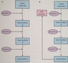
Explain the control of metabolic pathways by end-product inhibition
- Prevents the cell from wasting chemical resources and energy by making more of a substance than it needs
- Assembly-line
- When there is enough of the end-product, the line is shut done
- This is done by inhibiting the action of the enzyme in the first step
- As the existing end-product is used up by the cell, the first enzyme reactivates
- The enzyme that is inhibited and reactivated is an allosteric enzyme
- In high concentration, the end-product binds with the allosteric site and inhibits the action
- In low concentration, the end-product results in fewer bindings