Topic 6 - Bioenergetics Flashcards

1
Q

Key roles of mitochondria?

A
  1. A central player in bioenergetics
  2. Produce 90% of cellular energy
  3. Regulate cellular redox states
  4. Calcium homeostasis
  5. Produce reactive oxygen species
  6. Synthesis and degrade high energy biochemical intermediates
  7. regulate cell death –> programmed cell death
2
Q

What are the requirements for the existence of life?

A
  1. Information –> Nucleotides/DNA
  2. Energy –> ATP

Both of these requirements are needed because life is improbable as living systems are highly ordered –> doesn’t follow 2nd law of thermodynamics.

Information specifies what form the order should take and energy drives the reactions and processes for it to occur.

3
Q

How do we define energy?

A

In biochemistry –> The ability to cause specific change.

4
Q

What are the different categories of change?

A
  1. Synthetic –> changes in bonds –> i.e. to make macromolecules.
  2. Mechanical –> muscle contraction/flagellum/ribosome movement
  3. Concentration –> change in concentration across a membrane.
  4. Electrical –> movement of ions across membranes (neurons)
  5. Heat –> heat production –> maintain temperature.
5
Q

What are the different sources of energy?

A
  1. Solar radiation –> plants use to photosynthesize –> produce nutrients –> consumed by humans.
  2. Electrical discharge
  3. Chemical energy
6
Q

What are the two ways chemical energy can be released?

A
  1. Fermentation –> glycolysis
  2. Oxidation –> respiration
7
Q

Definition of thermodynamics?

A

The laws governing energy transactions that accompany most processes and reactions.

8
Q

Definition of bioenergetics?

A

Applied thermodynamics –> Application of thermodynamic principles to reactions and processes in biological worlds.

9
Q

Difference between classical and non-classical thermodynamics?

A
  1. Classical –> Equilibrium reactions in closed systems.
  2. Non-classical –> Does not occur at equilibrium, takes into account time and occurs in open systems.

Note –> Living systems do not operate at equilibrium –> hence we use non-classical thermodynamics.

10
Q

What do living systems do when they consume nutrients? (In terms of enthalpy and entropy)

A

Living systems consume high enthalpy/low entropy nutrients and convert them to low enthalpy/high entropy products.

  • Free energy from this is used to do work and thus allows organisms to create a high degree of organisation.
11
Q

Why is it important for an organism to stay in a non-equilibrium state?

A
  1. In order to perform useful work
  2. Equilibrium processes cannot be directed –> all regulatory functions require a non-equilibrium state.
  3. A non-equilibrium state is Inherently unstable –> gives rise to biochemical reactions –> allow us to degrade.
12
Q

Are regeneration and degradation constantly occurring?

A

Happens all the time –> requires a constant influx of energy.

13
Q

What is one thing that always occurs in non-equilibrium reactions?

A

In non-equilibrium reactions, something must always flow.

Flow –> change in spatial distribution of something (matter/heat/electrical charge/etc.)

Note –> All these flows are conjugate to their thermodynamic force.

Examples

  • Flow of matter in diffusion –> driven by conc. gradient
  • Transport of heat –> temperature gradient
  • Migration of electrical charge –> current –> due to voltage gradient
  • Chemical reaction –> difference in chemical potential.
14
Q

Can a thermodynamic force also promote a non-conjugate flow?

A

Yes, a thermodynamic force may also promote a non-conjugate flow.

For example, a concentration gradient of matter can give rise to a chemical reaction (or heat or electrical current)

I.e. ATP synthase uses an electrochemical gradient to create chemical bonds.

15
Q

What is energy transduction?

A

Energy transduction –> when a thermodynamic force stimulates a non-conjugate flow.

16
Q

What is metabolism?

A

Metabolism is the study of energy flow in biological systems –> overall process by which living systems acquire and utilize free energy.

17
Q

Definition of exergonic reaction?

A

An exergonic reaction refers to a reaction where energy is released –> ΔG is negative.

18
Q

Definition of endergonic reaction?

A

A chemical reaction in which energy is absorbed.

ΔG > 0

19
Q

What are the names of the different molecules after ATP hydrolysis?

A

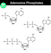
ATP –> ADP –> AMP –> Adenosine.

20
Q

Why does the hydrolysis of ATP release so much energy?

A

Phosphoanhydride bond is a high energy bond which releases a lot of energy.

21
Q

What is the ΔG for the hydrolysis of one phosphate and for two phosphates?

A

H2O + ATP —-> ADP + Pi (ΔG = -30.5 KJ mol-1)

ATP + H2O —-> AMP + PPi (ΔG = -45.6 KJ mol-1)

22
Q

What factors impact the ΔG of ATP hydrolysis?

A

Depends on pH, ionic strength and [Mg2+]

23
Q

Is it possible to have different nucleotide triphosphates?

A

Yes, some biosynthetic reactions are driven by the hydrolysis of other nucleoside triphosphates –> I.e. GTP.

24
Q

What are phosphoryl transfer reactions?

A

Refers to the transfer of a phosphate from one compound to another.

R1-O-PO32- + R2-OH —> R-OH + R2-O-PO32-

A very important reaction for biosynthesis of proteins, nucleic acids and carbohydrates.

25
Q

What is phosphoryl transfer potential?

A

Phosphoryl transfer potential –> examines the ease at which a phosphate group can be lost from a molecule. It is a relative measure (Phosphoryl transferred from a compound to water).

Greater phosphoryl transfer potential –> more energetically favourable for the hydrolysis of the phosphate group.

26
Q

Why does ATP have a high phosphoryl transfer potential?

A

High phosphoryl transfer potential is due to the structural differences between ATP and it hydrolysis products.

  1. Resonance stabilisation –> Products (inorganic Pi has greater resonance stabilisation)
  2. Electrostatic repulsion –> negative charges close together in ATP molecule.
  3. Entropy increase –> entropy of products is greater
  4. Stabilisation due to hydration –> H2O stabilizes products –> makes reverse reaction less favourable.
27
Q

What are the free energy sources in biological systems?

A
  1. Phosphate anhydride
  2. Enol Phosphate
  3. Some thioesters
  4. Some compounds containing N-P bonds –> phosphoguanidines.
28
Q

How do you show a high energy bond in a molecule?

A

~ –> indicates high energy bond.

29
Q

How to classify low energy and high energy phosphate compounds?

A

ATP ≈ -30 KJ mol-1

More than -30 KJ mol-1 (more negative) –> high energy phosphate compound

Less than -30 KJ mol-1 (more positive) –> low energy phosphate compound

30
Q

How can the synthesis of ATP occur from ADP if ΔG is positive?

A

Use high energy compounds and couple the reactions in order to synthesize ATP.

31
Q

What is substrate level phosphorylation?

A

The transfer of the phosphoryl group from a compound with a large ΔG value to ADP yielding ATP.

32
Q

Why is ATP important?

A
  1. ATP is intermediate in energy –> serves as an energy conduit between super high energy and low energy phospho-compounds.
  2. The highly exergonic phosphoryl transfer reactions of nutrient degradation are coupled to the synthesis of ATP from ADP + Pi
33
Q

Processes that require the consumption of ATP?

A
  1. Early stages of carbohydrate breakdown to produce low energy phosphate compounds.
  2. Interconversion of nucleoside triphosphates.
  3. Muscle contraction and active transport against the concentration gradient.
  4. Phosphoanhydride cleavage followed by pyrophosphate cleavage provides extra energy for fatty acyl-CoA synthesis and nucleic acid biosynthesis.
34
Q

What are the two ways in which ATP can be formed?

A
  1. Substrate level phosphorylation
  2. Oxidative phosphorylation
35
Q

What does adenylate kinase do?

A

Phosphotransferase enzyme that catalyzes the interconversion of adenine nucleotides (ATP, ADP, and AMP).

2ADP <—-> ATP + AMP

36
Q

What is the rate of ATP turnover?

A

Note –> Atp is a free energy transmitter, not a reservoir.

  • ATP pool supplies energy for about 1 minute
  • At rest –> the average human turns over ATP at a rate of approximately 1.5kg/hour
  • The brain only has a few seconds supply of ATP.
37
Q

What molecule acts as a reservoir of ATP?

A

Phosphocreatine acts as a reservoir for ATP formation (muscle and nerve cells).

ATP + creatine <—–> Phosphocreatine + ADP

38
Q

Why is the adenylate kinase reaction important in cells?

A

2ADP <—–> ATP + AMP

When we exercise large quantities of ATP are used up which results in high ADP concentration and low ATP concentrations.

Normally, ATP concentrations in cells are very low but when the reaction above is activated during exercise –> AMP concentrations increase –> this activates al energy producing pathways (glycolysis) –> Hence, AMP acts as a signal of low energy which stimulates the oxidation of nutrients.

39
Q

What are NAD+ , FAD and CoA?

A

They are all ATP derivatives.

Note –> All nucleotide triphosphates are energetically equivalent but ATP is the primary cellular energy carrier.

40
Q

How many electrons are needed to fully oxidise O2?

A

4 electrons

41
Q

What is the most important physical property of the inner mitochondrial membrane?

A

It is impermeable –> nothing can simply diffuse through.

42
Q

What are the 3 stages of energy extraction from food?

A
  1. Large molecules are broken down, absorbed and distributed –> no useful energy produced.
  2. Break down molecules into simple units –> acetyl unit of acetyl CoA.
  3. ATP is produced by complete oxidation of the acetyl unit.
43
Q

Why is phosphate so important in biological systems?

A
  • Phosphate esters are thermodynamically unstable but kinetically stable
  • Stability due to negative charges that make them resistant to hydrolysis without the action of enzymes –> enzymes can then manipulate energy release –> results in regulatory molecules (Kinases and phosphatases)
  • No other element has the same properties and it is also abundant.
44
Q

Equations for the reduction of NAD+ ?

A
45
Q

Equations for the reduction of FAD?

A
46
Q

What is the role of the electron carriers (NADH and FADH2)?

A

The electron carriers capture electrons and thus store the cell’s reducing power.

47
Q

What are the steps in glycolysis?

A
48
Q

Where does glycolysis take place?

A

Cytoplasm

49
Q

Can glycolysis take place in both aerobic and anaerobic conditions?

A

Yes, it can take place in both environments.

50
Q

What are the steps in the Krebbs cycle?

A

Before cycle –> Pyruvate dehydrogenase transfer pyruvate to CoA to form acetyl-CoA.

  1. Acetyl-CoA donates 2 carbons to the cycle –> from pyruvate added to oxaloacetate –> enzyme citrate synthase.
  2. a) Dehydration reaction (water released) –> Enzyme Aconitase (contains an Fe and S atom)
  3. b) Hydration (Water taken in) –> enzyme aconitase
  4. Oxidative decarboxylation (1st) (NADH+H+ released) –> enzyme isocitrate dehydrogenase –> requires NAD+ as a coenzyme.
  5. Oxidative decarboxylation (2nd) (NADH+H+ released) –> releases CO2 –> enzyme Isocitrate dehydrogenase –> Utilizes co-enzyme A.
  6. Substrate level phosphorylation (GDP to GTP which is latter enzymes convert to ATP later) –> Enzyme succincyl - coenzyme synthase –> Note enzyme CoA is regenerated.
  7. Dehydrogenation –> Succinate dehydrogenase (dependent on FAD –> hence FADH2 is created)
  8. Hydration (Water taken in) –> Fumerase
  9. Dehydrogenation –> Malate dehydrogenase –> reuqires to presence of NAD+ so NADH +H+ is released.

Note –> Steps and picture don’t always match.

51
Q

What are the main products of the Krebbs cycle?

A

Main products –> per pyruvate

3 x NADH

1 X FADH2

They generate reducing power.

52
Q

How is the citric acid-regulated?

A
  • Operates when ATP is needed
  • High levels of ATP and/or NADH inhibit the citrate synthetase (first step of cycle).
  • Conversely –> high levels of ADP and or NAD+ activate isocitrate dehydrogenase.
  • Low levels of ATP or high levels of acetyl CoA speed up the cycle to give energy.
53
Q

How does calcium impact the citric acid cycle?

A

Citric acid cycle is controlled by levels of Ca2+ concentration in the matrix.

For example…

High concentrations in the cytosol caused by an increase in muscle activity increase Ca2+ levels in the matrix which activates enzymes for the citric acid cycle.

54
Q

Why are redox reactions important in biological systems? What factor influences an organisms ability to carry out redox reactions?

A

Using redox reaction we can use the transfer of electrons between species to do useful work.

The ability of an organism to carry out redox reactions depends on the redox state of the environment or its reduction potential.

55
Q

What is reduction potential?

A

The reduction potential is a measure of the tendency of a chemical species to acquire electrons and thus be reduced.

It is measured in Volts, millivolts or E (1mv = 1 E)

The more positive the potential is –> the greater the species affinity for electrons is and thus has a greater tendency to be reduced.

56
Q

In made direction do electrons flow (reduction potential)?

A

Electrons flow from the low to high reduction potential.

57
Q

What is the standard reduction potential?

A

Standard reduction potential is measured under standard conditions and is defined relative to a standard hydrogen electrode –> which is arbitrarily given a potential of 0 volts.

58
Q

What does the electron transport chain look like?

A

Electron transport chain is located in the inner mitochondrial membrane.

  1. Complex 1

In the membrane, you find Quinine –> highly hydrophobic molecule that is dissolved and diffuses freely in the inner membrane space.

  1. Complex 2
  2. Complex 3

Note between Complex 3 and 4 there is a hydrophilic cytochrome c molecule located on the outside of the membrane.

  1. Complex 4
59
Q

What occurs in complex 1 (NADH dehydrogenase)?

A

1. NADH donates two electrons to complex 1. They are accepted FMN (FMN + 2H+ + 2e- —> FMNH2)

2. FMN donates one electron to FeS (it can only accept one electron). –> Results in the formation of quinone FMNH. (Note there are multiple FeS entres –> mammalian complex 1 has 7)

3. The FeS centres donate the electrons to quinine –> this is possible as there are quinine pools located in the non-polar region of the membrane.

4. Quinine gets reduced by complex 1 and acts as an electron carrier to complex 3 (Note –> it hangs around till it is fully reduced.)

60
Q

What is FMN?

A

What is FMN?

  • A prosthetic group of flavoproteins which is like FAD without the adenine nucleotide.
  • FMN can accept two electrons from NADH
  • But FMN can only give away one electron at a time –> results in the formation of a radical intermediate called a semiquinone.
61
Q

What is FeS?

A

What is FES?

Iron-sulfur centres which are prosthetic groups to proteins (each has 1-4 iron atoms). They can only transfer one electron but there are multiple centres as mentioned.

Note –> cytochromes have a haem prosthetic group which absorbs light –> absorption of light can determine whether oxi- or red- occurs.

62
Q

What is a quinine?

A

Quinine (also known as CoQ, Q, ubiqionone and Q10)

  • Reduced to quinol in a 2 electron reduction reaction

Q + 2e- + 2H+ —> QH2

  • It is an extremely hydrophobic molecule so it dissolves within the fatty acid part of the membrane —-> thus it can diffuse freely along the plane of the membrane.
63
Q

What happens in complex III?

A

Complex III –> CoQ, cytochrome c reductase

  1. Reduced Q donates 2 electrons into complex III
  2. One electron goes to FeS and the other goes to a low-affinity cytochrome b.
  3. a) FeS donates its electron to cyt C1
    b) The electron is transferred from the low to high-affinity cytochrome b and subsequently returned to Q.
  4. cyt C1 donates its electron to cytochrome C.
  5. Cytochrome C acts a mobile electron carrier thatis reduced by Complex III and oxidised by complex IV.
64
Q

What is cytochrome C?

A

Cytochrome C

It is a small water-soluble molecule with a haem group.

It attaches to the outside of the membrane on the side of the intermembrane space.

It acts as a mobile electron carrier between complexes III and IV.

65
Q

What occurs in complex IV?

A

Complex IV –> Cytochrome oxidase

The complex contains 3 copper atoms –> two in clusters CuA and one is CuB.

  1. Cytochrome C donates its electrons to complex IV –> accepted by CuA.
  2. The electrons are passed down to Cytc a and then Cytc a3 (Both are identical but they are found in different environments giving them different redox potentials).
  3. Oxygen accepts electrons from cytochrome a3 and along with protons produces water –> oxygen acts as the final electron acceptor.
  4. Complex iv translocates 8 protons (8 are taken up from the matrix) –> 4 are pumped into the intermembrane space, the others are chemical protons used for water formation.
66
Q

What happens in complex II?

A

Succinate 2 reductase

  • Succinate dehydrogenase
  • Situated between complex I and III but is not a transmembrane protein, only partially embedded in the matrix side of the bilayer
  • Oxidises FADH2 to FAD, producing two electrons –> The electrons are fed directly into Q.
67
Q

Explain the structure of complex I.

A

Complex 1

  • Complex 1 is L shaped with a large hydrophobic portion and a large hydrophilic portion
  • The Q chamber which is long, narrow and enclosed binds the two regions
  • Q chamber is linked to one channel through a funnel of charged residues which continue along the entire membrane domain.
  • The polar residues become surrounded by water to form a continuous hydrophilic axis spanning the entire domain
  • The hydrophobic region has many vertical half channels made of vertical helices
  • One set is exposed to the matrix, the other to the inner membrane space
  • Horizontal helices connect to vertical helices on matrix side and β hairpin (βH) helices link the vertical helices on the inner membrane space.
68
Q

Explain what happens to complex 1 when it gets reduced by NADH? (How does it move Protons across?)

A
  1. NADH donates electrons to FMN which then donates them to Q. (Hydrophilic part)
  2. Q2- is generated which interacts with the negatively charged amino acid residues.
  3. Creates conformation changes in the long α helices and β hairpins (vertical and horizontal helices).
  4. This causes changes in the pKa of the amino acids
  5. Allows protons from the matrix to bind to amino acids, dissociate in the water-filled channel and enter the intermembrane space.

Note

For every two electrons which are accepted by Quinone = 4H+

Q2- will take up protons from the matrix and form QH2

ANIONIC Q IS THE DRIVING FORCE

69
Q

What differentiates different cytochromes?

A

The substituents linked to the ring (prosthetic groups) –> Heme c, heme a, etc.

70
Q

What is the impact of having a build-up of H+ ions in the intermembrane space?

A
  1. Generates a pH gradient –> Higher pH in the matrix
  2. Generates a voltage gradient inside is -ve and outside is +ve.

Together both these forces are known as the electrochemical proton gradient which exerts a proton motive force (mV)

71
Q

Which complexes transport H+ across the inner mitochondrial membrane?

A

Complex I, III and IV can transport protons across the membrane.

Complex I –> Proton pump mechanism

Complex III –> Q cycle

Complex IV –> proton pump mechanism

72
Q

Explain the structure of ATP synthase.

A

ATP synthase

  1. ADP + Pi –> ATP –> Synthesis of a phospho-anhydride bond
  2. F1 (segment) is in the matrix and F0 (segment) is in the membrane.
  3. There is a stalk region which interconnects the subunits.
  4. F1 catalyses ATP hydrolysis
    - F1 (α3β3γδε)
    - Made up of homologous α &β (both have different conformations) subunits which alternate in a ring around the γ centre/stalk
    - Three nucleotide binding sites are present at αβ interfaces, mainly involving β subunit residues
    - γ creates asymmetry –> different interactions with α and β subunits
    - β contains the catalytic site for ATP synthesis.
    - Mg2+ will bind to the nucleotides in all the subunits
    - ATP synthase, without the presence of a proton gradient, will catalyse the reverse reaction and hydrolyse ATP to ADP

5. F0 (a1b2c9-10)

  • The membrane containing F0 is leaky to protons however inhibitors can interact with F0 to block the leaky properties
  • Number of c depends on the species
  • Also contains accessory peptides e.g. d, F6 and OSCP
73
Q

What are two things that are needed for the rotating movement of ATP synthase?

A

Rotation requires a rotor which rotates with respect to a stator.

Rotor –> γ,ε and C ring complex

Stator –> α, β and δ

74
Q

How does ATP synthesis occur?

A

ATP synthesis

  1. Translocation of protons by F0
  2. Catalysis of phosphoanhydride bond of ATP by F1
  3. Coupling of dissipation of proton gradient with ATP synthesis which requires both subunits interacting
75
Q

What is the binding changing mechanism?

A

How ATP is synthesized by the F1 subunit

  • 3 homologous subunits which are conformationally different.
  • Classified as Loose (bind to loosely -> no catalytic activity), tight (tight binding –> catalytically active) and open (Catalytically inactive)
  • Has γ in the centre

Process

  1. ATP is bound to tight
  2. ADP + Pi will bind to L and induce conformational changes in the three subunits
  3. ADP.Pi now are tightly bound in the T catalytic site and can form ATP
  4. ATP in T is now in O so is released

So….

L –> Initial binding of ADP + Pi

T –> Reaction is catalyzed to form ATP
O –> ATP released

76
Q

How exactly does the FO subunit rotate?

A

Rotation

Rotor= γε-C12 ring

Stator=ab2-α3β3δ

  • Rotational motion is due to the passage of protons from the outside (I.M.S) to the inside (Matrix)
  • Suggested that the b.δ complex prevents the α3β3 assembly from rotating with the γ subunit.
  • Coiled coils span the membrane with a central aspartic acid with a neutral charge (C subunits) –> Two proton path channels are formed in this subunit c
  • Subunit a has a cytosolic half channel and a matrix half channel –> they interact directly with the c subunit.

Process

  1. Aspartic acid is protonated (by proton from I.M.S), serine can rotate.
  2. Rotation –> aspartate moves into contact with the membrane and then the other half channel throughout the rotation.
  3. The proton then dissociates and leaves the matrix half channel.
77
Q

Why is the movement of protons and the rotation of F0 needed?

A

ATP will form and hang around the enzyme, it will only leave the catalytic site when protons flow through. The proton gradient is present to release ATP from ATP synthase rather than form the ATP itself.

78
Q

What are uncoupling proteins?

A

This occurs when there is still electron transfer but no ATP production. This is because the energy from the proton motive force is simply dissipated as heat.

79
Q

Examples of uncoupling agents?

A

UNCOUPLING REAGENTS

  • Need to be Lipid soluble and dissolve in the membrane.
  • Tend to be weak acids
  • Example –> Dinitrophenol –> proton dissociates from OH-.
80
Q

What are some examples of naturally occurring uncouplers?

A

Uncoupling proteins -> inner mitochondrial membrane

Example: Thermogenin -> Found in brown adipose tissue –> Found in all new-born mammals and all hibernating animals

  • Functions as a proton carrier blocking the electrochemical gradient formation so ΔG dissipates as heat –> results in ‘Non shivering thermogenesis’
  • Costly in terms of energy but the heat generated warms up the new-borns who are not yet able to shiver (lack muscles)
  • Inhibition by purine but the inhibition is overcome by free fatty acids but presence of the fatty acids activates it.
81
Q

Step by step process of how thermogenin is activated?

A
  1. Noradrenaline (controls fatty acid concentration in brown adipose) binds to a receptor.
  2. Adenylate cyclase increase cAMP
  3. cAMP activates cAMP dependant protein kinase
  4. The kinase phosphorylates triacylglycerol lipase –> activating it
  5. Lipase hydrolyses the triacylglycerol into fatty acids
  6. Activates and opens the Thermogenin proton channel.
  7. Protons do not enter ATP synthase so does not create the electrochemical gradient
  8. Heat is produced.
82
Q

How is ATP/ADP transported in and out of the mitochondria?

A

ATP and ADP can not pass inner-membrane –> highly charged.

Transported via a ATP/ADP translocase –> Can be used for either ATP or ADP –> when they bind there is a conformational change which moves them across the membrane.

  • Acts as an antiport –> exchange of ADP and ATP (ADP can only enter if ATP exists)
  • Note –> inner membrane has a positive membrane potential –> ATP has one more negative charge than ADP –> So ATP out and ADP in is favoured.
  • The exchange is driven and uses up the membrane potential –> hugely expensive process uses up a 1/4 of the proton-motive force generated by the respiratory chain.
83
Q

How can we transfer electrons from the cytosol to the mitochondria?

A

Malate-aspartate shuttle –> Common in heart and liver cells.

  • The function is to transfer electrons of cytosolic NADH to mitochondrial NAD + to form NADH.
  • Mitochondrial NAD + is reduced by cytosolic NAD+ through the reduction and regeneration of oxaloacetate.
  • Each NADH yields just over 2 ATP
  • The carrier proteins have no NADH interactions, only with electrons moving into the matrix.

Process

Cytosol –> Oxaloacetate reduced by malate NADH to form Malate (transported via protein into Mito)

Mitochondria –> Malate oxidised to form NADH and oxaloacetate.

  • The rest of the reactions in the cycle are present to regenerate oxaloacetate
84
Q

How is Calcium transported in and out of the mitochondria?

A

Influx –> protein channel –> rate of transport depends on cytosolic concentrations.

Efflux –> antiport with Na+ –> movement is driven by sodium gradient –> works at max velocity always –> not impacted by cytosolic concentrations.

  • Set point is where rate of influx = rate of efflux.

How exactly does it help maintain cytosolic levels?

  1. If Cytosolic [Ca2+] increases –> influx increases but efflux remains constant –> net is towards influx so cytosolic concentration is restored (more moves in).
  2. If cytosolic concentration is down, influx rate is reduced, and efflux remains constant. The net direction is efflux, so the concentration of the cytosol increases and restored (more net moves out)
85
Q

How does phosphate enter the matrix?

A

Phosphate enters the matrix with H+ by an electrochemical symport mechanism –> drive and uses up pH gradient.

86
Q

Why is it important to maintain Ca2+ concentrations?

A

Essential as it is a secondary messenger.

Mitochondria can store and release it, so it communicates with the ER (which can also do the same) in order to control cytosolic concentration.

87
Q

What two stages can the glycolytic pathway be divided into?

A

Stage 1 is the trapping and preparation phase. No ATP is generated in this stage.

In stage 1, glucose is converted into fructose 1,6-bisphosphate in three steps: phosphorylation, an isomerization, and a second phosphorylation reaction –> function: trap the glucose in the cell and form a compound that can be readily cleaved into phosphorylated three-carbon units.

Stage 2: ATP is harvested when the three-carbon fragments are oxidized to pyruvate.

88
Q

The function of hexokinase in glycolysis? (Step 1)

A

Glucose enters the cell via glucose transporters.

Hexokinase phosphorylates glucose to form glucose-6-phosphate.

Reason for this:

Glucose 6-phosphate cannot pass through the membrane because of the negative charges on the phosphoryl groups, and it is not a substrate for glucose transporters.

Addition of the phosphoryl group facilitates the eventual metabolism of glucose into three-carbon molecules with high-phosphoryl-transfer-potential.

89
Q

Explain how hexokinase catalyzes the phosphorylation of glucose?

A

Hexokinase, like adenylate kinase and all other kinases, requires Mg2+ (or another divalent metal ion such as Mn) for activity.

Glucose induces a large conformational change –> hexokinase consists of two lobes the move closer together when glucose binds –> this closes the cleft and surrounds the glucose molecule with protein —> except for -OH on carbon 6 –> the closing of the cleft makes the environment more non-polar –> favours reaction between -OH group and terminal phosphoryl group –> plus the closing of the cleft allows the enzyme to discriminate against water as water molecules are not near the active site –> this allows for phosphorylation of glucose.

90
Q

Explain the isomerisation of glucose-6-phosphate to fructose-6-phosphate in glycolysis? (step 2)

A

Isomerization of glucose 6-phosphate to fructose 6-phosphate.

Straight chain of glucose has an aldehyde on carbon 1 whereas the fructose has a ketone on carbon 2.

Hence, the isomerisation is a conversion between an aldose to a ketose.

Catalyzed by phosphoglucose isomerase –> takes several steps as it has to open the ring structure in order to catalyze the reaction.

91
Q

Explain the second phosphorylation reaction in glycolysis? (Step 3)

A

Fructose-6-phosphate is phosphorylated using ATP to form Fructose-1,6-bisphosphate.

This reaction is catalyzed by phosphofructokinase (PFK), an allosteric enzyme that sets the pace of glycolysis.

92
Q

Explain what happens in the first cleavage reaction in glycolysis? (Step 4)

A

Fructose 1,6-bisphosphate is cleaved into glyceraldehyde 3-phosphate (GAP) and dihydroxyacetone phosphate (DHAP), completing stage 1 of glycolysis.

This reaction, which is readily reversible, is catalyzed by aldolase –> enzyme derives its name from the nature of the reverse reaction, an aldol condensation.

93
Q

Describe the isomerisation reaction of DAHP in glycolysis? (Step 5)

A

Triosephosphate isomerase catalyzes the isomerization of DAHP into GAP.

This has to be done as DAHP cannot continue in the glycolytic pathway –> if not converted ATP will be lost.

The reaction is rapid and reversible –> normally at equilibrium there is 96% of DAHP –> but subsequent reactions in glycolysis remove the product –> driving the reaction to the RHS.

94
Q

What happens to the two GAP molecules in glycolysis? (Step 6)

A

Conversion of glyceraldehyde 3-phosphate into 1,3-bisphosphoglycerate (1,3-BPG), a reaction catalyzed by glyceraldehyde 3-phosphate dehydrogenase.

Sum of two processes: the oxidation of the aldehyde to a carboxylic acid by NAD+ and the joining of the carboxylic acid and orthophosphate to form the acyl-phosphate product.

This creates a molecule with a high phosphoryl-transfer potential.

95
Q

Explain the thermodynamics behind step 6 in glycolysis? (GAP –> 1,3-BPG)

A
  • The first reaction is thermodynamically favourable –> Free energy change of -50 KJ mol-1.
  • The second reaction is thermodynamically unfavourable –> Free energy change of +50 KJ mol-1

The two processes must be coupled so that the favourable aldehyde oxidation can be used to drive the formation of the acyl phosphate

How are they coupled?

The key is an intermediate, formed as a result of the aldehyde oxidation, that is linked to the enzyme by a thioester bond. Thioesters are high-energy compounds found in many biochemical pathways (Section 15.4). This intermediate reacts with orthophosphate to form the high-energy compound 1,3-bisphosphoglycerate.

The thioester intermediate is higher in free energy than the free carboxylic acid is. The favourable oxidation and unfavourable phosphorylation reactions are coupled by the thioester intermediate, which preserves much of the free energy released in the oxidation reaction. We see here the use of a covalent enzyme-bound intermediate as a mechanism of energy coupling.

96
Q

Describe what happens to the two 1,3-biphosphoglycerate molecules in glycolysis. (Step 7)

A

1,3-Bisphosphoglycerate is an energy-rich molecule with a greater phosphoryl-transfer potential than that of ATP. Thus, 1,3-BPG can be used to power the synthesis of ATP from ADP.

Phosphoglycerate kinase catalyzes the transfer of the phosphoryl group from the acyl phosphate of 1,3- bisphosphoglycerate to ADP.

The formation of ATP in this manner is referred to as substrate-level phosphorylation because of the phosphate donor.

Background info –> energy released of the oxidation of glyceraldehyde-3- phosphate to 3-phosphoglycerate is temporarily trapped as 1,3-bisphosphoglycerate –> energy powers the transfer of a phosphoryl group from 1,3-bisphosphoglycerate to ADP to yield ATP.

97
Q

What happens to 3-phosphoglycerate in glycolysis? (Step 8)

A

Rearrangement reaction –> The position of the phosphoryl group shifts in the conversion of 3-phosphoglycerate into 2-phosphoglycerate, a reaction catalyzed by phosphoglycerate mutase.

Generally –> Mutase is an enzyme catalyzes the intramolecular shift of a chemical group, such as a phosphoryl group.

98
Q

What happens to 2-phosphoglycerate in glycolysis? (Step 9)

A

Dehydration of 2-phosphoglycerate introduces a double bond, creating an enol.

Enolase catalyzes this formation of the enol phosphate phosphoenolpyruvate (PEP).

Dehydration reaction increases the transfer potential of the phosphoryl group –> because the phosphoryl group traps the molecule in its unstable enol form –> when the phosphoryl group has been lost –> molecule forms the more stable ketone (pyruvate).

Hence, the difference in stability between the enol and the ketone form creates the high phosphoryl transfer potential.

99
Q

What happens to phosphoenolpyruvate (PEP) in glycolysis? (Step 10)

A

Substrate level phosphorylation –> ADP to ATP

Irreversible transfer of a phosphoryl group from phosphoenolpyruvate to ADP is catalyzed by pyruvate kinase.

Extra –> The formation of pyruvate from 2-phosphoglycerate is, in essence, an internal oxidation-reduction reaction; carbon 3 takes electrons from carbon 2 in the conversion of 2-phosphoglycerate into pyruvate. Compared with 2-phosphoglycerate, C-3 is more reduced in pyruvate, whereas C-2 is more oxidized.

100
Q

How is NAD+ regenerated for the use in glycolysis?

A

The cell has limited amounts of NAD+ so the cell must regenerate it for glycolysis to proceed.

  1. During anaerobic conditions –> The regeneration of NAD+ in the reduction of pyruvate to lactate or ethanol sustains the continued process of glycolysis under anaerobic conditions.

Fermentation (ethnaol) –> 1. decarboxylation first to form acetaldehyde (In H+ / Out CO2) 2. reduction from acetaldehyde to ethanol (In NADH / Out NAD+)

Fermentation (latic acid) –> 1. Reduction of pyruvate to form lactate (In NADH / Out NAD+)

  1. TCA cycle/E.T.C –> The NAD+ required for this reaction and for the oxidation of glyceraldehyde 3-phosphate is regenerated when NADH ultimately transfers its electrons to O2 through the electron transport chain in mitochondria.
101
Q

How does fructose enter the glycolytic pathway?

A
  1. A major area of fructose metabolism is the liver –> Fructose is phosphorylated –> fructose to fructose 1-phosphate by fructokinase. Fructose 1-phosphate is then split into glyceraldehyde and dihydroxyacetone phosphate an intermediate in glycolysis.
  2. In other tissues, such as adipose tissue –> fructose can be phosphorylated to fructose 6-phosphate by hexokinase –> enter the glycolytic pathway.
102
Q

How does galactose enter the glycolytic pathway?

A

Galactose is converted into glucose 6-phosphate in four steps.

  1. Galactose to galactose-6-phosphate –> galactokinase.
  2. Galactose-6-phosphate then attains a uridyl group from uridine diphosphate glucose (UDP-glucose) –> Enzyme: galactose 1-phosphate uridyl transferase. Products: UDP-galactose and glucose 1-phosphate
  3. Enzyme UDP-Galactose-4-epimerase –> galactose moiety of UDP-galactose is then epimerized to glucose
  4. Finally, glucose 1-phosphate, formed from galactose, is isomerized to glucose 6-phosphate by phosphoglucomutase.
103
Q

Two main roles of the glycolytic pathway?

A

The glycolytic pathway has a dual role: it degrades glucose to generate ATP and it provides building blocks for biosynthetic reactions.

104
Q

How does the citric acid cycle transform fuel molecules into ATP? (Generally speaking)

A

Fuel molecules are carbon compounds that are capable of being oxidized that is, of losing electrons.

The citric acid cycle includes a series of oxidation-reduction reactions that result in the oxidation of an acetyl group to two molecules of carbon dioxide. This oxidation generates high-energy electrons that will be used to power the synthesis of ATP.

Note –> The citric acid cycle doesn’t produce a lot of ATP by itself –> instead removes electrons from acetyl CoA and uses these electrons to reduce NAD+ and FAD to form NADH and FADH2.

105
Q

What happens to pyruvate when it enters the mitochondria?

A

Pyruvate is oxidatively decarboxylated by the pyruvate dehydrogenase complex to form acetyl CoA.

Pyruvate + CoA + NAD+ –> acetyl CoA + CO2 + NADH + H2

The pyruvate dehydrogenase complex is a large, highly integrated complex of three distinct enzymes.

106
Q

Describe the first step in the citric acid cycle –> condensation of OAA to citrate.

A

Condensation of a four-carbon unit, oxaloacetate, and a two-carbon unit, the acetyl group of acetyl CoA. Oxaloacetate reacts with acetyl CoA and H2O to yield citrate and CoA.

The first step is an aldol reaction (produces citryl CoA –> energy-rich due to thioester bond) which is followed by a hydrolysis reaction (driving reaction –> pushes reaction forward) –> both catalyzed by citrate synthase.

107
Q

How does citrate synthase prevent undesirable reactions from occurring?

A

Because citrate synthase catalyses the first reaction that initiates the citric acid cycle it is important that side reactions (hydrolysis of acetyl CoA to acetate and CoA) be minimized.

Citrate synthase exhibits sequential, ordered kinetics: oxaloacetate binds first, followed by acetyl CoA. The reason for the ordered binding is that oxaloacetate induces a major structural rearrangement leading to the creation of a binding site for acetyl CoA –> catalyzes condensation reaction.

Newly formed citryl CoA induces additional structural changes in the enzyme, causing the active site to become completely enclosed.

Hence, how is waste hydrolysis prevented? How does it distinguish between acetyl-CoA and Citryl CoA.

  • Acetyl CoA can only bind when OAA is bonded
  • residues crucial for hydrolysis are only properly aligned until Citryl CoA is present.
108
Q

Describe the second step of the citric acid cycle –> citrate to isocitrate.

A

Hydroxyl group is not properly situated for oxidative decarboxylation in the next step –> citrate has to undergo isomerization.

Isomerization of citrate is accomplished by a dehydration step followed by a hydration step. The result is an interchange of an H+ and an OH_.

The enzyme catalyzing both steps is called aconitase because cis-aconitate is an intermediate.

109
Q

Describe the third step of the citric acid cycle –> isocitrate to alpha-ketoglutarate.

A

Oxidative decarboxylation

First of four oxidation-reduction reactions in the citric acid cycle.

The oxidative decarboxylation of isocitrate is catalyzed by isocitrate dehydrogenase.

  1. Oxidation –> Isocitrate to Oxalosuccinate
  2. Decarboxylation –> Oxalosuccinate to alpha-ketoglutarate

The intermediate in this reaction is oxalosuccinate, an unstable beta-ketoacid. While bound to the enzyme, it loses CO2 to form alpha-ketoglutarate.

Produces first high-transfer-potential electron carrier –> NADH.

110
Q

Describe the fourth step of the citric acid cycle –> alpha-ketoglutarate to Succinyl CoA

A

Oxidative decarboxylation reaction, the formation of succinyl CoA from alpha-ketoglutarate.

Catalyzed by the a-ketoglutarate dehydrogenase complex –> an organized assembly of three kinds of enzymes

111
Q

Describe the fifth step of the citric acid cycle –> Succinyl CoA –> succinate.

A

Cleavage reaction of the thioester bond –> removes CoA

Coupled with the phosphorylation of a purine nucleoside diphosphate, usually ADP.

This reaction, which is readily reversible, is catalyzed by succinyl CoA synthetase (succinate thiokinase).

Note –> ADP or GDP can be used for the phosphorylation

Depends on the needs of the cells/tissue –> large amounts of cellular respiration, (skeletal and heart muscle) the ADP-requiring isozyme predominates. In tissues that perform many anabolic reactions, such as the liver, the GDP-requiring enzyme is common.

Note –> nucleoside diphosphokinase enzyme can be used to interconvert between GTP and ATP.

112
Q

Describe the fifth step of the citric acid cycle –> succinate –> fumarate.

A

Succinate is oxidized to fumarate by succinate dehydrogenase. The hydrogen acceptor is FAD rather than NAD+.

FAD is the hydrogen acceptor in this reaction because the free-energy change is insufficient to reduce NAD+ .

113
Q

Describe the sixth step of the citric acid cycle –> fumarate –> L-Malate.

A

Hydration of fumarate to form L-malate. Fumarase catalyzes a stereospecific trans addition of H+ and OH_ . The OH group adds to only one side of the double bond of fumarate; hence, only the L isomer of malate is formed.

114
Q

Describe the sixth step of the citric acid cycle –> L-Malate –> oxaloacetate

A

Malate is oxidized to form oxaloacetate

This reaction is catalyzed by malate dehydrogenase and NAD+ is again the hydrogen acceptor.

The standard free energy for this reaction, unlike that for the other steps in the citric acid cycle, is significantly positive (+29.7 KJ mol-1) –> oxidation of malate is driven by the use of the products—oxaloacetate by citrate synthase and NADH by the electron transport chain.

115
Q

Draw the entire citric acid cycle.

A
116
Q

Are the carbons that enter the cycle the same that leave?

A

Isotope-labeling studies have revealed that the two carbon atoms that enter each cycle are not the ones that leave. The two carbon atoms that enter the cycle as the acetyl group are retained during the initial two decarboxylation reactions and then remain incorporated in the four-carbon acids of the cycle.

The two carbons that enter the cycle as the acetyl the group will be released as CO2 in subsequent trips through the cycle.

117
Q

Are enzymes in the citric acid cycle closely associated together?

A

The evidence is accumulating that the enzymes of the citric acid cycle are physically associated with one another. The close arrangement of enzymes enhances the efficiency of the citric acid cycle because a reaction product can pass directly from one active site to the next through connecting channels, a process called substrate channelling.

118
Q

How is the pyruvate dehydrogenase regulated in the citric acid cycle?

A

Pyruvate dehydrogenase converts pyruvate into acetyl CoA

  • This is an irreversible step –> can’t get pyruvate from acetyl CoA.
    1. High concentration of products –> decreases the reaction rate –> Acetyl CoA + NADH –> informs the enzyme that the energy needs of the cell have been met or that FAs are being degraded to produce energy –> prevents wasting glucose.
    2. Phosphorylation of pyruvate dehydrogenase by pyruvate dehydrogenase kinase (PDK) switches off the activity of the complex –> this is revered (switching complex back on) –> dehydrogenase phosphatase (PDP)

Summary –> Muscle cells

At rest –> High NADH/Acetyl CoA/ATP –> decreases reaction rate + phosphorylation switches of complex. (visa-versa)

119
Q

What is the first control point in the citric acid cycle?

A

The first control site is isocitrate dehydrogenase. The enzyme is allosterically stimulated by ADP, which enhances the enzyme’s affinity for substrates.

In contrast, ATP is inhibitory. The reaction product NADH also inhibits isocitrate dehydrogenase by directly displacing NAD+ .

120
Q

What is the second control point in the citric acid cycle?

A

A second control site in the citric acid cycle is alpha-ketoglutarate dehydrogenase, which catalyzes the rate-limiting step in the citric acid cycle.

Alpha-Ketoglutarate dehydrogenase is inhibited by succinyl CoA and NADH, the products of the reaction that it catalyzes. In addition, alpha-ketoglutarate dehydrogenase is inhibited by a high energy charge. Thus, the rate of the cycle is reduced when the cell has a high level of ATP

121
Q

The difference in permeability between the outer and inner mitochondrial membrane?

A

The outer membrane is quite permeable to most small molecules and ions because it contains mitochondrial porin –> voltage-dependent anion channel (VDAC) –> controls flux of metabolites –> usually anionic species such as phosphate, chloride, organic anions, and the adenine nucleotides.

The inner membrane –> impermeable to nearly all ions and polar molecules –> large family of transporters shuttles metabolites such as ATP, pyruvate, and citrate across the inner mitochondrial membrane.

122
Q

What are the different complexes found in the E.T.C?

A

Complex I –> NADH-Q oxidoreductase

Complex II –> succinate-Q reductase

Complex III –> Q-cytochrome C oxidoreductase

Complex IV –> Cytochrome C oxidase

123
Q

Describe the role of Coenzyme Q/ubiquinone in the E.T.C.

A

Coenzyme Q or ubiquinone

Ubiquinone is a hydrophobic quinone that diffuses rapidly within the inner mitochondrial membrane –> it carriers electrons from complex I to complex III by reduced Q.

FADH2 gets oxidized at complex II –> electrons get passed to Q which then transfers them to complex III.

Ubiquinone is soluble in the membrane, a pool of Q and QH2 , the Q pool, is thought to exist in the inner mitochondrial membrane –> however recent research has shown that Q pool is confined to the respirasome (supercomplex containing all the complexes)

124
Q

Explain the structure of Ubiquinone.

A

Coenzyme Q is a quinone derivative with a long tail consisting of five carbon isoprene units that account for its hydrophobic nature –> number of isoprene units differs on the species –> mammalian form contains 10 isoprene units (coenzyme Q10)

It may exist in several oxidation states.

  • Fully oxidized –> two keto groups
  • Oxidation of one electron and one proton –> semiquinone (QH)
  • Semiquinone can lose a proton to form a semiquinone radical anion (Q .-)
  • Fully reduced state –> 2 e- and 2 H+ –> ubiquinol (QH2)
125
Q

Explain the role of Cytochrome C in the E.T.C.

A

The second special electron carrier is a protein. Cytochrome c, a small soluble protein, shuttles electrons from Complex III to Complex IV.

126
Q

What are the different iron-sulfur clusters found in biological systems?

A

Iron–sulfur clusters or iron–sulfur proteins (also called nonheme iron proteins) play a critical role in a wide range of reduction reactions in biological systems.

Several different types are known:

  1. Single iron ion is tetrahedrally coordinated to the sulfhydryl groups of four cysteine residues of the protein.
  2. Second kind, denoted by 2Fe-2S, contains two iron ions, two inorganic sulfides, and usually four cysteine residues.
  3. Third type, designated 4Fe-4S, contains four iron ions, four inorganic sulfides, and four cysteine residues
127
Q

Which iron-sulfur clusters does complex I have?

A

Contains both 2Fe-2S and 4Fe-4S clusters. Iron ions in these Fe-S complexes cycle between Fe2+ (reduced) and Fe3+ (oxidized) states.

128
Q

What happens to the electrons from NADH when it gets oxidised at complex 1?

A
  1. Binding of NADH –> transfer of its two high potential electrons to the flavin mononucleotide (FMN) prosthetic group –> creates reduced form FMNH2 .

Note –> The electron acceptor of FMN, the isoalloxazine ring, is identical with that of FAD.

  1. Electrons are then transferred from FMNH2 to a series of iron–sulfur clusters the second type of prosthetic group in complex I. ]
  2. Eventually, these electrons get used to reduce Coenzyme Q –> QH2 .
129
Q

Explain the structure of complex I in the E.T.C.

A

Protein is L-shaped –> region sticking out into the matrix (hydrophilic) and an inner-membrane region (hydrophobic)

Membrane region

Has four proton half-channels consisting, in part, of vertical helices –> One set of half channels are on the matrix side whereas the others are on the inner membrane space side.

The Vertical helices on the matrix side are linked by a long horizontal helix –> connects the matrix half channels.

The cytoplasmic half-channels are joined by a series of beta-hair-pin-helix connecting elements (BH).

An enclosed Q-chamber –> site where Q accepts electrons exists between the junction of the hydrophilic and hydrophobic region of the protein

Finally, a hydrophilic A.A. funnel connects the Q chamber to a water-lined channel (into which the half channels open) that extends the entire length of the membrane-embedded portion.

130
Q

How exactly does complex 1 transport protons across?

A
  1. When Q accepts two electrons from NADH, generating Q2-.
  2. Negative charges on Q2- interact electrostatically with negatively-charged amino acid residues in the membrane-embedded arm, causing conformational changes in the long horizontal helix and the BH elements.
  3. Changes in conformation of the vertical helices –> which changes the pKa of the amino acids.
  4. This allows protons to bind to the amino acids –> and then dissociate into the water-lined channel and finally enter the intermembrane space.

Hence –> flow of two electrons from NADH to NAD+ –> pumps four protons into the inner-membrane space (proton motive force) + reduced Q2- accepts two protons to form QH2 –> leaves protein complex so that more Q molecules can bind.

131
Q

Explain how electrons from FADH2 enter the electron transport chain.

A

FADH2 enters the electron-transport chain at the second protein complex (integral membrane protein) of the chain.

In citric acid cycle –> oxidation of succinate to fumarate by succinate dehydrogenase –> produces FADH2

Succinate dehydrogenase is also part of complex II.

  1. Electrons from FADH2 are transferred to Irons-sulfur centres.
  2. Subsequently, they are passed on to Q to form QH2.

Note –> no protons pumped –> so more ATP is produced from NADH oxidation.

132
Q

Explain the different electron transferring groups that are present in complex III.

A

Electrons from QH2 are passed on to complex III –> flow of electrons through this complex leads to the transport of 2H+ –> due smaller thermodynamic driving force.

Contains two types of cytochromes –> b and c1 .

Note –> cytochrome is an electron-transferring protein that contains a heme prosthetic group.

The two types of cytochromes contain a total of three heme groups:

  • two hemes with cytochrome b –> bL (low affinity) and bH (high affinity)
  • one heme group with cytochrome c1.

These identical heme groups have different affinities due to the different polypeptide environments that they are found in.

The protein also has a Iron-sulfur center (2Fe-2S) which is coordinated by two histidine rather than cysteine residues –> stabilizes the center and increases its reduction potential –> so that it can readily be reduced by QH2

133
Q

Explain the Q-cycle in complex III.

A

QH2 passes two electrons to complex III but it only can accept one.

Overall –> Two QH2 molecules bind to the complex consecutively, each giving up two electrons and two H2 –> These Protons are released on the cytoplasmic side of the membrane.

  1. First QH2 binds to first Q binding site (Qo) –> releases it electrons which travel through the compelx to different destinations.
    - One electron –> 2Fe-2S cluster –> cytochrome c1 –> then to Cytochrome C turning it into its reduced form. –> free to diffuse to complex IV.
    - The second electron passes through two heme groups (low to high affinity) –> used to reduce oxidized ubiquinone in second binding site to form semiquinone radical anion.
  2. Now fully oxidized Q in first binding site leaves to enter Q pool.
  3. A second molecule of QH2 binds to the Qo of complex III and reacts in the same way as the first –> one e- to Cyto C whereas, the other is used to reduce Q radical to an anion (second binding site).
  4. On addition of the second electron, the anion takes up two protons from the matrix side –> The removal of these two protons from the matrix contributes to the formation of the proton gradient

As a whole –> Four protons are released on the cytoplasmic side, and two protons are removed from the mitochondrial matrix.

134
Q

Summary of what happens in the one Q-cycle?

A

In one Q cycle, two QH2 molecules are oxidized to form two Q molecules, and then one Q molecule is reduced to QH2 . The problem of how to efficiently funnel electrons from a two-electron carrier (QH2) to a one-electron carrier (cytochrome c) is solved by the Q cycle.

The cytochrome b component of the reductase is, in essence, a recycling device that enables both electrons of QH2 to be used effectively.

135
Q

Explain the structure of complex IV.

A

Last of the three proton-pumping assemblies of the respiratory chain –> catalyzes the transfer of electrons from the reduced form of cytochrome c to molecular oxygen.

The complex consists of 13 subunits –> it also has two heme A groups and three copper ions (arranged in two copper centers)

136
Q

Explain how electrons flow through complex IV.

A

An electron flows from cytochrome c to CuA/CuA, to heme a, to heme a3, to CuB, and finally to O2

Heme a3 and CuB are directly adjacent. Together, heme a3 and CuB form the active centre at which O2is reduced to H2O.

137
Q

Explain how electrons from cytochrome C are used to convert O2 into H2O.

A
  1. 2 Cytochrome C is oxidized and releases two electrons –> they flow down an electron-transfer pathway within cytochrome c oxidase, one stopping at CuB and the other at heme a3 .
  2. Both centres are in the reduced state so they can now bind to an oxygen molecule.
  3. When it binds the O2 obtains the 2 electrons from the centres in order to form a peroxide bridge between CuB and Heme a3 .
  4. Two more molecules of cytochrome C are oxidised and the released electrons travel to the active center.
  5. Addition of two electrons plus 2 H+ is used to reduce the oxygens to -OH (forms CuB2+-OH and Fe3+-OH).
  6. Reaction with two more H+ ions allows the release of two molecules of H2O and resets the enzyme to its initial, fully oxidized form.

Note –> the four protons come from the matrix –> directly contribute to proton gradient.

Also note that during this entire process the complex transports another 4 protons into the inner-membrane space –> total of 8 protons removed from matrix –> mechanism not clear.

138
Q

How many electrons are needed to reduce one molecule of O2 into H2O?

A

4 electrons are needed for this reduction which equates to four molecules of cytochrome c.

139
Q

How does complex IV prevent the release of unwanted radicals?

A

Partial reduction generates hazardous compounds. In particular, the transfer of a single electron to O2 forms superoxide ion, whereas the transfer of two electrons yields peroxide.

Prevented by…

the catalyst does not release partly reduced intermediates. Cytochrome c oxidase meets this crucial criterion by holding O2 tightly between Fe and Cu ions.

140
Q

How does the cell deal with reactive oxygen species (ROS)?

A

Superoxide dismutase –> This enzyme scavenges superoxide radicals by catalyzing the conversion of two of these radicals into hydrogen peroxide and molecular oxygen.

The hydrogen peroxide formed by superoxide dismutase and by other processes is scavenged by catalase, a ubiquitous heme protein that catalyzes the dismutation of hydrogen peroxide into water and molecular oxygen.

141
Q

What is the proton motive force composed of?

A

Proton motive force (Δp) = Chemical gradient (ΔpH) + charge gradient (ΔΨ)

142
Q

Explain the structure of ATPase.

A

Can be described as a ball on a stick.

Stick = F0 subunit –> inner mitochondrial membrane

Ball = F1 subunit –> produdes into matrix –> catalytic activity of ATPase.

F1 subunit –> hydrophilic segment

Five types of polypeptide chains (α3 , β3, γ, δ, ε) with the indicated stoichiometry

α and β subunit make up the bulk of the subunit –> form hexameric ring –> arranged alternately –> homologous to each other –> Both bind nucleotides but only the β subunits are catalytically active.

Below the α and β subunits is a central stalk consisting of the γ and ε proteins –> γ long helical coiled coil that extend into the center of the hexamer –> γ breaks the symmetry of the α and β subunits –> forms different interactions –> different conformation.

F0 subunit –> hydrophobic

F0 contains the proton channel of the complex

Channel –> a ring comprising from 8 to 14 c subunits that are embedded in the membrane.

A single a subunit binds to the outside of the ring.

General

The F0 and F1 subunits are connected in two ways: by the central stalk and by an exterior column. The exterior column consists of one a subunit, two b subunits, and the ε subunit.

143
Q

Does ATPase form dimers?

A

ATP synthases associate with one another to form dimers, which then associate to form large oligomers of dimers.

This association stabilizes the individual enzymes to the rotational forces required for catalysis and facilitates the curvature of the inner mitochondrial membrane.

144
Q

What is the exact reaction catalyzed by the F1 subunit of ATP synthase?

A

ADP3- + HPO42- + H+ <—> ATP4- + H2O

Terminal oxygen atom of ADP attacks the Pi –> forms a pentacovalent intermediate –> dissociates to form ATP and H2O.

145
Q

What is the role of the proton gradient in ATP synthesis using ATPase?

A

The role of the proton gradient is not to form ATP but to release it from the synthase.

146
Q

How many active sites are there on the F1 subunit? What are the different active sites?

A

Since there are 3 Beta subunits –> Beta subunits are the only sites that are catalytically active –> Hence, there are three active sites on the enzyme, each performing one of three different functions at any instant.

At any one moment…

Loose active site –> This conformation ADP + Pi bind (note –> can not release once bound)

Tight active site –> Bind to ATP with high avidity –> it converts ADP + Pi into ATP. (note –> can not release once bound)

Open active site –> has a more open conformation and can bind or release adenine nucleotides.

Note –> rotation of the γ subunit by 120 degrees –> changes the conformation of the Beta subunits –> thus also changing their function (L, T or O).

147
Q

What is the stator and the rotor in ATPase?

A

(1) the moving unit, or rotor, consists of the c ring and the stalk (Gamma, epsilon subunits)
(2) The stationary unit, or stator, is composed of the remainder of the molecule.

148
Q

Explain the process by which the F1 subunit catalyzes the phosphorylation of ADP + Pi into ATP.

A

General steps are

(1) ADP and Pi bind
(2) ATP synthesis
(3) ATP release.

Basically…

  1. ADP + Pi bind to the O- form site
  2. The stalk rotates by 120 degrees which converts the O site into an L site –> traps the substrates –> can’t leave.
  3. Another rotation converts it into the T site –> catalyzes the conversion into ATP.
  4. One more rotation converts it back into the O form which allows the ATP to be released.
149
Q

Describe the structure of the Fo subunit.

A

Stationary a subunit is located next to the ring consisting of 8-14 subunits.

A subunit

  • a subunit contains two hydrophilic half-channels that do NOT span the membrane –> protons can move into either of these channels but they are unable to pass the membrane.
  • a subunit is positioned so that each half channel can interact with one c subunit in the ring structure.

C subunits

  • Structure of c subunit –> each polypeptide forms a pair of alpha-helices that span the membrane –> glutamic acid is found in the middle of the polypeptide.
  • When glutamate is charged –> not protonated –> it will not move into the membrane.
  • Key to proton movement –> Proton-rich environment –> cytoplasmic side (Inner membrane space) –> proton will enter and bind to glutamate. BUT in the proton-poor environment –> matrix side –> the half-channel releases a proton.
150
Q

Explain the movement of protons through the Fo subunit.

A
  1. The cytoplasmic half channel is deprotonated.
  2. Proton from the Intermembrane space enters the half channel and protonates the glutamate residue.
  3. The ring structure rotates clockwise –> movement of protons through the half channels from the high proton concentration of the cytoplasm to the low proton concentration of the matrix powers the rotation of the c ring.

Note –> a subunit remains stationary throughout the entire process.

  1. The protonated glutamic acid residue is now in the matrix half-channel.
  2. Glutamic acid gets deprotonated –> releases H+ into the matrix.
151
Q

How does the rotation of the c ring lead to the synthesis of ATP?

A

C ring is tightly linked to the subunits in the stalk –> hence as the c ring turns –> the stalk rotates as well –> allows ATP synthesis through the binding-change mechanism (allows the process to continue –> otherwise no ATP will be released or no new ADP + Pi will bind).

Note –> the two b chains on the exterior are used to prevent the hexamer ring rotating in sympathy with the c ring structure.

152
Q

Why are there a different number of C-subunits in the ring structure?

A

Range from 8-14 –> changes between species.

This number is significant because it determines the number of protons that must be transported to generate a molecule of ATP.

i.e. 8 subunits –> 8 protons for 360 degree turn –> each turn produces 3 ATP molecules. If you had 14 subunits –> 14 protons for a full turn –> less efficient.

Vertebrates have 8 subunits –> most efficient.

153
Q

How much ATP is generated from NADH and FADH2

A

Electrons from NADH pump enough protons to generate 2.5 molecules of ATP, whereas those from FADH2 yield 1.5 molecules of ATP.

154
Q

How do electrons from cytoplasmic NADH (i.e. glycolysis) enter the mitochondria?

A

The inner mitochondrial membrane is impermeable to NAD+ and NADH.

Solution –> transport electrons from NADH not NADH itself –> Glycerate 3-phosphate shuttle.

  1. Transfer of a pair of electrons from NADH to dihydroxyacetone phosphate –> to form glycerol 3-phosphate –> catalyzed by a glycerol 3-phosphate dehydrogenase in cytoplasm
  2. Glycerol 3-phosphate dehydrogenase moves into the inner-membrane space of the mitochondria.
  3. Reoxidized back to dihydroxyacetone phosphate –> catalyzed by an enzyme that is on the surface of the inner mitochondrial membrane (glycerol 3-phosphate dehydrogenase).
  4. Electron pair from glycerol-3-phosphate is transferred to a FAD prosthetic group on the enzyme -> converted to FADH2.
  5. Electrons then used to reduce Q to form QH2 .

Note –> 1.5 ATP is produced rather than the expected 2.5 from NADH because FAD is the electron acceptor.

This method of electron transfer is predominant in muscles –> allows the cell to maintain high levels of oxidative phosphorylation.

155
Q

How many ATP molecules are generated by the complete oxidation of glucose to CO2?

A

About 30 molecules of ATP are formed when glucose is completely oxidized to CO2 .

The calculation showed below.

156
Q

How is the rate of the electron-transport chain controlled?

A

When ADP concentration rises, as would be the case in active muscle, the rate of oxidative phosphorylation increases to meet the ATP needs of the muscle. The regulation of the rate of oxidative phosphorylation by the ADP level is called respiratory control or acceptor control.

The rate of oxygen consumption by mitochondria increases markedly when ADP is added and then returns to its initial value when the added ADP has been converted into ATP.

157
Q

How does ADP concentration impact the citric acid cycle?

A
  1. At low concentrations of ADP, as in a resting muscle, NADH and FADH2 are not consumed by the electron-transport chain. The citric acid cycle slows because there is less NAD+ and FAD to feed the cycle.
  2. As the ADP level rises and oxidative phosphorylation speeds up, NADH and FADH2 are oxidized, and the citric acid cycle becomes more active –> more NAD+ and FAD to feed into the cycle.
158
Q

How is ATPase controlled?

A

Inhibitory factor 1 (IF1), specifically inhibits the potential hydrolytic activity of the F0F1 of ATPase.

Scenario –> tissue deprived of oxygen

  • This will stop the electron transport chain/generation of proton motive force –> there is no terminal electron acceptor
  • Consequently –> ATPase will hydrolyze ATP instead —> role of IF1 is to prevent the wasteful hydrolysis of ATP.