Thursday [07/10/2021] Flashcards
What would be a sign that kidney failure was chronic and not acute? [1]
Hypocalcaemia
Why are calcium levels reduced in chronic renal failure? [1]
ia. This is because renal failure can result in reduced levels of metabolised vitamin D/1,25(OH)2D. This results in reduced calcium reabsorption in the kidneys.
What is the best way to differentiate between chronic and acute renal failure? [1]
Renal ultrasound scan -> most patierns with CRF have bilateral small kidneys
Exceptions to bilateral small kidneys in CRF on USS? []
autosomal dominant polycystic kidney disease
diabetic nephropathy
amyloidosis
HIV-associated nephropathy
How common is DDH? [1]
Developmental dysplasia of the hip (DDH) is gradually replacing the old term ‘congenital dislocation of the hip’ (CDH). It affects around 1-3% of newborns.
RFs for DDH [7]
female sex: 6 times greater risk
breech presentation
positive family history
firstborn children
oligohydramnios
birth weight > 5 kg
congenital calcaneovalgus foot deformity
How to screen for DDH [3]
the following infants require a routine ultrasound examination
- first-degree family history of hip problems in early life
- breech presentation at or after 36 weeks gestation, irrespective of presentation at birth or mode of delivery
- multiple pregnancy
all infants are screened at both the newborn check and also the six-week baby check using the Barlow and Ortolani tests
How to examine DDH [3]
Barlow test: attempts to dislocate an articulated femoral head
Ortolani test: attempts to relocate a dislocated femoral head
other important factors include:
symmetry of leg length
level of knees when hips and knees are bilaterally flexed
restricted abduction of the hip in flexion
What is generally used to confirm Dx of DDH [2]
ultrasound is generally used to confirm the diagnosis if clinically suspected
however, if the infant is > 4.5 months then x-ray is the first line investigation
Mx of DDH [3]
most unstable hips will spontaneously stabilise by 3-6 weeks of age
Pavlik harness (dynamic flexion-abduction orthosis) in children younger than 4-5 months
older children may require surgery
What is organophosphate insecticide poisoning? [2]
This question involves a classic presentation of organophosphate poisoning. Organophosphate poisoning tends to be seen in the context of exposure to organophosphate pesticides, as is likely to be the case for this gardener, or, rarely, secondary to bioterrorism attacks with organophosphate ‘nerve agents’ such as VX and sarin
MoA of organophosphate insecticide poisoning [2]
One of the effects of organophosphate poisoning is inhibition of acetylcholinesterase leading to upregulation of nicotinic and muscarinic cholinergic neurotransmission. In warfare, sarin gas is a highly toxic synthetic organophosphorus compound that has similar effects.
Features of organ. insecticide poinsoning [6]
Features can be predicted by the accumulation of acetylcholine (mnemonic = SLUD)
Salivation
Lacrimation
Urination
Defecation/diarrhoea
cardiovascular: hypotension, bradycardia
also: small pupils, muscle fasciculation
Mx of insecticide poisoning [2]
atropine
the role of pralidoxime is still unclear - meta-analyses to date have failed to show any clear benefit
At birth, what can lead to elevated bilirubin levels in a newborn bonr by forceps delivery? [1]
Bruising can lead to hemolysis
When is jaundice always pathological? [1]
Jaundice in the first 24h is always pathological
What are the 4 causes of jaundice in the first 24h of being born? [4]
rhesus haemolytic disease
ABO haemolytic disease
hereditary spherocytosis
glucose-6-phosphodehydrogenase
When is jaundice in the neonate commonly physiological? [1]
Jaundice in the neonate from the c. 2-14 days is common (up to 40%) and usually physiological. It is more commonly seen in breastfed babies
Which tests are done if jaundice is prolonged after 14d? [5]
conjugated and unconjugated bilirubin: the most important test as a raised conjugated bilirubin could indicate biliary atresia which requires urgent surgical intervention
direct antiglobulin test (Coombs’ test)
TFTs
FBC and blood film
urine for MC&S and reducing sugars
U&Es and LFTs
Causes of prolonged jaundice [5]
biliary atresia
hypothyroidism
galactosaemia
urinary tract infection
breast milk jaundice
congenital infections e.g. CMV, toxoplasmosis
RFs for gestational diabetes [3]
BMI of > 30 kg/m²
previous macrosomic baby weighing 4.5 kg or above
previous gestational diabetes
first-degree relative with diabetes
family origin with a high prevalence of diabetes (South Asian, black Caribbean and Middle Eastern)
Screnning for gestational diabetes [2]
women who’ve previously had gestational diabetes: oral glucose tolerance test (OGTT) should be performed as soon as possible after booking and at 24-28 weeks if the first test is normal. NICE also recommend that early self-monitoring of blood glucose is an alternative to the OGTTs
women with any of the other risk factors should be offered an OGTT at 24-28 weeks
Diagnostic thresholds for gestational diabetes [2]
fasting glucose is >= 5.6 mmol/L
2-hour glucose is >= 7.8 mmol/L
If fasting glucose is below 7, what should be offered to pregnant mother? [3]
if the fasting plasma glucose level is < 7 mmol/l a trial of diet and exercise should be offered
if glucose targets are not met within 1-2 weeks of altering diet/exercise metformin should be started
if glucose targets are still not met insulin should be added to diet/exercise/metformin
gestational diabetes is treated with short-acting, not long-acting, insulin
If fasting glucose is above 7mmol/l, what hsould be offered to pregnnat mother? [1]
if at the time of diagnosis the fasting glucose level is >= 7 mmol/l insulin should be started
if the plasma glucose level is between 6-6.9 mmol/l, and there is evidence of complications such as macrosomia or hydramnios, insulin should be offered
Mx of pre-existing gestational diabetes [5]
weight loss for women with BMI of > 27 kg/m^2
stop oral hypoglycaemic agents, apart from metformin, and commence insulin
folic acid 5 mg/day from pre-conception to 12 weeks gestation
detailed anomaly scan at 20 weeks including four-chamber view of the heart and outflow tracts
tight glycaemic control reduces complication rates
treat retinopathy as can worsen during pregnancy
A 26-year-old gentleman presents to the GP with a two week history of a tongue lesion. It is not painful. He has a past medical history of asthma, gonorrhoea and syphilis. He does not smoke. On examination, the lesion is a white, streaky plaque that is present only the side of the tongue. It cannot be scraped off.
What is the most appropriate next step?
HIV test:
Hairy leukoplakia is an EBV-associated lesion on the side of the tongue, and is considered indicative of HIV. Has history of unprotected sex.
Malignancies associated with EBV infection [4]
Burkitt’s lymphoma*
Hodgkin’s lymphoma
nasopharyngeal carcinoma
HIV-associated central nervous system lymphomas
What is the monospot test done for? [1]
Infectious mononucleosis
A 40-year-old man presents to his GP with dysuria and urinary frequency since yesterday. He has also noticed his urine is cloudy and foul-smelling. He has no flank pain and is systemically well. He has never experienced similar symptoms before.
His urinalysis is positive for nitrites and leucocytes.
What is the most appropriate first-line treatment?
Has lower UTI, men with lower UTI should have nitrofurantoin/trimethoprim for 7d unless prostatis is suepcted
A 78-year-old nursing home resident with a long term catheter presents to general practice with a positive urine culture. This reveals an E coli sensitive to amoxicillin, trimethoprim and nitrofurantoin. He is otherwise well and denies any dysuria. He is apyrexial with normal vital signs.
What is the best management of this patient?
Do not treat asymptomatic bacteria in catheterised patients
Metabolic SE of antipsychtoics [3]
Dysglycaemia, dyslipidaemia, DM
MoA of typical antipyschotics [1]
Dopamine D2 receptor antagonists, blocking dopaminergic transmission in the mesolimbic pathways
MoA of atypical antipyschotics [1]
Act on a variety of receptors (D2, D3, D4, 5-HT)
Adverse effects of typical vs atypical antipsychotics [2]
Typical
- Extrapyramidal side-effects and hyperprolactinaemia common
Atypical
- Extrapyramidal side-effects and hyperprolactinaemia less common
- Metabolic effects
Exmaples of typical AP [2]
Haloperidol
Chlopromazine
Example atpypical AP [3]
Clozapine
Risperidone
Olanzapine
What are ESPEs? [4]
Parkinsonism
acute dystonia
- sustained muscle contraction (e.g. torticollis, oculogyric crisis)
- may be managed with procyclidine
akathisia (severe restlessness)
tardive dyskinesia (late onset of choreoathetoid movements, abnormal, involuntary, may occur in 40% of patients, may be irreversible, most common is chewing and pouting of jaw)
Other SE of antipsychotics [5]
antimuscarinic: dry mouth, blurred vision, urinary retention, constipation
sedation, weight gain
raised prolactin
may result in galactorrhoea
due to inhibition of the dopaminergic tuberoinfundibular pathway
impaired glucose tolerance
neuroleptic malignant syndrome: pyrexia, muscle stiffness
reduced seizure threshold (greater with atypicals)
prolonged QT interval (particularly haloperidol)
Increase risk of antipyshcotics in elderley patietns why? [2]
increased risk of stroke
increased risk of venous thromboembolism
Which are UTIs more in common in in paediatrics? [2]
Urinary tract infections (UTI) are more common in boys until 3 months of age (due to more congenital abnormalities) after which the incidence is substantially higher in girls. At least 8% of girls and 2% of boys will have a UTI in childhood
Presentation of UTI in infants vs younger childre vs older childre vs upper UTI [4]
infants: poor feeding, vomiting, irritability
younger children: abdominal pain, fever, dysuria
older children: dysuria, frequency, haematuria
features which may suggest an upper UTI include: temperature > 38ºC, loin pain/tenderness
Urine collection method for UTI [4]
clean catch is preferable
if not possible then urine collection pads should be used
cotton wool balls, gauze and sanitary towels are not suitable
invasive methods such as suprapubic aspiration should only be used if non-invasive methods are not possible
Mx of UTI [4]
infants less than 3 months old should be referred immediately to a paediatrician
children aged more than 3 months old with an upper UTI should be considered for admission to hospital. If not admitted oral antibiotics such as cephalosporin or co-amoxiclav should be given for 7-10 days
children aged more than 3 months old with a lower UTI should be treated with oral antibiotics for 3 days according to local guidelines, usually trimethoprim, nitrofurantoin, cephalosporin or amoxicillin. Parents should be asked to bring the children back if they remain unwell after 24-48 hours
antibiotic prophylaxis is not given after the first UTI but should be considered with recurrent UTIs
Age should infant be referred to hospital for UTI Sx [1]
less than 3m
What is shoulder dystocia associated with? [1]
Shoulder dystocia is a cause of both maternal and fetal morbidity. It is associated with postpartum haemorrhage and perineal tears with respect to the former, and brachial plexus injury with respect to the latter, amongst other complications. Neonatal death occasionally occurs.
Key RFs for shoulder dystocia [3]
Key risk factors for shoulder dystocia include fetal macrosomia, high maternal body mass index, diabetes mellitus and prolonged labour.
How to Mx shoulder dystocia [2]
help should be called as soon as shoulder dystocia is identified and McRoberts’ manoeuvre should be performed
How does McRObert’s manoeuvre work? [2]
This manoeuvre entails flexion and abduction of the maternal hips, bringing the mother’s thighs towards her abdomen. This rotation increases the relative anterior-posterior angle of the pelvis and often facilitates a successful delivery.
What should doctor do if patient comes in ill at the time of his influenza vaccine? [1]
The seasonal flu vaccine should be postponed if the patient is acutely unwell until they have recovered
What are the three types of influenza? [1]
A, B and C
Why are there different CI for children and elderly for the influenza vaccine? [2]
Remember that the type of vaccine given routinely to children and the one given to the elderly and at risk groups is different (live vs. inactivated) - this explains the different contraindications
How is the influenza vaccein delivery to children, and when is it given? [2]
it is given intranasally
the first dose is given at 2-3 years, then annually after that
it is a live vaccine (cf. injectable vaccine below)
CI to the influenza vaccine [5]
immunocompromised
aged < 2 years
current febrile illness or blocked nose/rhinorrhoea
current wheeze (e.g. ongoing viral-induced wheeze/asthma) or history of severe asthma (BTS step 4)
egg allergy
pregnancy/breastfeeding
if the child is taking aspirin (e.g. for Kawasaki disease) due to a risk of Reye’s syndrome
SE to influenza vaccein [3]
blocked-nose/rhinorrhoea
headache
anorexia
Who is given the influenza vaccine in adulthood? [5]
The Department of Health recommends annual influenza vaccination for all people older than 65 years, and those older than 6 months if they have:
chronic respiratory disease (including asthmatics who use inhaled steroids)
chronic heart disease (heart failure, ischaemic heart disease, including hypertension if associated with cardiac complications)
chronic kidney disease
chronic liver disease: cirrhosis, biliary atresia, chronic hepatitis
chronic neurological disease: (e.g. Stroke/TIAs)
diabetes mellitus (including diet controlled)
immunosuppression due to disease or treatment (e.g. HIV)
asplenia or splenic dysfunction
pregnant women
adults with a body mass index >= 40 kg/m²
Features of the influenza vaccine [5]
it is an inactivated vaccine, so cannot cause influenza. A minority of patients however develop fever and malaise which may last 1-2 days
should be stored between +2 and +8ºC and shielded from light
contraindications include hypersensitivity to egg protein.
in adults the vaccination is around 75% effective, although this figure decreases in the elderly
it takes around 10-14 days after immunisation before antibody levels are at protective levels
Obstructive lung disease pulmonary function test results [3]
FEV1 - significantly reduced
FVC - reduced or normal
FEV1% (FEV1/FVC) - reduced
Restrictive disease pulmonary function test results [3]
FEV1 - reduced
FVC - significantly reduced
FEV1% (FEV1/FVC) - normal or increased
Obstructive lung disease diseases [3]
Asthma
COPD
Bronchiectasis
Bronchiolitis obliterans
Restrictive lung disease diseases [3]
Pulmonary fibrosis
Asbestosis
Sarcoidosis
Acute respiratory distress syndrome
Infant respiratory distress syndrome
Kyphoscoliosis e.g. ankylosing spondylitis
Neuromuscular disorders
Severe obesity
AS
Conditions which all pregnant women should be offered screening [5]
Anaemia
Bacteriuria
Blood group, Rhesus status and anti-red cell antibodies
Down’s syndrome
Fetal anomalies
Hepatitis B
HIV
Neural tube defects
Risk factors for pre-eclampsia
Syphilis
The following should be offered depending on the history:
Placenta praevia
Psychiatric illness
Sickle cell disease
Tay-Sachs disease
Thalassaemia
Conditions for which screening should not be offered? [5]
Bacterial vaginosis
Chlamydia
Cytomegalovirus
Fragile X
Hepatitis C
Group B Streptococcus
Toxoplasmosis
What is the most important component of Mx of haemorrhoids? [1]
Fibre supplementation
Clinical features of haemorrhoids [4]
painless rectal bleeding is the most common symptom
pruritus
pain: usually not significant unless piles are thrombosed
soiling may occur with third or forth degree piles
What are haemorrhoids an enlargement of? [2]
Haemorrhoidal tissue is part of the normal anatomy which contributes to anal continence. These mucosal vascular cushions are found in the left lateral, right posterior and right anterior portions of the anal canal (3 o’clock, 7’o’clock and 11 o’clock respectively). Haemorrhoids are said to exist when they become enlarged, congested and symptomatic
DIfferentiate between external and internal haemorrhoids [2]
External
originate below the dentate line
prone to thrombosis, may be painful
Internal
originate above the dentate line
do not generally cause pain
Compare grades of internal haemorrhoids [4]
Grade I Do not prolapse out of the anal canal
Grade II Prolapse on defecation but reduce spontaneously
Grade III Can be manually reduced
Grade IV Cannot be reduced
Mx of haemorrhoids [5]
soften stools: increase dietary fibre and fluid intake
topical local anaesthetics and steroids may be used to help symptoms
outpatient treatments: rubber band ligation is superior to injection sclerotherapy
surgery is reserved for large symptomatic haemorrhoids which do not respond to outpatient treatments
newer treatments: Doppler guided haemorrhoidal artery ligation, stapled haemorrhoidopexy
Presentartion of acutely thrombosed external haemorrhoids [3]
typically present with significant pain
examination reveals a purplish, oedematous, tender subcutaneous perianal mass
if patient presents within 72 hours then referral should be considered for excision. Otherwise patients can usually be managed with stool softeners, ice packs and analgesia. Symptoms usually settle within 10 days
What is the vaccination Mx for HF? [2]
offer annual influenza vaccine
offer one-off pneumococcal vaccine
adults usually require just one dose but those with asplenia, splenic dysfunction or chronic kidney disease need a booster every 5 years
First-line Mx for HF [3]
The first-line treatment for all patients is both an ACE-inhibitor and a beta-blocker
generally, one drug should be started at a time. NICE advise that clinical judgement is used when determining which one to start first
beta-blockers licensed to treat heart failure in the UK include bisoprolol, carvedilol, and nebivolol.
ACE-inhibitors and beta-blockers have no effect on mortality in heart failure with preserved ejection fraction
Second-line Mx of HF [2]
Second-line treatment is an aldosterone antagonist
these are sometimes referred to as mineralocorticoid receptor antagonists. Examples include spironolactone and eplerenone
it should be remembered that both ACE inhibitors (which the patient is likely to already be on) and aldosterone antagonists both cause hyperkalaemia - therefore potassium should be monitored
Third-line Tx for HF [5]
Third-line treatment should be initiated by a specialist. Options include ivabradine, sacubitril-valsartan, hydralazine in combination with nitrate, digoxin and cardiac resynchronisation therapy
ivabradine
criteria: sinus rhythm > 75/min and a left ventricular fraction < 35%
sacubitril-valsartan
criteria: left ventricular fraction < 35%
is considered in heart failure with reduced ejection fraction who are symptomatic on ACE inhibitors or ARBs
should be initiated following ACEi or ARB wash-out period
digoxin
digoxin has also not been proven to reduce mortality in patients with heart failure. It may however improve symptoms due to its inotropic properties
it is strongly indicated if there is coexistent atrial fibrillation
hydralazine in combination with nitrate
this may be particularly indicated in Afro-Caribbean patients
cardiac resynchronisation therapy
indications include a widened QRS (e.g. left bundle branch block) complex on ECG
What is hyperthyroidism associated with in terms of periods? [1]
Hyperthyroidism is associated with oligomennorhoea, or amennorhoea
WHat is hypothyoidism associated with in terms of periods? [1]
hypothyroidism is associated with menorrhagia
What are the 3 types of hypothyroidism? [3]
primary hypothyroidism: there is a problem with the thyroid gland itself, for example an autoimmune disorder affecting thyroid tissue (see below)
secondary hypothyroidism: usually due to a disorder with the pituitary gland (e.g.pituitary apoplexy) or a lesion compressing the pituitary gland
congenital hypothyroidism: due to a problem with thyroid dysgenesis or thyroid dyshormonogenesis
Most common cause of hypothyroidism [4]
most common cause in the developed world
autoimmune disease, associated with type 1 diabetes mellitus, Addison’s or pernicious anaemia
may cause transient thyrotoxicosis in the acute phase
5-10 times more common in women
Most common cause of thyrotoxicosis [2]
most common cause of thyrotoxicosis
as well as typically features of thyrotoxicosis other features may be seen including thyroid eye disease
Other causes of hypothyroidism [5]
causes Subacute thyroiditis (de Quervain’s)
associated with a painful goitre and raised ESR
Riedel thyroiditis
fibrous tissue replacing the normal thyroid parenchyma
causes a painless goitre
Postpartum thyroiditis
Drugs
lithium
amiodarone
Iodine deficiency
the most common cause of hypothyroidism in the developing world
Other causes of hyperthyoidism [2]
Toxic multinodular goitre
autonomously functioning thyroid nodules that secrete excess thyroid hormones
Drugs
amiodarone
What would high T4, low TSH indicate? [1]
Thyrotoxicosis
What would high TSH and low T4 indicate? [1]
Primary hypothyroidism
What would low TSH and low T4 indicate? [2]
Either secondary hypothyroidism or sick euthyroid syndrome
WHat is sick euthyroidism syndrome? [1]
Common in hospital inpatients. Changes are reversible upon recovery from the systemic illness and no treatment is usually needed
What would high TSH and normal T4 indicate? [2]
Either subclinical hypothyroidism or poor compliance with thyroxine
Features of subclinical hypothydoism [2]
This is a common finding and represents patients who are ‘on the way’ to developing hypothyroidism but still have normal thyroxine levels. Note how the TSH levels, as mentioned above, are a more sensitive and early marker of thyroid problems
Features of poor compliance with thyroxine [2]
Patients who are poorly compliant may only take their thyroxine in the days before a routine blood test. The thyroxine levels are hence normal but the TSH ‘lags’ and reflects longer term low thyroxine levels
Tx for thyrotoxicosis [3]
propranolol: this is often used at the time of diagnosis to control thyrotoxic symptoms such as tremor
carbimazole: blocks thyroid peroxidase from coupling and iodinating the tyrosine residues on thyroglobulin → reducing thyroid hormone production. Agranulocytosis is an important adverse effect to be aware of
radioiodine treatment
A 60-year-old male is admitted to A&E with a fall. He lives with his wife and still works as a restaurant manager. He has a past history of benign prostatic hypertrophy and is currently taking tamsulosin. He is otherwise fit and healthy. On examination there is right hip tenderness on movement in all directions. A hip x-ray confirms an intertrochanteric fracture.
The correct answer is: Dynamic hip screw67%
The blood supply to the femoral head may be intact and the fracture should heal with compression type devices such as gamma nails or dynamic hip screws. The latter device being the most commonly performed therapeutic intervention.
An 86-year-old retired pharmacist is admitted to A&E following a fall. She complains of right hip pain. She is known to have hypertension and is currently on bendrofluazide. She lives alone and mobilises with a Zimmer frame. Her right leg is shortened and externally rotated. A hip x-ray confirms a displaced intracapsular fracture.
Hemiarthroplasty71%
Hemiarthroplasty is offered to older, less mobile individuals compared to fracture reduction and fixation in younger patients.
A 74-year-old male is admitted to A&E with a fall. He is known to have rheumatoid arthritis and is on methotrexate and paracetamol. He lives alone in a bungalow and enjoys playing golf. He is independent with his ADLs. He complains of left groin pain, therefore has a hip x-ray which confirms a displaced intracapsular fracture.
Total hip replacement68%
This patient has pre-existing joint disease, good level of activity and a relatively high life expectancy, therefore THR is preferable to hemiarthroplasty.
Features of a hip fracture [3]
pain
the classic signs are a shortened and externally rotated leg
patients with non-displaced or incomplete neck of femur fractures may be able to weight bear
What are the two locations for hip fractures? [2]
intracapsular (subcapital): from the edge of the femoral head to the insertion of the capsule of the hip joint
extracapsular: these can either be trochanteric or subtrochanteric (the lesser trochanter is the dividing line)
Which system is used to classify hip fractures? Go through the 4 types [4]
The Garden system is one classification system in common use.
Type I: Stable fracture with impaction in valgus
Type II: Complete fracture but undisplaced
Type III: Displaced fracture, usually rotated and angulated, but still has boney contact
Type IV: Complete boney disruption
In which fracture types is blood supply disrupted? [2]
Types 3 and 4
Mx of undisplaced fracture [2]
internal fixation, or hemiarthroplasty if unfit.
Mx of displaced fracture [3]
NICE recommend replacement arthroplasty (total hip replacement or hemiarthroplasty) to all patients with a displaced intracapsular hip fracture
total hip replacement is favoured to hemiarthroplasty if patients:
were able to walk independently out of doors with no more than the use of a stick and
are not cognitively impaired and
are medically fit for anaesthesia and the procedure.
Mx of extracapsular fracture
stable intertrochanteric fractures: dynamic hip screw
if reverse oblique, transverse or subtrochanteric fractures: intramedullary device
How common is induction of labour? [1]
Happens around 20% pregnancies
Indications for induction of labour [5]
prolonged pregnancy, e.g. 1-2 weeks after the estimated date of delivery
prelabour premature rupture of the membranes, where labour does not start
diabetic mother > 38 weeks
pre-eclampsia
rhesus incompatibility
What is the Bishop score? [1]
The Bishop score is used to help assess the whether induction of labour will be required
What does a Bishop score of 3 indicate? [1]
a score of < 5 indicates that labour is unlikely to start without induction
What does a Bishop score 8 indicate? [1]
a score of ≥ 8 indicates that the cervix is ripe, or ‘favourable’ - there is a high chance of spontaneous labour, or response to interventions made to induce labour
Methods of induction of labour [5]
Membrane sweep, PGE2, materanl oxytocin infusion, amniotomy [breaking the waters], cervical ripening balloon
Go through what a membrane sweep is? [4]
involves the examining finger passing through the cervix to rotate against the wall of the uterus, to separate the chorionic membrane from the decidua
can be done by a midwife at the antenatal clinic. Nulliparous women are typically offered this at the 40- and 41-week antenatal visit, whereas parous women are offered it at the 41-week visit
membrane sweeping is regarded as an adjunct to induction of labour rather than an actual method of induction
prior to formal induction of labour, women should be offered a vaginal examination for membrane sweeping
When is PGE2 preferred mehtod? [1]
NICE state that vaginal PGE2 is the preferred method of induction of labour, unless there are specific clinical reasons for not using it
What is one of the main Cx of induction of labour? [1]
uterine hyperstimulation
refers to the prolonged and frequent uterine contractions -> sometimes callewd tachysystole
Consequences of uterine hyperstimulation [2]
potential consequences
intermittent interruption of blood flow to the intervillous space over time may result in fetal hypoxemia and acidemia
uterine rupture (rare)
Mx of uterine hyperstimulation [2]
removing the vaginal prostaglandins if possible and stopping the oxytocin infusion if one has been started
tocolysis with terbutaline
A 58-year-old woman with a previous history of tuberculosis in her youth, presents with small volume haemoptysis. She has no other symptoms currently. Her rheumatoid arthritis is well controlled on methotrexate. She is a non-smoker. Her father died of mesothelioma. Examination identifies dullness to percussion at the right upper zone. Observations are within normal limits. Chest X-ray shows a partially-filled cavity with a crescent of air.
What is the most likely diagnosis?
Aspergilloma -> lung cavity developed secondary to previous TB. Upper zone of the lungs.
What is aspergilloma? [2]
An aspergilloma is a mycetoma (mass-like fungus ball) which often colonises an existing lung cavity (e.g. secondary to tuberculosis, lung cancer or cystic fibrosis).
Features of aspergilloma [2]
cough
haemoptysis (may be severe)
Ix for aspergilloma [2]
chest x-ray containing a rounded opacity. A crescent sign may be present
high titres Aspergillus precipitins
What is the Ix of choice when diagnosing reflux nephorpathy? [1]
Micturating cystography
What is VUR, what does it predispose chidlren to, nad why is it important to Ix? [3]
Vesicoureteric reflux (VUR) is the abnormal backflow of urine from the bladder into the ureter and kidney. It is a relatively common abnormality of the urinary tract in children and predisposes to urinary tract infection (UTI), being found in around 30% of children who present with a UTI. As around 35% of children develop renal scarring it is important to investigate for VUR in children following a UTI
PP of VUR [3]
ureters are displaced laterally, entering the bladder in a more perpendicular fashion than at an angle
therefore shortened intramural course of the ureter
vesicoureteric junction cannot, therefore, function adequately
Possible presentairton of VUR [3]
antenatal period: hydronephrosis on ultrasound
recurrent childhood urinary tract infections
reflux nephropathy
term used to describe chronic pyelonephritis secondary to VUR
- commonest cause of chronic pyelonephritis
- renal scar may produce increased quantities of renin causing hypertension
ix VUR [2]
VUR is normally diagnosed following a micturating cystourethrogram
a DMSA scan may also be performed to look for renal scarring
How would EBV present? [2]
Epstein Barr virus is incorrect. This can also cause fever and sore throat. While a rash can be present, it is a less prominent feature. There is often significant cervical lymphadenopathy. Koplik spots would not be present. Palatal petechiae may be seen early in infection
How would rubella present? [2]
Rubella is incorrect. This is another cause of a maculopapular rash affecting the face. There may be post-auricular and sub-occipital lymphadenopathy. However, Koplik spots will not be visible. Fever tends to be less prominent.
How would scarlet fever present? [2]
Scarlet fever is incorrect. The rash of scarlet fever caused by a streptococcal infection, which usually starts on the abdomen and spreads to the back and limbs. Sore throat is prominent and there may be tonsillar exudate. Cough is not a typical feature and there may be a ‘strawberry tongue’.
How would parvovirus B19 present? [2]
Parvovirus B19 is incorrect. This can cause a rash on the cheeks and occasionally a red, lacy rash that can be mistaken for measles. Koplik spots will not be present.
What is measles caused by? Incubation period [4]
RNA paramyxovirus
spread by droplets
infective from prodrome until 4 days after rash starts
incubation period = 10-14 days
Features of measles [4]
prodrome: irritable, conjunctivitis, fever
Koplik spots (before rash): white spots (‘grain of salt’) on buccal mucosa
rash: starts behind ears then to whole body, discrete maculopapular rash becoming blotchy & confluent
diarrhoea occurs in around 10% of patients
Ix for measles [1]
IgM antibodies can be detected within a few days of rash onset
Mx for measles [3]
mainly supportive
admission may be considered in immunosuppressed or pregnant patients
notifiable disease → inform public health
Cx for measles [5]
otitis media: the most common complication
pneumonia: the most common cause of death
encephalitis: typically occurs 1-2 weeks following the onset of the illness)
subacute sclerosing panencephalitis: very rare, may present 5-10 years following the illness
febrile convulsions
keratoconjunctivitis, corneal ulceration
diarrhoea
increased incidence of appendicitis
myocarditis
what is the most common complication in measles? [1]
otitis media
What is the most common cause of death from measles? [1]
pneumonia
Mx of contacts with measles [2]
if a child not immunized against measles comes into contact with measles then MMR should be offered (vaccine-induced measles antibody develops more rapidly than that following natural infection)
this should be given within 72 hours
You are reviewing a 75-year-old male patient with hypertension. He takes 10mg once a day of ramipril and 10mg once a day of amlodipine. His blood pressure remains uncontrolled and you want to start a third agent. His K+is 4.3 mmol/l.
According to the NICE guidelines, what would be the most appropriate third-line agent for this man?
Indapamide:
- Poorly controlled hypertension, already taking an ACE inhibitor and a calcium channel blocker - add a thiazide diuretic
When is spironolactone added as a hypertensive medication? [1]
Spironolactone is used as a fourth agent in resistant hypertension if the K+ is <4.5 mmol/l. [after A + C + D]
What is the MoA of Dabigatran? [1]
Direct thrombin inhibitor
What is Rivaroxaban MoA? [1]
Rivaroxaban is a direct factor Xa inhibitor. Apixaban is also a direct factor Xa inhibitor.
How does heparin work? [1]
Heparin activates antithrombin III.
How does warfarin act? [1]
Warfarin inhibits clotting factors II, VII, IX and X.
Steps on the analgesic pain ladder? [3]
Initially peripherally acting drugs such as paracetamol or non-steroidal anti-inflammatory drugs (NSAIDs) are given.
If pain control is not achieved, the second part of the ladder is to introduce weak opioid drugs such as codeine or dextropropoxyphene together with appropriate agents to control and minimise side effects.
The final rung of the ladder is to introduce strong opioid drugs such as morphine. Analgesia from peripherally acting drugs may be additive to that from centrally-acting opioids and thus, the two are given together.
When is spinal anaeshtesia indicated? [1]
rovides excellent analgesia for surgery in the lower half of the body and pain relief can last many hours after completion of the operation if long-acting drugs containing vasoconstrictors are used.
SE of spinal anaesthesia [3]
Side effects of spinal anaesthesia include: hypotension, sensory and motor block, nausea and urinary retention.
When is epidural anaesthesia indicated? [2]
An indwelling epidural catheter inserted. This can then be used to provide a continuous infusion of analgesic agents. It can provide excellent analgesia. They are still the preferred option following major open abdominal procedures and help prevent postoperative respiratory compromise resulting from pain.
Disadvantages of epidural anesthesia [2]
Disadvantages of epidurals is that they usually confine patients to bed, especially if a motor block is present. In addition an indwelling urinary catheter is required. Which may not only impair mobility but also serve as a conduit for infection. Epidural haematoma is a recognised complication. They are contraindicated in coagulopathies.
Neuropathic pain first, second and third line [3]
First line: Amitriptyline (Imipramine if cannot tolerate) or pregabalin
Second line: Amitriptyline AND pregabalin
Third line: refer to pain specialist. Give tramadol in the interim (avoid morphine)
Diabetic neuropathic pain [1]
Duloxetine
How do NSAIDs work? [2]
Inhibition of prostaglandin synthesis by the enzyme Cyclooxygenase which catalyses the conversion of arachidonic acid to the various prostaglandins that are the chief mediators of inflammation. All NSAIDs work in the same way and thus there is no point in giving more than one at a time. .
Ix of choice for ectopics [1]
TVU
[also pregnancy test will be +ve]
What are the 3 ways of managing an ectopic pregnancy? [3]
Expectant Mx, medical management, surgical Mx
Depends on factors like size [35mm over or under] symtomatic etc. of ectopic
What is nephoritc syndrome? [3]
Proteinuria (> 3g/24hr) causing
- Hypoalbuminaemia (< 30g/L) and
- Oedema
A 45-year-old female with nephrotic syndrome develops renal vein thrombosis. What changes in patients with nephrotic syndrome predispose to the development of venous thromboembolism?
Loss of antithrombin-III, proteins C and S and an associated rise in fibrinogen levels predispose to thrombosis. Loss of thyroxine-binding globulin lowers the total, but not free, thyroxine levels.
What is Barrett’s oesophagus? [2]
Barrett’s refers to the metaplasia of the lower oesophageal mucosa, with the usual squamous epithelium being replaced by columnar epithelium. There is an increased risk of oesophageal adenocarcinoma, estimated at 50-100 fold. There are no screening programs for Barrett’s - it’s typically identified when patients have an endoscopy for evaluation of upper gastrointestinal symptoms such as dyspepsia.
How can Barrett’s oesophagus be subdvidied? [2]
Barrett’s can be subdivided into short (<3cm) and long (>3cm). The length of the affected segment correlates strongly with the chances of identifying metaplasia. The overall prevalence of Barrett’s oesophagus is difficult to determine but may be in the region of 1 in 20 and is identified in up to 12% of those undergoing endoscopy for reflux.
RFs for Barrett’s oesophagus [4]
gastro-oesophageal reflux disease (GORD) is the single strongest risk factor
male gender (7:1 ratio)
smoking
central obesity
is alcohol an independent RF for Barrett’s
Interestingly alcohol does not seem to be an independent risk factor for Barrett’s although it is associated with both GORD and oesophageal cancer.
Mx of Barrett’s oesoephagus [2]
endoscopic surveillance with biopsies
high-dose proton pump inhibitor: whilst this is commonly used in patients with Barrett’s the evidence base that this reduces the change of progression to dysplasia or induces regression of the lesion is limited
Endoscopic surveillance of Barrett’s oesophagus [1]
for patients with metaplasia (but not dysplasia) endoscopy is recommended every 3-5 years
If dysplasia is noted on endoscopic surviellance, what is offered? [2]
If dysplasia of any grade is identified endoscopic intervention is offered. Options include:
endoscopic mucosal resection
radiofrequency ablation
A 72-year-old woman presents to the emergency department with severe shortness of breath. She complained of a productive cough which started yesterday. Her past medical history includes hypertension and two recent episodes of myocardial infarction. On examination, she appears to be anxious, breathless and sweaty. Jugular venous pressure is increased. Auscultation of the chest reveals widespread end-inspiratory crackles. Her pulse rate is 120 beats per minute, respiratory rate is 33 breaths per minute and oxygen saturation is 88% on room air.
Based on the likely diagnosis, which of the following is the best pharmacological treatment for this patient?
IV diuretics:
- acute pulmonary oedema is a complication of MI
The most likely diagnosis in this patient is acute pulmonary oedema or heart failure due to past history of myocardial infarction. Intravenous diuretics such as furosemide is the best pharmacological treatment for this patient as this method of administration has better bioavailability since the patient is severely dyspnoeic with very poor vital signs. IV diuretics are also recommended by NICE guidelines for the treatment of acute heart failure. Nitrates are not routinely offered. Oral antibiotics are not required as there are no signs of infection and the clinical presentation is in keeping with acute pulmonary oedema.
A 1-year-old child is brought into your surgery for a routine examination. His parents are worried that he is too small for his age. On further questioning his parents explain he is difficult to feed, and eats a milk and soft food based diet. He is otherwise asymptomatic.
On general examination he looks healthy but is on the 3rd centile for weight. Cardiac examination reveals a systolic murmur in the pulmonary area and a fixed splitting to the second heart sound. Pulses are all palpable and within normal range
What is the most likely diagnosis?
ASD
The majority of atrial septal defects (ASDs) are asymptomatic in children. If these congenital hearts defects are not picked up prenatally then symptomatic patients with severe ASD may experience shortness of breath, lethargy, poor appetite and growth and increased susceptibility to respiratory infections. On examination you would typically hear a ejection systolic murmur and fixed splitting of the second heart sound.
Sound of coarctation of the aorta [1]
Crescendo-decrescendo murmur in the upper left sternal border
Murmur heard in PDA
Diastolic machinery murmur in the upper left sternal border
Murmur heard in pumonary stensosi [1]
Ejection systolic murmur in the upper left sternal border
Acynatoic CHD [5]
ventricular septal defects (VSD) - most common, accounts for 30%
atrial septal defect (ASD)
patent ductus arteriosus (PDA)
coarctation of the aorta
aortic valve stenosis
Cyanotic CHD [3]
tetralogy of Fallot
transposition of the great arteries (TGA)
tricuspid atresia
Which drug interacts with PDE 5 inhibitors and cannot be given? Why? [2]
PDE 5 inhibitors (e.g. sildenafil) - contraindicated by nitrates and nicorandil
Important for meLess important
Patients taking nitrates cannot take sildenafil concurrently as this may potentiate the vasodilating effects of such drugs
SE of PDE 5
visual disturbances
blue discolouration
non-arteritic anterior ischaemic neuropathy
nasal congestion
flushing
gastrointestinal side-effects
headache
priapism
The blue pill, Viagra (sildenafil), causes blue discolouration of vision
A 29-year-old man presents with a 12 day history of watery diarrhoea that developed one week after returning from India. He had travelled around northern India for two months. On examination he is apyrexial and his abdomen is soft and non-tender. What is the most likely causative organism?
Giardasis
-The incubation period and prolonged, non-bloody diarrhoea point towards giardiasis
Features of Giarasis [5]
often asymptomatic
lethargy, bloating, abdominal pain
flatulence
non-bloody diarrhoea
chronic diarrhoea, malabsorption and lactose intolerance can occur
stool microscopy for trophozoite and cysts are classically negative, therefore duodenal fluid aspirates or ‘string tests’ (fluid absorbed onto swallowed string) are sometimes needed
Tx of Giardasis [1]
Metronidazole
Which AED is most associatedd with weight gain? [1]
Sodium valproate may cause weight gain
How does sodium valproate work? [2]
Sodium valproate is used in the management of epilepsy and is first-line therapy for generalised seizures. It works by increasing GABA activity.
Adverse effects of sodium valproate [10]
Adverse effects
teratogenic
maternal use of sodium valproate is associated with a significant risk of neurodevelopmental delay in children
guidance is now clear that sodium valproate should not be used during pregnancy and in women of childbearing age unless clearly necessary. Women of childbearing age should not start treatment without specialist neurological or psychiatric advice.
P450 inhibitor
gastrointestinal: nausea
increased appetite and weight gain
alopecia: regrowth may be curly
ataxia
tremor
hepatotoxicity
pancreatitis
thrombocytopaenia
hyponatraemia
hyperammonemic encephalopathy: L-carnitine may be used as treatment if this develops
What is DI characterised by? [2]
Diabetes insipidus (DI) is a condition characterised by either a decreased secretion of antidiuretic hormone (ADH) from the pituitary (cranial DI) or an insensitivity to antidiuretic hormone (nephrogenic DI).
Causes of cranial DI [5]
idiopathic
post head injury
pituitary surgery
craniopharyngiomas
histiocytosis X
DIDMOAD is the association of cranial Diabetes Insipidus, Diabetes Mellitus, Optic Atrophy and Deafness (also known as Wolfram’s syndrome)
haemochromatosis
Causes of nephrogenic DI [4]
genetic: the more common form affects the vasopression (ADH) receptor, the less common form results from a mutation in the gene that encodes the aquaporin 2 channel
electrolytes: hypercalcaemia, hypokalaemia
lithium
lithium desensitizes the kidney’s ability to respond to ADH in the collecting ducts
demeclocycline
tubulo-interstitial disease: obstruction, sickle-cell, pyelonephritis
Features of DI [2]
Plyuria, polydipsia
Ix of DI [3]
high plasma osmolality, low urine osmolality
a urine osmolality of >700 mOsm/kg excludes diabetes insipidus
water deprivation test
Mx of DI [1]
nephrogenic diabetes insipidus: thiazides, low salt/protein diet
- central diabetes insipidus can be treated with desmopressin
What is CMPA? [1]
Cow’s milk protein intolerance/allergy (CMPI/CMPA) occurs in around 3-6% of all children and typically presents in the first 3 months of life in formula-fed infants, although rarely it is seen in exclusively breastfed infants.
Two types of CMPA [2]
Both immediate (IgE mediated) and delayed (non-IgE mediated) reactions are seen. The term CMPA is usually used for immediate reactions and CMPI for mild-moderate delayed reactions.
Features of CMPA [5]
regurgitation and vomiting
diarrhoea
urticaria, atopic eczema
‘colic’ symptoms: irritability, crying
wheeze, chronic cough
rarely angioedema and anaphylaxis may occur
Dx of CMPA [2]
skin prick/patch testing
total IgE and specific IgE (RAST) for cow’s milk protein
If Sx are severe for CMPA, Tx? [1]
If the symptoms are severe (e.g. failure to thrive) refer to a paediatrician.
Mx of formula-fed CMPA [3]
extensive hydrolysed formula (eHF) milk is the first-line replacement formula for infants with mild-moderate symptoms
amino acid-based formula (AAF) in infants with severe CMPA or if no response to eHF
around 10% of infants are also intolerant to soya milk
Mx of breastfed CMPA [3]
continue breastfeeding
eliminate cow’s milk protein from maternal diet. Consider prescribing calcium supplements for breastfeeding mothers whose babies have, or are suspected to have, CMPI, to prevent deficiency whilst they exclude dairy from their diet
use eHF milk when breastfeeding stops, until 12 months of age and at least for 6 months
Prognosis of CMPA [3]
in children with IgE mediated intolerance around 55% will be milk tolerant by the age of 5 years
in children with non-IgE mediated intolerance most children will be milk tolerant by the age of 3 years
a challenge is often performed in the hospital setting as anaphylaxis can occur.
What is seen on fundoscopy of a patient with papilloedema? [1]
Blurring of the optic disc margin
WHat is papilloedema caused by? [1]
Papilloedema describes optic disc swelling that is caused by increased intracranial pressure. It is almost always bilateral.
Other features of papilloedema on fundoscopy [5]
venous engorgement: usually the first sign
loss of venous pulsation: although many normal patients do not have normal pulsation
blurring of the optic disc margin
elevation of optic disc
loss of the optic cup
Paton’s lines: concentric/radial retinal lines cascading from the optic disc
Causes of papilloedema and raised ICP? [5]
space-occupying lesion: neoplastic, vascular
malignant hypertension
idiopathic intracranial hypertension
hydrocephalus
hypercapnia
rare causes of papilloedema [2]
hypoparathyroidism and hypocalcaemia
vitamin A toxicity
A 55-year-old female is referred to the gastroenterology clinic by her GP with a 3-month history of unintentional weight loss, lower abdominal discomfort, bloating and loss of appetite. The blood tests sent in primary care unfortunately haemolysed so you elect to repeat them in clinic.
Which test would be most important to send alongside routine full blood count, urea and electrolytes and liver function?
The correct answer is CA 125. The patient being referred to the gastroenterologist is a red herring in this vignette; she has many presenting features that should raise concerns of ovarian cancer above all other diagnoses. Accordingly, CA 125, a tumour marker for ovarian cancer, would be important in ruling this in or out as a differential.
What is alfa-fetoprotein a marker for? [1]
Alfa-fetoprotein (AFP) is incorrect. AFP is a marker of hepatocellular carcinoma, and a history of right upper quadrant pain, jaundice and weight loss would be more suggestive of this diagnosis.
What is CEA a marker for? [1]
CEA is incorrect; CEA is a marker of colorectal carcinoma. Although in practice this is a reasonable test to send, in the absence of hematochezia or change in bowel habit, ovarian cancer is the most fitting diagnosis to rule out here.
What is CA 15-3 a marker for? [1]
CA 15-3 is incorrect. This is a marker of breast cancer, and without report of a breast lump, breast cancer would not be a top differential given these symptoms.
what is CA 19-9 a marker for? [1]
CA 19-9 is incorrect. CA 19-9 is a marker for pancreatic cancer and suggestive symptoms to look for in a vignette would include upper abdominal pain and jaundice alongside weight loss and appetite loss.
What are tumour markers divided into? [4]
Tumour markers may be divided into:
monoclonal antibodies against carbohydrate or glycoprotein tumour antigens
tumour antigens
enzymes (alkaline phosphatase, neurone specific enolase)
hormones (e.g. calcitonin, ADH)
Dsiadvantgae of tumour markers? [1]
It should be noted that tumour markers usually have a low specificity
Types of monoclonal antibodies [3]
CA 125, CA 19-9, CA 15-3
types of tumour antigens
PSA, AFP, CEA
A 56-year-old female enters the pre-operative assessment clinic. She has worsening chronic kidney disease secondary to diabetes and will require dialysis in the near future. An elective arteriovenous (AV) fistula insertion is planned in the next few days.
From now, how long will it take for the fistula to be fully functioning?
The time taken for AV fistula for develop is 6-8 weeks
When are AV fistulas performed surgically? [1]
To allow access for haemodialysis. They are now regarded as the preferred method of access for haemodialysis due to the lower rates of complications.
What are AV fistulas? [2]
Arteriovenous fistulas are direct connections between arteries and veins. They may occur pathologically but are generally formed surgically to allow access for haemodialysis.
Potential Cx of AV fistulas [4]
infection
thrombosis
may be detected by the absence of a bruit
stenosis
may present with acute limb pain
steal syndrome
Following SaH, what are most intracranial aneurysms now Tx? [1]
Coiling by interventional radioologist
Confirmation of SAH [3]
CT
LP
Referral to neurosurgery ASAP
Tx of SAH [3]
The treatment in spontaneous SAH is in accordance with the causative pathology
Intracranial aneurysms are at risk of rebleeding and therefore require prompt intervention, preferably within 24 hours
Most intracranial aneurysms are now treated with a coil by interventional neuroradiologists, but a minority require a craniotomy and clipping by a neurosurgeon
Until the aneurysm is treated, the patient should be kept on strict bed rest, well-controlled blood pressure and should avoid straining in order to prevent a re-bleed of the aneurysm
Vasospasm is prevented using a 21-day course of nimodipine (a calcium channel inhibitor targeting the brain vasculature) and treated with hypervolaemia, induced-hypertension and haemodilution**
Hydrocephalus is temporarily treated with an external ventricular drain (CSF diverted into a bag at the bedside) or, if required, a long-term ventriculo-peritoneal shunt
When do vaspasms typically occur in SAH and how are these prevented? [1]
Prevented with nimodipine 21-day course, typically occur 7-14d after onset of Sx
how is alcoholic ketoacidosis Mx? [1]
Infusion of 0.9% saline and IV thiamine
How can acloholic ketacidosis develop? [2]
Alcoholic ketoacidosis is a non-diabetic euglycaemic form of ketoacidosis. It occurs in people who regularly drink large amounts of alcohol. Often alcoholics will not eat regularly and may vomit food that they do eat, leading to episodes of starvation. Once the person becomes malnourished, after an alcohol binge the body can start to break down body fat, producing ketones. Hence the patient develops a ketoacidosis.
Sx of alcoholic ketoacidosis [4]
Metabolic acidosis
Elevated anion gap
Elevated serum ketone levels
Normal or low glucose concentration

Scabies

Crsuted scabies

Leadpipe UC
What do the yellow and red arrows indicate? [2]


Compare UC to Corhn’s

How does a barium enema appear on an AXR? [3]

Compare different types of thyoird pathology [7]

Compare types of thyroid diseases [3]

How to Tx stone burden less than 2cm in aggregate vs complex renal calculi [2]

What are the three types of presentations of HTN during pregnancy, compare them [3]


Sigmoid volvulus: note signs of large bowel obstruction alongside coffee bean sig


Rosacea?

HIV associated toxoplasmosis

primary CNS lymphoma


erythema multiforme major

ECG of cardiac tamponade

Tuberous sclerosis - subungal firbomata
Compare neurofibromatosis to tuberous scleorosis


gestational pemphigoid?
Compare IgA nephropathy to post-streptococcal glomuerlonephritis [3]


Adenocarcinoma
ECG changes in the territories of the heart

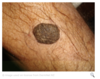
Seborrhoiec kerratosis

Pharyngeal pouch:


Kartagener syndrome:
This patient has x-ray findings consistent with dextrocardia and bronchiectasis (tram-track opacities). Hyperinflation is also seen in this film.

Plaque psoriasis
What is a Bishop score used to indicate? [5]


Grading of VUR


Loplik spots and measles
Compare Mx of ecoptics

Compare glomerulopathies


Barrett’s


