Theme 2- core clinical immunology- week 1 Flashcards
What is the atopic triad?
Triad- rhinitis, dermatitis, asthma
What does asthma cause?
Airway inflammation
What is rhinitis?
Rhinitis, also known as coryza, is irritation and inflammation of the mucous membrane inside the nose.
What does rhinitis cause?
Blocked / runny / itchy nose, sneezing - often with eye symptoms (Itching / burning / watery eyes, redness)
What does rhinitis lead to?
Leads to allergic and non-allergic subdivisions
What does the allergic response to rhinitis lead to?
Allergic leads to seasonal and perennial:
Seasonal- pollen and moulds
Perennial- house dust mite, animal dander
What does the non-allergic response of rhinitis lead to?
Non-allergic leads to vasomotor, infective, structural, drugs, hormonal and polyps
What is the treatment of rhinitis?
Treatment – Antihistamines and intranasal steroids
What is asthma?
Disease of inflammation & hyper-reactivity of small airways
Who does asthma mostly affect?
In childhood - aero-allergic stimuli - house dust mite key pathogenic importance
What is asthma mediated by?
Immediate symptoms are IgE-mediated
What does asthma cause?
Damage to airways due to late phase response
Damaged airways are hyper-reactive to non-allergic stimuli e.g. Fumes
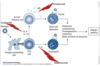
What is the pathogenesis of asthma?
Mechanism- allergen presenting by APC –> T cell proliferation and differentiation into a TH2 cell releasing cytokines- IL4, IL13 and IL5–> B cell plasma cell forming mast cells, basophils and TH2 cell forms eosinophils–>Mediator- histamines, leukotrienes, prostaglandins, cytokines and basic protein and enzymes–> Leads to asthma
What is dermatitis?
CLINICALLY - Intense itching, blistering/weeping, cracking of skin

What is the major trigger in atopic disease of dermatitis?
HOUSE DUST MITE now thought to be MAJOR TRIGGER in atopic disease
What is the treatment for dermatitis?
Topical Steroids & Moisturisers
What is atropy?
Atopy refers to the genetic tendency to develop allergic diseases such as allergic rhinitis, asthma and atopic dermatitis (eczema).
What is non-atopic?
Some conditions are not dependent on IgE but still involve an abnormal immune response to a wide variety of external environmental agents. These conditions are known as non-atopic (non-IgE-mediated).
What causes an itch?
Filaggrin protein- main barrier function- allergic reaction leads to distruption and leads to a Th1 response then Th2 response leading to secretion of IgE. Leads to increased IL-31 causing itch.

How does sentisation lead to exposure?
IgE exposure on B cells and APC–> activates T cells- Th1 to TH2 cells via cytokines–> Th2 main allergic response–>sends to B cells to secrete IgE then differentiate into plasma cells to release IgE–> Crosslinking of mast cell and basophil cell-surface –> release of vasoactive amines–> lipid mediators, chemokines and cytokines
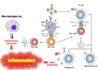
Summary of Late Phase Inflammation
- Allergic inflammation (late phase of the allergic reaction).
- Following migration to sites of allergen exposure under the influence of chemokines and other cytokines, allergen-specific T cells are reactivated and clonally expand.
- Local IgE-facilitated antigen presentation by dendritic cells (DCs) increases T-cell activation.
- Local IgE production is seen in allergic rhinitis and asthma but not in allergic skin inflammation (the main example of which is atopic dermatitis).
- Eosinophils are one of the main inflammatory cells
- TH1 cells, which produce interferon- (IFN) and tumour-necrosis factor (TNF), contribute to the activation and apoptosis of keratinocytes (in the skin), bronchial epithelial cells and pulmonary smooth-muscle cells.
- Activation of mast cells and basophils, which release histamine, chemokines and other cytokines
What is specific IgE testing?
An allergen-specific immunoglobulin E (IgE) test measures the levels of different IgE antibodies.
What occurs in specific IgE testing?
Allergen binds to different things add patients serum, antibodies will bind then need to confirm if antibody IgE type, need to bind an antibody that binds to the IgE then need a mechanism for identification which will change colour.
Specific IgE Testing (>0.35 KuA/L)
What is the skin prick test?
Skin prick test (>2mm wheal)- prick the skin so can trigger IgE reaction- will form a wheal over 2mm of the negative control (usually 0mm) for an allergic reaction
What is the intra-dermal skin test?
Intra-dermal test- more invasive- get more allergen than in the skin prick test. Inject tiny amount into the dermis and then mark the size of the circle that has been drawn around the bleb. No control needed. Size need to increase 3mm from site of injection. Takes longer than skin prick test. Primarily done for brochoallegies.
What is the basophil activation test?
The basophil activation test (BAT) is a flow-cytometry-based functional assay that assesses the degree of cell activation after exposure to a stimuli.
How does the basophil activation test work?
Antibody binds to basophil. Then add to add allergen. This triggers the basophils, makers on the basophil increase expression.

What is the graded challenge test?
Graded challenge test- exposure to patient slowly and watch if any reactions
Gold standard test for allergy- graded challenge test
What are the advantages and disadvantages of specific IgE testing?
Specific IgE Testing
Safe
False negatives
False positives
What are the advantages and disadvantages of the skin prick test?
Skin Prick Test
Quick
Patient satisfaction
False negatives
False positives
Antihistamines
Slight risk
Treatment of common allergies?
Symptomatic
Antihistamines, Steroids, Adrenaline
What is specific- immunotherapy (subcutaenous or sublingual- under the tongue) used for?
Indications:
Life threatening reactions to Wasp & Bee sting
Severe Hay fever
Animal dander allergy
Not Helpful:
Multiple allergies
Food allergy
Eczema
Spontaneous Urticaria
What does allergic-specific immunotherapy allow?
Allergen-specific immunotherapy (SIT) improves the quality of life of treated individuals and has been shown to reduce both symptoms of allergy and medication use in controlled clinical trials.
What is the mechanism of SIT?
- reduces mediator release
- reduces number of cytokines in the blood, increased cytokines in tissues
- decrease in allergen specific IgE
- increase in ILs
- prevention of new sensitisation and reduces symptoms of allergic reaction
What are major food allergens?
- Water soluble glycoproteins 10 - 60 kd
- Cow’s milk
- Egg
- Legumes - peanut; soybean; tree nuts
- Fish
- Crustaceans / molluscs
- Cereal grains
What are the adverse reactions to foods?
- Gastrointestinal- vomiting, diarrhoea, oral symptoms
- Respiratory (upper & lower)- rhinitis, bronchospasm
- Cutaneous- urticaria, angioedema, role of food in atopic dermatitis unclear
- Anaphylaxis
Why does someone reaction if they have no IgE detected?
Different sensitivities
When taking someones history for a drug what should you find out?
- Indication for the drug
- Detailed description of the reaction
- Time between drug intake and onset of symptoms
- Number of doses taken before onset
- Aware of pharmacological effects and non-immunological ADR
When managing someones drug allergy what should you do?
Management
- Intradermal testing (maybe contraindicated in some situations)
- Graded challenge
- Desensitization
What cells are seen in the innate immunity?
- Macrophages
- Dendritic cells
- Mast Cells
- Neutrophils
- Complement
What cells are seen in adaptive immunity?
- T cells
- B cells
What is the innate response? Recognition of what? Memory? Regulation? Response time? Duration?
- Pattern recognition against broad classes of antigen
- No memory
- No amplification
- Little regulation
- Fast response (hours – days)
- Short duration
What is the adaptive response? Recognition? Memory? Regulated? Response time? Duration?
- Highly specific (T and B cell receptors)
- Strong memory and amplification component (e.g. vaccines, previous infection)
- Many regulatory mechanisms
- Slow response (days to weeks for initial exposure)
- Responses may last months - years
How does the innate response occur?
Innate immune cells directly detect and attack antigenic targets (e.g. microbes) Occurs at sites of infection – e.g. barrier organs
- Phagocytosis
- Cytotoxicity- complement sticks to cell walls and destroys them
- Inflammatory mediators and chemokines to attract other cells
- Dendritic cells present antigen to T cells
- Cross talk between DCs, T cells and B cells
- Immune memory to determine specific learned responses
- Occurs in lymphoid tissues

How does the adaptive immune system respond?
Adaptive immune cells activate innate immune cells, directing tissue inflammation to specific targets
T cell cytokines activate monocytes, macrophages
B cell antibodies activate complement

What are phagocytic cells?
Phagocytic cells
Neutrophils: eat and destroy pathogens
Macrophages: also produce chemokines to attract other immune cells
Dendritic cells: also present antigen to adaptive immune system
What are histamine producing cells?
Histamine-producing cells
Mast cells, basophils, eosinophils: produce histamine and other chemokines and cytokines
Vasodilatation, attract other immune cells
Defence against parasites, wound healing but also allergy and anaphylaxis
What are complements in innate immune system?
Complement
Directly attacks pathogens via alternative and lectin pathways
May be activated by adaptive immune system via antibodies
What are cytokines?
Cytokines
Signal between different immune cells (e.g. innate to adaptive, adaptive to innate)
What are chemokines?
Chemokines
Attract other immune cells to sites of inflammation
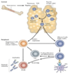
How do T cells cause inflammation by inflammatory cytokines or by helping B cells make autoantibodies?
APC picks up antigen, goes to T cell can presents it (immune synapse- one cell talking to the other), T cells can convert naïve T helper cell to a T helper 1 cell, which makes inflammatory cytokines.
Another thing T helper 2 cells- cross talk with B cells into memory B cells which make plasma cells and produce lots of autoantibodies.
Net results of this is inflammation.
What do autoantibodies do?
Auto antibodies- Some autoantibodies directly interfere with normal physiological function rather than causing inflammation and tissue damage.

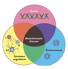
Definition of autoimmunity?
Defining characteristic: the adaptive immune system recognises and targets the body’s own molecules, cells and tissues (instead of infectious agents and malignant cells)
How does autoimmunity occur?
Main characteristics:
- T cells that recognise self antigens
- B cells and plasma cells that make autoantibodies
- Inflammation in target cells, tissues and organs is secondary to actions of T cells, B cells and autoantibodies
What is the definition of autoinflammation?
The term introduced in 1999 following identification of the genetic basis of periodic fever syndromes (FMF, TRAPS, CAPS and HIDS)
To distinguish from autoimmune diseases- don’t have the adaptive immune component
What are the main characteristics of autoinflammation?
Main characteristics:
- seemingly spontaneous attacks of systemic inflammation
- no demonstrable source of infection as precipitating cause
- absence of high-titre autoantibodies and antigen specific autoreactive T cells
- No evidence of auto-antigenic exposure
Table of comparison of autoinflammation and autoimmunity

What are the components of autoimmunity?
- Theoretical concept
- Breakdown of self-tolerance
- Many cells of the immune system have capacity for autoimmune functions
- Overlap with normal immune functions such as anti-tumour immunity
- Some people have autoantibodies without any symptoms
What is autoimmune disease caused by? What does it lead to?
- Distinct clinical entities
- Environmental factors acting on favorable genetic background
- Wide variety of pathogenic mechanisms between diseases
- Autoimmunity leading to inflammation, organ dysfunction and damage
Where are T cells selected?
T cell selection in the thymus
What are B cells produced by?
B cell selection in the bone marrow
What happens when autoimmune T cells and B cells are produced?
When make T cells and B cells they are selected so any autoimmune ones are deleted and ones that aren’t are kept.
What types of cells select for certain self-antigens?
- Certain HLA (MHC) types select for certain self-antigens
- Other genes that regulate immune functions
What are antigenic factors?
- Infections that trigger autoimmune responses
- Environmental triggers: UV light, smoking
- Alterations in self-proteins that increase their immunogenicity
What are the causes of the autoimmune disease?
- There is usually some genetic predisposition
- The immune system has many regulatory functions to shut down immune functions – tolerance of self is a dynamic state
- But we are not born with autoimmunity – there must be environmental triggers
As T cells are randomly generated in the thymus they are tested against self antigens. What happens to them and what do they produce?
Deletion
Effector CD4 and CD8 T cells
Regulatory T cells
What is the process in the thymus and bone marrow?
- This is a process in the thymus for T cells and in the bone marrow in B cells
- In thymus have progenitors of T cells
- Need lots of T cells receptors that recognize any antigen that you might meet including bacteria/ viruses that doesn’t exist yet
- This is done by when it is made they rearrange their genes
- This variety will react to self-antigens so T cells are tested against self-antigens in the thymus and will get deleted, if doesn’t react to a self-antigen is useful and won’t harm you
- T cells can recognize self-antigen weakly, thymus will turn that into a regulatory T cells which when instead of the attacking the antigen it will protect it and turn things off
What is MHC Class 1 encoded by? Where are they found? What does it present to?
- Encoded by genes in HLA-A, HLA-B, HLA-C
- On all nucleated cells- if cell infected of virus it falls on that cell to report to the immune system
- Presents antigen to CD8+ T cells- cytotoxic T cells

What is MHC class 2 encoded by? On what cells? Presented by what cells?
- Encoded by genes in HLA-DP, HLA-DQ, HLA-DR
- On dedicated APCs
- Presents antigen to CD4+ T cells- T helper cell which directs a whole immune response- innate

What are causative associations other than genes for autoimmune diseases?
Sex (hormonal influence)- women >> men
Age- autoimmunity more common in elderly- more environmental influences as you are older
Sequestered Antigents- May be recognised as foreign by the immune system (e.g. cell nucleus, eye, testis)- antigen not seen in the immune system as it is inside a cell more likely to be recognized as foreign
Environmental triggers
- Infection
- Trauma-tissue damage
- smoking
What can cause autoimmunity as well as normal immune response?
- Infections activate the immune system generally
- Molecular mimicry-E.g. in rheumatic fever (inflammation of heart valves) antibodies against M protein of Streptococcus also react against the glycoproteins of the heart
How can changes in amount or nature or autoantigens may cause autoimmunity?
- Citrullination of proteins make them more immunogenic (leads to rheumatoid arthritis)- citrulline switches with alanine
- Tissue transglutamase alters gluten to help it bind to HLA-DQ (leads to coeliac disease)
- Failure to clear apoptotic debris increases availability of sequestered antigens inside the cell (leads to systemic lupus erythamatosus)
Problems with T cell selection in the thymus and B cell selection in the bone marrow leads to what?
Failure of Central tolerance leads to autoimmunity
How does autoimmunity occur from genetic predisposition?
Certain HLA (MHC) types select for certain self-antigens
Other genes that regulate immune functions
How do antigenic factors lead to autoimmunity?
Infections that trigger autoimmune responses
Environmental triggers: UV light, smoking
Alterations in self-proteins that increase their immunogenicity
What is organ specific autoimmunity?
- Affect a single organ
- Autoimmunity restricted to autoantigens of that organ
- Overlap with other organ specific diseases
- Autoimmune thyroid disease is typical- antigen specific to the thyroid, immune system attacking it so get autoimmunity
What is systemic autoimmunity?
- Affect several organs simultaneously
- Autoimmunity associated with autoantigens found in most cells of body
- Overlap with other non-organ specific diseases
- Connective tissue diseases are typical
What age of onset of autoimmunity most common?
More common in the elderly and women
Cause of these symptoms?
A 28-year-old woman with recent tiredness and difficulty concentrating had experienced a decline in memory over the last several months. She also noted decreased frequency of bowel movements and an increased tendency to gain weight. She felt chilled without light sweater, even in warm weather.
Examination
She was 5’5” tall and weighed 125 lb. Her pulse rate was 58 beats per minute and her blood pressure was 138/88. She had a slightly puffy face and her eyebrows were sparse, especially at the lateral margins. The deep tendon reflexes were normally contractive, but showed delayed relaxation.
Diagnosis- hypothyroidism
Cause of these symptoms?
Case 2
History
30 yrs old women presents with anxiety, weight loss, diarrhea and palpitations. Although it is winter she hardly notices cold weather. She finds it almost impossible to sleep but despite this she seems to have abundance of energy.
Examination
She had swollen red eyes, rapid pulse and sweaty hands. She has rapid reflexes.
Diagnosis- hyperthyroidism
What is Hashimotos thyroiditis? What does it lead to?
Destruction of thyroid follicles by autoimmune process
Associated with autoantibodies to thyroglobulin and to thyroid peroxidase
Leads to hypothyroidism
What is Grave’s disease? What does it result to?
Inappropriate stimulation of thyroid gland by anti-TSH-autoantibody- mimics TSH
Leads to hyperthyrodism
Difference between type 2 and 3 hypersensitivity?
Type 2 hypersensitive- antibody sticks to the cell of the organ that gets the inflammation
Type3- antibody causes inflammation in another organ that isn’t the thyroid
What is myoasthenia gravis?
Myasthenia gravis-
NM disease- autoantibodies block the ACh receptor
What is pernicious anaemia?
Pernicious anaemia
If have an iron deficiency- have smaller cells as less inside
Pernicious anaemia- deficiency in B12- have a problem in making the cell- lot of Hb but not enough cells so end up with MCV being large
How do you get pernicious anaemia?
To absorb B12- can’t just absorb it naturally, need intrinsic factor that sticks to the B12 and allows it to be absorbed. Made by parietal cells. If have autoimmune condition that affects these cells, won’t get IF and B12 thus getting pernicious anaemia.

What are the symptoms of SLE?
- Photosensitive malar rash
- Multiple mouth ulcers on roof of mouth
- Arthalgia
- Alopecia
How does SLE occur?
Autoantibodies form against many different molecules in the cell nucleus
- ddDNA
- dsDNA
- Ribosomes
- Histones
Normally cell shouldn’t see things inside the cells, releasing nucleus content forming a rash i.e. sun burn damages cells
Classically antibody deposition and inflammation are seen in the skin
How does autoimmunity occur from failed clearence of nuclear antigens?
- Nuclear self-antigens are normally always intracellular, so shielded from the immune system – sequestered antigens
- In cell death by apoptosis nuclear material is digested and cleared
- In cell death by necrosis nuclear antigens may not be cleared and may then act as antigens to the immune system
- UV light causes DNA damage, and cell death
- Some people have genetic defects in mechanisms to clear cell debris
What do antibodies against antigens form? Where do they deposit?
Antibodies against antigens in the nucleus combine with their targets to form IMMUNE COMPLEXES in the circulation
Immune complexes deposit in any organ – activate complement and cause inflammation
What is lupus nephritis?
Normal glomerulus should be lacey and blood flows through and moves into collecting system. If have immune complex landing in there causes inflammation.
Lupus nephritis is inflammation of the kidney that is caused by systemic lupuserythematous (SLE).
What are types of connective tissue disease?
- Systemic lupus erythematosus
- Scleroderma
- Polymyositis
- Sjogrens syndrome
- Ubiquitous antigens (components of the cell nucleus) cause multisystem inflammation
What are the two ways adaptive immune system may cause disease?
- Directly (e.g. autoantibodies blocking a receptor in myaesthenia gravis)
- By causing inflammation (e.g. autoantibodies causing inflammation and organ damage in hashimoto’s thyroiditis)
What are the two types of autoimmune disease?
Organ specific (often due to an autoantigen specific to that organ, e.g. hashimoto’s thyroiditis)
Or non-organ specific (usually due to a ubiquitous antigen, e.g. anti-nuclear antibodies and widespread immune complexes)
51 yo lady
18month history of SOB on exertion
Saw respiratory team and diagnosed with pulmonary fibrosis
Fatigue, aches and pains
Thickening of skin on hands and changes in the skin around her mouth
Examination
Sclerodactyly both hands- hardening of skin on hand
Livedo reticularis on the legs (spasms of BV- make skin mottled)
Cool feet on palpation
Diagnosis?
Clinical diagnosis of scleroderma (diffuse systemic sclerosis)
What is scleroderma?
Scleroderma is a long-lasting disease that affects your skin, connective tissue, and internal organs. It happens when your immune system causes your body to make too much of the protein collagen, an important part of your skin.
How to work out if a patient has an autoimmune disease?
To work this out we
- take the clinical history
- examine the patient
- then perform some blood testing
Different autoimmune diseases can have similar symptoms
Achieve correct diagnosis
Leads to correct management
Tests required to confirm autoimmune disease
ANA- antinuclear antibody
ANCA
CK- creatinine kinase
Rheumatoid Factor
Anti-CCP antibody
Complement
FBC U&Es LFTs CRP
General principles of diagnostic testing
Use to answer specific questions and/or to support a clinical diagnosis,
But not as screening tools
The ability of the tests to correctly discriminate between health and disease is improved when they are used in the appropriate population
Definition of sensitivity
Sensitivity [a/(a+c)]
measure of how good the test is in identifying people with the disease
a=True positive
c= False negative

Definition of specificity
Specificity
[d/(b+d)]
measure of how good the test is at correctly defining people without the disease
b= False positive
d= True negative

Positive predictive value defintion
Positive predictive value
[a/(a+b)]
The proportion of people with a positive test who have the target disorder

Defintion of negative predictive value
Negative predictive value = d/(c+d) The proportion of people with a negative test who do not have the target disorder

What does this diagram show?

ND- top diagram shows ND, if don’t have disease should test negative and vice versa, depending where we set cut off of test- want to make sure when we assign a value to the patients results that we are definitely saying they have the disease or not
Bottom diagram- can change cut off- more people testing negative when have disease vice versa
Types of diagnostic test
Non specific- Inflammatory markers
Disease specific:
Autoantibody testing
HLA typing
Non-specific markers of systemic inflammation?
- ESR
- CRP
- Ferritin
- Fibrinogen
- Haptoglobin
- Albumin
- Complement
What are antinuclear antibodies?
Antibodies in the patient’s blood that bind to the cell nucleus
We can be then more specific and identify subtypes of antibody that bind to different bits of the cell nucleus
How Antinuclear antibody testing works?
1st part – put an antigen on a plate. Separate the patient’s serum (antibodies in). Put antibodies fluid over the plate. If antibody present for antigen, it should bind.
2nd part – add an antibody (with a light up label) to the antibody.
3rd part – put under fluorescent microscope to see if it’s lit. Different patterns signify different diseases.

What do DsDNA and ENA tests do?
Other Techniques if positive to work out what antibodies
Immunoblots- strip of paper with specific antigens
Individual ELISA’s- add enzyme
>100 different antibodies described in SLE
What is a microbead-based immunoassay?
Bead coated with antigen and then bead gives off different colours and is read my machine how much each antibody is present
3 month history
Patchy alopecia
Painful and swollen joints of the hands
Mouth ulcers
Diagnosis?
SLE
67 year old man
Presents to GP with joint stiffness and pain mainly affecting the small joints of both hands.
Stiffness worst in the morning and improves over 1-2 hours
Joint X ray- peri-articular swelling with effusion of MCP joints, osteopenia, joint space narrowing and erosions
CXR normal
Diagnosis?
RA
What is rhemuatoid factor?
Antibody (IgM, IgG or IgA) directed against the Fc portion of IgG
Commonly found in rheumatoid arthritis but not diagnostic of the diseases (sensitivity and specificity around 70%)
Can be seen with other diseases in which polyclonal stimulation of B cells is seen (chronic infections)
What is another marker for RA?
Anti-CCP Antibody (ACPA)
ACPA more specific (95%) for RA then RF
Similar sensitivity to RF
Useful prognostic marker
ACPA positive patients tend to have more severe and erosive disease
How to diagnose RA?
The diagnosis of Rheumatoid Arthritis is clinicalCombination of symptoms, Xrays, blood tests
High RhF, CCP and elevated ESR and CRP add to the probability of a definitive diagnosis
Early treatment can prevent joint damage and significant morbidity
What is ordered when patient has vasculitis?
Anti neutrophil cytoplasmic antibodies (ANCA)
53yo man
Presented to ED with 3 week history of fever fatigue muscle pain, night sweats and weight loss 6kg.
Urine dip contained blood
CXR showed evidence of lung granuloma formation
Diagnosis?
Diagnosis of granulomatosis with polyangitis (formally known as Wegener’s granulomatosis)
What is granulomatosis?
Granulomatosis with polyangiitis is an uncommon disorder that causes inflammation of the blood vessels in your nose, sinuses, throat, lungs and kidneys. Formerly called Wegener’s granulomatosis, this condition is one of a group of blood vessel disorders called vasculitis. It slows blood flow to some of your organs.
What identifies vasculitis?
Cytoplasmic (c)ANCA
Perinuclear (p)ANCA
What antigens do cytoplasmic (c)ANCA target?
PR3
What antigens does perinuclear (p)ANCA target?
MPO
Cytoplasmic (c)ANCA under microscope

Perinuclear (p)ANCA under a microscope

What does a positive ANCA mean?
Positive ANCA; useful in suggesting the diagnosis in the proper clinical setting
Does a negative ANCA assay exclude AASV?
Negative ANCA assays do not exclude AASV (vasculitis)
10%-50% of patients may be ANCA neg
Reemergence of ANCA pos in a patient who was ANCA neg whilst in remission suggests what?
suggests a risk of disease flare.
Time to flare uncertain however
What is organ specific autoimmune testing?
Theme of this testing similar to the other tests with flourescence- instead have cells on slides- tissues from rats or monkeys form stomach of liver

How to test for autoimmune liver disease?
Part of non-invasive liver screen
Perform if liver tests deranged
Autoimmune liver disease- Anti-mitochondrial Ab specific for what?
primary biliary sclerosis
Anti-smooth muscle and anti-liver/kidney/microsomal (LKS) Abs, found in what?
autoimmune hepatitis
How are antibodies detected in autoummune liver disease screening?
Antibodies detected by IF screening using rodent tissue block (oesophagus, liver and kidney) and antigen specific ELISA
What are the autoantibodies in type 1 disease?
Several types:
- islet cell antibodies
- anti-GAD65 anti-GAD67
- anti-insulinoma antigen 2 (IA-2)
- insulin autoantibodies (IAAs)
Disappear with progression of disease and total destruction of β islet cells
Disease confirmation
to identify relatives and patients at risk of developing autoimmune diabetes
What is Addison’s disease? Where do you detect autoantibodies?
Addison’s disease, also called adrenal insufficiency, is an uncommon disorder that occurs when your body doesn’t produce enough of certain hormones. In Addison’s disease, your adrenal glands, located just above your kidneys, produce too little cortisol and, often, too little aldosterone
Clinical features
Detect auto antibodies targeting the adrenal cortex
Impairs production of Cortisol
What is pernicious anaemia? What antigen is located in the parietal cells?
Pernicious anemia (per-NISH-us uh-NEE-me-uh) is a condition in which the body can’t make enough healthy red blood cells because it doesn’t have enough vitamin B12.
Antigen: H+K+-ATPase located in the gastric parietal cells of rodent stomach.
Clinical Antibody present in more than 90% of patients with Pernicious anaemia.
Autoimmune gastritis leads to pernicious anaemia which is characterised by antibodies to GPC and intrinsic factor
How do we test for anti-nuclear antibodies?
Indirect immunofluorescence, ELISA, microbead immunoassay, immunoblot
What disease is associated with anti neutrophil cytoplasmic antibodies (ANCA)?
Vasculitis
