The Black Lung Flashcards
Tidal Volume (TV)
amount of air exchanged w/ each breath = 7 ml/kg normally
Residual Volume (RV)
volume of gas left in lung after a complete expiration = 25% of total lung capacity (TLC)
Inspiratory Reserve Volume (IRV)
How do you calculate?
volume of air that can be inspired from end of tidal inspiration to total lung capacity = IC – TV

Expiratory Reserve Volume (ERV)
volume of air that we can exhale from end of a tidal expiration to residual volume –> forcibly exhale
Total Lung Capacity (TLC)
= total volume of gas in lung at end of maximal inspiration = sum of all above volumes (RV + ERV + TV + IRV)
volume at which inward (expiratory) recoil of the lung and chest wall > strength of inspiratory muscles
Vital Capacity
= TLC – RV = volume of air that can be exhaled from total lung capacity to residual volume = 75% of total lung capacity measurement – inspire completely and then expire VC into spirometer
Functional Residual Capacity (FRC)
RV + ERV
volume of gas in the lungs after normal expiration
Inspiratory Capacity (IC)
= TLC – FRC = TV + IRV = amount of gas we can inspire from functional residual capacity to total lung capacity.
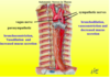
Elastic Properties of the Lung (3)
transpulmonary pressure = alveolar pressure – pleural pressure
compliance
recoil forces: tissue (elastin/collagen) and surface tension
How does lung compliance change with Greater lung volume and growth
HIGH volume in lung (inspiration) –> LOW compliance
HIGH lung volume (size of lung) –> HIGH absolute compliance
*an adult is more compliant than a child
Function of surfactant
What happens when there is none?
Surfactant: prevents collapse of alveoli and allows them to stretch open more easily, thus stabilizing the lung
Small alveolus (small radius), surfactant breaks up (reduces) the surface tension
Large alveolus (large radius), surface tension is greater because the surfactant is diluted by the larger surface area
Premature infants have no surfactant –> alveoli collapse. Cannot generate enough inspiratory pressure to overcome surface tension –> respiratory distress syndrome
Elastic properties of the chest wall
- resting volume of chest wall = 70% of TLC
- outward recoil force on lung at FRC (40% TLC) aids inspiration
- inward recoil of force at >70% TLC aids expiration

Elastic properties of the total respiratory system (chest wall + lungs)
- lung always wants to deflate
- chest wall wants to expand (except at highest volumes)
- total lung capacity (TLC) = point at which muscles can no longer overcome expiratory recoil of lung + expiratory recoil of chest wall
- functional residual capacity (FRC) = balance of outward recoil of chest against inward recoil of lung
How does gravity work in causing regional ventilation differences b/w apex and base of lung?
-
pleural pressure gradient (more P at base) – weight of lung increases pressure on plural space as you go down
- more negative pressure (less pressure) at apex –> High transpulmonary pressure (PL) –> alveoli are bigger (higher percent of max size) and stiffer
-
regional ventilation
- High size of base alveoli –> High compliance –> High ΔP during inhalation –> High ventilation
- matches High perfusion at base of lung

What is pulmonary hypertension
pulmonary arteriole HTN?
Resting MAP of >25 mmHg (right heart catheterization)
PAH adds to the criteria that pulmonary venous pressure (or capillary wedge) must be <15 mmHg
Equation for pulmonary arteriole pressure
PA mean = (CO x PVR) + PCWP
Determined by:
Right-sided CO
Pulmonary vascular resistance
Mean pulmonary venous pressure/left atrial pressure (PCWP)
What medical conditions lead to increases in Pulmonary venous pressure? (pulmonary HTN)
LV dystolic/systolic dysfunction
mitral valve disease –> increase pulmonary venous pressure
What medical conditions lead to increases in Pulmonary vascular resistance? (pulmonary HTN)
any condition that decreases the area of the pulmonary vascular bed (pulmonary emboli, C.T. diseases, interstitial lung disease, COPD)
or induce hypoxic vasoconstriction (any lung disease producing hypoxia) increases pulmonary vascular resistance
What medical conditions lead to increases in Right-sided cardiac output? (pulmonary HTN)
Left-to-right atrial septal defects (ASD)
Left-to-right ventricle septal defects (VSD)
other systemic-to-pulmonary shunts
overall, increase right-sided CO by increasing RV volume
What are the 3 abnormal signaling pathways in pulmonary HTN?
Prostacyclin: Arachidonic acid –> cAMP –I Ca2+ entry into sm. muscle cell –> vasodilation, antiproliferation.
NO: forms NO from arginine –> guanylate cyclase(GTP–>cGMP) –I Ca2+ entry into sm. muscle cell –> vasodilation, antiproliferation
Endothelin: forms ETA and ETB –> vasoconstriction, proliferation
Pulmonary HTN: decreased Prostacyclin, NO and Increased Enothelin
Ways to treat pulmonary HTN based off of the 3 altered pathways
Calcium channel blockers
Prostacyclin analogs (Low prostacyclin synthase)
Sildenifil (PDE5 inhibitors): promote activity of NO pathway
Endothelin receptor antagonists block the effect of endothelin at sm. muscle cell receptors
What are the general types of pneumonia
Community-acquired pneumonia: 95% viral
Nosocomial pneumonia: hospital-acquired (gram negative)
Aspiration pneumonia: mix of anaerobic/aerobic bacteria
Pnemonia in immunosuppressed patient: HIV, leukemia, lymphoma, chemotherapy, iatrogenic immunosuppresion
May be categorized based off of organism
Pathogenesis of Pneumonia (5)
Loss of defense:
- inhibition of the normal cough reflex (neuromuscular disease, drug overdose, intubation, coma)
- injury of mucociliary apparatus (viral destruction, smoking, genetic disease - immotile cilia syndrome)
- interference of phagocytic or bactericidal action of alveolar macrophages (alcohol, tobacco smoke, anoxia)
- bronchial obstruction (neoplasm, mucus plugging)
- decreased immunity (immunodeficiency, viral infections, leukemia, lymphoma, immunosuppressive therapy, chemotherapy)

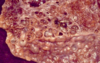
Bacterial pneumonia: Gram-positive cocci
Alveolar spaces are filled by neutrophils, fibrin, RBCs, and macrophages. Alveolar septa are typically hyperemic and congested but not inflamed.