Testing/Imaging Flashcards

1
Q

How can chromosomal abnormalities cause disease?

A

Altering the concentration of particular gene products

  • Duplication or amplification (typically oncogenes)
  • Deletion or gene interruption (typically tumor suppressor genes)

Altering the gene product by producing a new fusion gene with altered expression and/or a new fusion protein with altered function.

  • Example: fuse regulatory region from one gene with functional portion of another such that expression levels, timing and/or distribution within or among cells is altered.
  • Example: Alter the function of a protein by substituting one functional domain with another (e.g. substitute one DNA binding site with another).
2
Q

CML accounts for ____% of all leukemia and occurs most frequently in ____ year olds.

A

CML accounts for 15-20% of all leukemia and occurs most frequently in 40-50 year olds.

3
Q

CML was the first cancer to be associated with a ________.

A

specific recurrent chromosome abnormality

4
Q

CML is characterised by a ___ chromosome that had formed as a result of a ___ translocation

A

CML is characterised by a Philadelphia chromosome that had formed as a result of a 9;22 translocation

5
Q

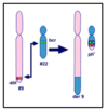
The _____ or Philadelphia chromosome contains a fusion gene composed of the ______ serine/threonine kinase gene on 22 and the _____ tyrosine kinase gene on chromosome 9.

A

The der(22) or Philadelphia chromosome contains a fusion gene composed of the BCR (breakpoint cluster region) serine/threonine kinase gene on 22 and the ABL1 (Abelson1) tyrosine kinase gene on chromosome 9.
6
Q

The new fusion BCR/ABL1 tyrosine kinase produced by the fusion gene is permanently _____ and activates a number of ______. (CML)

A

The new fusion BCR/ABL1 tyrosine kinase produced by the fusion gene is permanently turned on and activates a number of signal pathways. (CML)

7
Q

Permanent _____ of the BCR/ABL1 fusion kinase leads to _____ by interfering with basic cellular processes.

A

Permanent activation of the BCR/ABL1 fusion kinase leads to malignant transformation by interfering with basic cellular processes.

8
Q

95% of patients with CML carry a _____ translocation

A

Recriprocal 9;22

9
Q

Explain the probe placement used for BCR-ABL1 sequencing in CML patients:

A

An orange/yellow probe is placed at the bottom of chromosome 9

A green probe is placed spanning the BCR region of chromosome 22 and upstream on 22 as well.

10
Q

What would a FISH assay look like for a patient with CML (who has a Philadelphia chromosome)?

A

Top is normal - see two greens and two reds. Can see in interphase, aka non-dividing cells as well.

In mutant - see red and green signals on normal 9 and 22, and also see a yellowy/white signal on the derivative 9 and derivative 22 as well. See abnormal pattern in interphase, non-dividing cells too.

11
Q

_____ is the first-line drug of choice for the treatment of most patients with CML in chronic phase. WHY?

A

Imatinib.

ATP is normally bound to BCR-ABL, resulting in the phosphorylation of a tyrosine on the substrate (cancer protein). The substrate is then able to interact with an effector protein and multiple cancer pathways are stimulated. Right panel, imatinib is bound to BCR-ABL in place of ATP. The tyrosine of the substrate is not phosphorylated, and the substrate can no longer interact with the effector protein.

Because the BCR-ABL1 tyrosine kinase enzyme exists only in cancer cells and not in healthy cells, imatinib works as a form of targeted cancer cells are killed through the drug’s action. Well tolerated and induces long-lasting remissions.

12
Q

Imatinib is a ____ inhibitor developed to treat ____

A

tyrosine kinase inhibitor

CML treatment

13
Q
A
14
Q

What is the major cause of relapse in cancer and leukemia?

A

Minimal residual disease - small numbers of cancer cells that remain in the patient during treatment, or after treatment when the patient is in remission.

15
Q

How are patients treated with Imatinib monitored post-treatment?

A

Monitored at 3- 6 month intervals until cytogenetic remission is obtained then yearly to monitor for marrow dysplasia or the emergence of new clones with additional chromosome abnormalities [+8, i(17q), additional copies of der(22)] associated with disease progression. Molecular monitoring is also performed.

In the lab, do this with INTERPHASE FISH. Allows us to look at many cells quickly. Can see zoomed-in verson and see difference in signaling.

16
Q

How is karyotyping helpful in identifying additional clones that signal disease progression in CML patients?

A

Allows us to identify additional clones that would signal disease progression.

This patient has remaining 9 cells that carry translocation that also carry an extra 8 and 17. This suggests progression and worsening, with a clone that has more abnormalities.

FISH would have identified the 9;22 translocation, but not the other abnormalities that are signaling/suggesting disease progression.

17
Q

Chromosome abnormalities involving the MLL (KMT2A) gene have been implicated in at least 10% of _____ (e.g. __ and __)

A

Chromosome abnormalities involving the MLL (KMT2A) gene have been implicated in at least 10% of acute leukemias (e.g. AML and ALL)

18
Q

The MLL (KMT2A) gene mutation is located at __ (site) and encodes a transcriptional regulatory factor essential for _____ and self-renewal of ______ and _____ progenitors

A

The MLL (KMT2A) gene mutation is located at 11q23.3 (site) and encodes a transcriptional regulatory factor essential for embryonic body plan formation and self-renewal of hematopoietic stem cells and immature progenitors

19
Q

Why is the MLL gene associated with a poor prognosis?

A

Currently no targeted therapies for patients with MLL rearrangements.

Some exceptions do exist however, such as a deletion or inversion of 11q23

20
Q

Recurrent ____ translocations are associated with at least 80 partner genes

A

MLL (KMT2A)

The mechanism whereby the multiple MLL fusion partners initiate cellular transformation is largely unknown.

Fusion protein acts as a dominant gain-of-function mutation (possibly increased multimer formation and/or stability of fusion protein).

21
Q

Can traditional G-banding be used to identify MLL (KMT2A) rearrangements?

A

Yes

This patient carries a 4-11 translocation. Fuses the MLL gene to AF4 gene. Look for a short chromosome 4 with a little 11 hanging off it, and a long 11 with a lot of 4 hanging off it.

22
Q

Given there are multiple pairing partners in patients with the MLL mutation, ____ is used rather than dual fusion FISH.

A

Break Apart

Use a green probe that hybridizes to 5’ end and upstream sequences, and a red probe that fuses to 3’ end and downstream sequences.

In any case with a MLL rearrangement, there will be a breakpoint in the MLL gene, so the red probe will migrate to new partner location.

PROS: this allows us to detect basically all the MLL rearrangements

CONS: it doesn’t tell us who the partner chromosome is.

23
Q

Which of the following samples would be most appropriate for chromosome microarray analysis?

A. A bone marrow sample from a patient suspected of having ALL secondary to a t(4;11)(q21;q23)

B. A sample from a patient suspected of having neuroblastoma with amplification of the MYCN gene as well as surrounding genes

C. A follow-up bone marrow sample looking for minimal residual disease in a patient with CLL and the following karyotype at diagnosis: 46,XY,-11,del(11)(q23),+12,del(21)(q22)

A

B. A sample from a patient suspected of having neuroblastoma with amplification of the MYCN gene as well as surrounding genes

A is balanced, which isn’t detectible

C shows minimal residual disease, so it’s not detectible.

24
Q

What are limitations of chromosome microarray analysis in cancer patients?

A

•Can’t detect balanced rearrangements

  • No gain or loss present to identify the balanced t(9;22) associated with CML or the t(4;11)(q21;q23) seen in ALL

•Low level mosaicism/clonal evolution

  • in setting of minimal residual disease the data from the normal cells would mask the data from the abnormal cells
  • minor clones that provide information about disease progression may be difficult to detect
  • Abnormalities present in less than 10-15% of cells are typically NOT detected
25
Q

Benefits of chromosome microarray analysis in cancer patients:

A

Microarray testing will detect some cytogenetic abnormalities that would be missed by traditional cytogenetic including:

  • Cryptic rearrangements that are below level of G-band detection especially those for which there are no commercially available FISH probes
  • Copy number neutral loss of heterozygosity (LOH) or acquired homozygosity which has been identified as a major mechanism of tumor suppressor gene inactivation
    • SNP microarray analysis required
26
Q

Neutral loss of heterozygosity or acquired homozygosity:

A

Major mechanism of tumor suppressor gene inactivation.

Is a common finding in all types of leukemia (as well as solid tumors)

  • Acute myelogenous leukemia (AML): ~29%
  • Acute lymphoblastic leukemia (ALL): ~29%
  • Chronic lymphocytic leukemia (CLL): ~20%
  • Myelodyplastic syndrome (MDS): ~9%
  • Multiple Myeloma (MM): ~24%
27
Q

Monitoring is typically done using a combination of _____ and ____. ____ testing is NOT typically used because low level clones would not be readily identifiable.

A

Monitoring is typically done using a combination of karyotyping and FISH. CMA testing is NOT typically used because low level clones would not be readily identifiable.

28
Q

The FISH assay strategy used (dual fusion vs break apart) varies depending on whether you are testing for a single very specific rearrangement (e.g. the t(9;22) in CML - you would use a ____ assay) or a rearrangement that involves multiple partners (e.g. MLL - you would use a ___ assay).

A

The FISH assay strategy used (dual fusion vs break apart) varies depending on whether you are testing for a single very specific rearrangement (e.g. the t(9;22) in CML - you would use a dual fusion assay) or a rearrangement that involves multiple partners (e.g. MLL - you would use a break apart assay).

29
Q

What are the 7 modalities of imaging used in radiology?

A

•X-ray/plain film/radiography: shoot X-rays at patient, which get blocked to varying degrees

•Fluoroscopy: real-time x-rays

•Angiography: x-rays of the vessels using IV contrast

  • Computed tomography (CT): X-ray machine that swivels around the patient and gives a 3D image
  • Magnetic resonance (MR, MRI): images using magnets to excite protons

•Ultrasound (US)/sonography: images using echoes from sound waves

  • Echocardiography (‘echo’) is a heart ultrasound

•Nuclear medicine or gamma imaging: inject something radioactive and look at the radiation emitted from the patient

  • This includes SPECT and PET
30
Q

On an x-ray, subjects that are ____ or have a ____ atomic number are lighter in color.

A

On an x-ray, subjects that are denser or have a higher atomic number are lighter in color.

  • Air (Black)
  • Soft Tissue (Gray)
  • Bone (White)
  • Metal (Extremely White)
31
Q

What is fluoroscopy?

A

–Real-time X-Ray

–Barium or other contrast agent used to fill viscus and provide contrast

–Good for GI investigations

  • Esophagus, stomach, bowel
32
Q

What is angiography?

A

–Real-time X-Ray of the vascular system

–Iodinated contrast agent injected to opacify vessels and provide contrast

–Used for vascular investigations coupled with intervention

  • Angioplasty
  • Coronary Catheterization
  • Vascular Embolization
  • Endovascular Aneurysm Treatment

–Biliary structures can also be seen

33
Q

What is a CT? What is it used for?

A

–Uses X-rays; multiple mobile detectors

–Extremely fast

–Greater range of densities:

  • Air (Black)
  • Fat (Blackish)
  • Fluid (Gray)
  • Soft Tissue (Dark Gray)
  • Bone (White)
  • Metal (Extremely White)

–Performed with or without IV and/or Oral contrast

–Good for most body parts

34
Q

What is an MRI? What is it used for?

A

–No Radiation; Magnetic Field and Radiofrequency Pulses; based on Hydrogen Proton Orientation

–Huge grayscale range (best soft tissue contrast resolution)

–Performed with or without IV contrast (gadolinium)

–Good for all soft tissues

–Not great for lungs (air lacks Hydrogen Protons); no ferromagnetic material allowed in or around scanner; magnet is always ON

35
Q

What is an ultrasound? What is it used for?

A

–No Radiation; Sound waves (echoes) used to create image

–Portable and cheap

–Great for structures that contain fluid:

  • Gallbladder, Kidneys, Bladder, Uterus, Ovaries, Gestations

–Great for superficial parts

  • Thyroid, Testes, Tendons

–Not good for lungs (air); bowel (air); lots of fat (scatter); bones (bounce back)

36
Q

What is Nuclear Medicine? What is it used for?

A

–A different kind of imaging: physiologic and anatomic data gained simultaneously

Radiotracer administered intravenously, radiotracer emits energy, camera detects energy emission and forms image

  • Bone Scan: Technetium-labeled Phosphate
  • Thyroid Scan: Radioactive Iodine
  • Myocardial Scan: Technetium-labeled Sestamibi
  • Positron Emission Tomography (PET): Radiolabeled Glucose (FDG-Glucose) and others
37
Q

What are x-rays good for and not good for?

A

Good for: heart, lungs, bowel with air

Not good for: soft tissues

38
Q

What is fluoro good for and not good for?

A

Good for: GI investigations

Not good for: anything outside of the investigated tract

39
Q

What is angiography good for and not good for?

A

Good for: vessels and interventions

Not good for: anything outside of the investigated vessel

40
Q

What are CTs good for and not good for?

A

Good for: most body parts

Not good for: soft tissues with little change in density

41
Q

What are MRIs good for and not good for?

A

Good for: soft tissues, young/pregnant people

Not good for: lungs, anything with metal, BEWARE OF MAGNET

42
Q

What are ultrasounds good for and not good for?

A

Good for: fluid-filled structures, superficial parts, young/pregnant

Not good for: lungs, air, lots of fat, bones

43
Q

What is nuclear medicine good for and not good for?

A

Good for: functional imaging; whatever the specific target of the study is

Not food for: small objects, anything other than the specific target of the study

44
Q

What imaging methods have the best spatial resolution?

A

X-rays or plain films

fluoroscopy

angiography

45
Q

What imaging method has the best soft tissue contrast resolution?

A

MR

46
Q

What is the best imaging method for physiologic imaging?

A

nuclear medicine

47
Q

What imaging methods use ionizing radiation?

A

x-rays

fluoro

angio

nuclear medicine

48
Q

what imaging methods use sound waves?

A

ultrasound

49
Q

What imaging methods use magnetic field and radiofrequency pulses?

A

MR

50
Q

Transmission technique of imaging:

A

Source of energy is outside the patient, passes through the patient, and then is captured to make the image

51
Q

Emission technique:

A

Energy source is a radiotracer injected into the patient; energy emitted from the patient is captured to make the image

(e.g. nuclear medicine)

52
Q

Radiation therapy causes ___ damage - how?

A

DNA damage

2/3 of effect is indirect, via free radicals. free radicals live longer with oxygen.

1/3 is direct action via photon.

53
Q

___ causes free radicals to live longer, enhancing cell death via radiation

A

oxygen

54
Q

The 4 R’s of radiation biology:

A

Reoxygenation (more oxygen, more radiosensitive)

Redistribution (different phases of the cell cycle)

Repair (DNA damage repair)

Repopulation (tumor cell regrowth)

55
Q

Surgery is better to treat ____ disease, whereas radiation is better to threat ____ disease

A

Surgery is better to treat gross disease, whereas radiation is better to threat microscopic disease

56
Q

What is performance status? What two methods are used to measure it?

A

Performance status measures a patient’s performance ability in terms of daily activity

Karnofsky Performance Score (70% and up is good)

ECOG/Zubrod (2 is cutoff - 0 is asymptomatic, 5 is dead)

57
Q

Radiation always necesitates ____ prior to starting treatment

A

simulations

58
Q

Types of radiation:

A

Photons vs. particles

  • Physical characteristics

Low vs. high energy

  • Depth you want to treat

External (x-ray) vs. internal (brachytherapy)