Telencephalon and Diencephalon Flashcards

1
Q

describe the folding of the neural tube

A
  • prosencephalon (forebrain)
  • mesencephalon (midbrain)
  • rhombencephalon (hindbrain)
  • developed from the back forward
2
Q

what are the derivatives of the prosencephalon?

A

forebrain

  • telencephalon: cerebral hemispheres, surrounds the lateral ventricles
  • diencephalon: epithalamus (pineal), thalamus, hypothalamus, optic cup/nerves, surrounds the 3rd ventricle
3
Q

describe the ventricles of the brain

A
  • R and L lateral ventricles, third ventricle, fourth ventricle
  • connected by foramina
  • filled with CSF, contains choroid plexus (produces CSF, is located in every ventricle)
4
Q

describe ventricle development and association to key brain regions

A
5
Q

what is CSF?

A
  • continuously produced in the ventricles
  • high in sodium, low in potassium (these create chemical gradients)
  • no protein (the presence of protein is usually indicative that there is a problem)
6
Q

what are the 3 main purposes of CSF?

A
  • buoyancy
  • protection (positive pressure, cushion)
  • chemical stability (high sodium, nutrients, etc.)
7
Q

describe the volume of CSF

A
  • total adult volume is ~100-150ml
  • replaced about 4 times each day (~500ml daily)
8
Q

describe the flow of CSF

A
9
Q

what are meninges?

A
  • a system of membranes which envelops the centeral nervous system
  • 3 layers - dura mater, arachnoid mater, pia mater
  • protect organs from rubbing against bones of the skull and spine
10
Q

describe the meningeal layers

A

dura mater - dura

leptomeninges (layers and a space):

  • arachnoid - a barrier
  • subarachnoid space - CSF
  • pia - vessels run in this layer and penetrate the cortex
11
Q

what is obstructive hydrocephalus?

A
  • obstruction of the CSF flow that causes buildup of CSF in ventricles
  • can be reversed with the use of a splint
12
Q

which two main arteries supply blood to the brain?

A
  • internal carotid artery
  • vertebral arteries
13
Q

name the branches of the internal carotid artery and vertebral arteries that supply the brain

A
14
Q

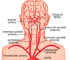
describe vertebrobasilar and carotid circulation

A
15
Q

find the posterior cerebral a., basilar a., internal carotid a., anterior spinal a., and vertebral a.

A
16
Q

which arteries supply each colored area of this brain?

A
  • yellow - anterior cerebral artery
  • red - middle cerebral artery
  • blue - posterior cerebral artery
17
Q

what is the blood brain barrier?

A
  • composed of high-density cells connected by tight junction that restrict passage of substances from the bloodstream
  • it allows diffusion of small hydrophobic molecules (O2, CO2, hormones)
  • cells actively transport metabolic products such as glucose across the barrier with specific proteins
18
Q

describe characteristics of an epidural hematoma

A
  • between skull and dura mater
  • dura peels off skull, space fills with arterial blood
  • compresses brain
  • usually a result of a skull fracture and torn middle meningial artery
19
Q

describe characteristics of a subdural hematoma

A
  • occurs below the dura mater in the subdural space
  • dura still attached to skull
  • venous blood fills subdural space and compresses brain
  • result of a torn bridging cerebral vein
20
Q

what is a subarachnoid hemorrhage?

A
  • commonly occur as a result of rupture of an a. in the circle of willis
  • between arachnoid mater and pia mater; subarachnoid space
21
Q

what is the telencephalon? what are its 4 main divisions?

A
  • cerebral cortex
  • frontal lobe, parietal lobe, temporal lobe, occipital lobe
22
Q

what 2 structures are inside the telencephalon?

A

basal ganglia and limbic system

23
Q

what are Brodmann’s areas?

A

regions of the cerebral cortex that are defined by a number

24
Q

what is a commissure?

A

reciprocal connections

25
what is a chiasma?
crossing and/or intersection of two tracts
26
what is decussation?
cross-over of independent tracts at the same site (form an X)
27
what are "crossing fibers"?
ill-defined fibers whose principal direction may not be defined
28
what is a fascicle?
a bundle
29
what is a fasciculus?
a bundle of fibers
30
describe the diencephalon
* surrounds the third ventricle * major divisions: epithalamus, thalamus, hypothalamus, "sub thalamus"
31
describe the epithalamus
* pineal (serotonin/sleep) * habenula (negative feedback to reward) * posterior commissure - pupillary light reflex
32
describe the thalamus
* major "relay" nuclei * on either side of 3rd ventricle
33
describe the hypothalamus
* endocrine/physiological stasis * surrounds the anterior portion of the third ventricle
34
label these portions of the hypothalamus
35
what are the 4 subregions of the hypothalamus?
* preoptic area * suprachiasmotic (anterior) region * tuberal (middle) region * mammillary (posterior) region
36
what does the hypothalamus control?
* autonomic * endocrine * emotional * somatic
37
describe the basal ganglia
* sits on top of (dorsal to) the diencephalon * extrapyramidal motor system * modulates the initiation, termination, and amplitude of intentional movements
38
describe the two main components of the modulatory cortical loop
* basal ganglia afferents - robust input from almost all parts of the cortex * basal ganglia efferents - ventral thalamic relay to cortex; others - superior colliculus, forebrain, limbic * somatic motor loop: somatomotor control; occulomotor loop (eye movements), frontal loop (cognitive functions), and limbic loop (emotional and visceral functions
39
what are the 5 major nuclei of the basal ganglia?
* caudate nucleus * putamen * globus pallidus * subthalamic nucleus * substantia nigra
40
what forms the striatum?
the caudate and the putamen
41
what forms the lentiform nucleus?
putamen and globus pallidus
42
what are the two subdivisions of the substantia nigra?
* pars compacta * pars reticulata
43
describe the blood supply to the basal ganglia
* branches of the anterior and middle cerebral artery (remember these come from the internal carotid artery)
44
what is hyperkinesia?
excessive abnormal movements, often with increasing dementia example: Huntington's disease * chorea * hemiballism
45
describe Huntington's disease
* type of hyperkinesia * cause: genetic * loss of striatal projection neurons * anatomy: striatal atrophy atrophy of putamen: chorea atrophy of caudate: dementia
46
what is chorea?
involuntary, quick, jerky, irregular movements: * major chorea * minor chorea (Sydenham's chorea): associated with rheumatic fever
47
what is hemiballism?
* repetitive, but constantly varying, large amplitude involuntary movements of the proximal parts of the limbs * decrease activity of the subthalamic nucleus
48
describe hypokinesia
overall decreased bodily movements example: Parkinson's disease
49
describe Parkinson's disease
* hypokinesia * cause: genetic/environmental/idiopathic loss of substantia nigra dopamanergic projection neurons to the striatum * symptoms: bradykinesia, rigidity, tremor at rest
50
what is the circuit of Papez?
part of the limbic system
51
what is wernicke-korsakoff syndrome?
* limbic amnesia * cause: vit B1 deficiency and alcoholism * site: mammillo-thalamic tract disruption (no damage to hippocampus) * symptoms: inability to form new memories loss of memory, can be severe hallucinations confabulation (make up stories)
52
describe alzheimer's disease
* limbic amnesia * bilateral medial temporal lobe damage * common hippocampal deficits: amnestic/inability to form new memory reduced motivation troubles navigating familiar places general cognitive impairment (often mild)