Systems to Cells Flashcards

You may prefer our related Brainscape-certified flashcards:
1
Q

How many ppl in Uk hae diabetes?

A

4.7 million ppl have diabetes currently

2
Q

What is the 1st law of thermodynamics?

A

The First Law of Thermodynamics (Conservation) states that energy is always conserved, it cannot be created or destroyed. In essence, energy can be converted from one form into another.

3
Q

Diagram of Energy balance

A
4
Q

what is energy needed for?

A
  • Cell growth and division
  • Building new molecules and replacing old ones
  • Movement (muscle contraction is ATP-dependant)
  • Breathing, thinking
5
Q

What is our energy currency?

A

ATP is an energy currency formed by substrate-level and oxidative phosphorylation.

6
Q

Memorise ATP energy release diagram

A
7
Q

List facts about ATP NOW!

A

Uhh okay,

  • The average human body has 100-250g of ATP
  • Daily requirement of ATP is 50-70kg. You make more than your body weight of ATP everyday
  • ATP is re-formed from ADP around 1000x a day.
  • It is essential for life
  • We need to keep replenishing this energy.
8
Q

what does the complete oxidation og glucose give?

A

ΔG° = -2840kJ/mol

9
Q

How is glucose broken down?

A

Broken down to pyruvate by glycolysis. Under aerobic conditions the pyruvate is converted to Acetyl-CoA and this enters the Krebs Cycle

Under anaerobic conditions, it’s converted into lactate.

can be efficiently stored as starch/glycogen

10
Q

Is glucose a key energy source?

A

Glucose is a key energy source.

The brain and nerves have an absolute requirement for glucose for enrgy. So do erythrocytes, tstes and kidney medulla.

11
Q

What is Whole blood glucose homeostasis?

A

Whole Body Glucose Homeostasis

  • Blood sugar levels kept the same by a range of homeostatic mechanisms
  • When in excess, glucose is stored as glycogen in liver and muscle cells or triglytcerides in adipose tissue.
  • When levels are low, these tissues become net exporters of glucose/fatty acids
12
Q

What is Hyperglycaemia vs hypoglycaemia?

A

Hyperglycaemia is high blood glucose while hypoglycaemia is low blood sugar

13
Q

How are metabolic pathways involved in glucose metabolism organised?

A

They’re organised at multiple levels

  • System - Human, migrating bird, hibernating brown bear
  • Tissue/Organ - Brain, liver, gut
  • Cellular - Liver and muscle respond differenlty to high/low glucose
  • Subcellular - mitochondria, lipid droplet, cystol
  • Genetic - Cells/tissues can change patterns of gene expression in response to nutritional status.
14
Q

How is glucose level controlled?

A

Blood glucose is controlled by complex mechanisms

Insulin - rel;eased from pancreatic beta cells when blood glucose inreases

Glucagon - released from pancreatic alpha cells when blood glucose falls.

15
Q

What does insulin do?

A

Increasses glucose uptake into fat and muscle cells

Increases uptake of glucose by liver and increases glycogen synthesis in the liver

Inhibits gluconeogenesis in liver

Signals the fed state and the removal of glucose from the blood

16
Q

What is gluconeogeneis?

A

Gluconeogenesis is a metabolic oathway that results in the generation of glucose from non-carb carbon substrates such as lactate or amino acids.

17
Q

What does Glucagon do?

A
  • Stimulates gluconeogenesis
  • Inhibits glycogen synthesis in the liver
  • Triggers lipid breakdown
  • Glucagon signals the release of glucose into the blood.
18
Q

Homeostasis of Blood Glucose diagram

A
19
Q

Where is glycogen mainly stored?

A

Glycogen is mainly stored in liver and muscle cells

20
Q

Memorise metabolic pathway of glucose to glycogen

A
21
Q

How does Insulin integrate into the glycogenesis pathway.

A

Insulin turns ‘on’ glycogen synthase and switches off glycogen phosphorylase. Glucagon does the opposite.

Both reactions are energetically favourable - they happen spontaneously

  • Allows regulation of enzymes as neeeded
  • Allows system to quickly react to changes in blood sugar levels
22
Q

How do mammals regulate enzymes?

A
  • Changing the rate of biosynthesis/degradation of an enzyme levels by (takes time though)
  • Changing the activity of the enzyme (the most common way)
  • changing the location of the enzyme
23
Q

What is reversible covalent modification and how does it regulate key mammalian enzymes?

A

Commonly used way to quickly regulate enzyme activity in response to a signal (e.g. hormones) is used to use reversible covalent modification. Although this can be include many forms (prenylation, ubiquitation, glycosylation etc.) But most common is phosphorylation

24
Q

What does phosphorylation involve?

A

Phosphorylation involves the covalent addition of a phosphate, transferred from ATP by the action of a class of enzymes called kinases.

This is reversible, and the removal of the phosphate is catalysed by a group of enzymes called phosphatases.

25
Q

What does phosphorylation do?

A

Phosphorylation may turn an enzyme on/off,

It alters the 3D conformation of the target protein because of the high charge density of the protein-bound phosphoryl group, -2 at physiological pH (quite a large charge). These often make salt bridges with nearby Arginine and Lysine residues which are positively charged

26
Q

What are the different tyoes of kinases?

A

Two main classes of kinase: those that phosphorylate Tyrosine residues and those that phosphorylate Serine/Thereonine residues

27
Q

Memorise the 20 amino acids and their side chains

A
28
Q

How does glucagon cause glycogen degradation?

A

Glucagon increases glycogen degradation by activating Protein Kinase A which goes on to phosphorylate Glycogen synthase a (active form) into Glycogen synthase b (inactive form).

Simultaneously, Glycogen phosphorylase b (inactive form) is phosphorylated into Glycogen phosphorylate a (active form).

Conversion of both Glycogen synthase a and Glycogen phosphorylase b into their alternate forms uses one molecule of ATP

  • ATP + H2O = ADP + Pi + H+ + ENERGY
29
Q

How does insulin increase glycogen production?

A

Insulin increases glycogen synthesis by activating Protein phosphotase-1 which goes on to dephosphorylate Glycogen synthase b (inactive form) into Glycogen synthase a (active form).

Simultaneously, Glycogen phosphorylase a (active form) is phosphorylated into Glycogen phosphorylate b (inactive form).

Conversion of both Glycogen synthase b and Glycogen phosphorylase a into their alternate forms uses a molecule of water and produces a Phosphate ion.

ATP + H2O = ADP + Pi + H+ + ENERGY

30
Q

What is gluconeogenesis?

A

Gluconeogenesis is the metabolic process by which organisms produce sugars (namely glucose) for catabolic reactions from non-carbohydrate precursors. Glucose is the only energy source used by the brain (with the exception of ketone bodies during times of fasting), testes, erythrocytes, and kidney medulla.

31
Q

What are the general themes of metabolic pathways?

A
  • Biosynthetic and degradative pathways are almost always distinct. this means that both pathways can be thermodynamically favourable.
  • The rates of metabolic pathways are governed by the activities of key enzymes (and not by mass action).
    • Glycogen formation/breakdown is a perfect example of this
  • Specific hormones induce soecific events in cells/tissues.
    • Allosteric modulation of enzyme activity. Reversible covalent modification is a well-used example.
  • In cases where the direction of a metabolic pathway has to be reversed, the pathway is controlled at an irreversible step.
    • Where glycogen needs to be reversed back into Glucose 1-P for example. Glycogen synthase cannot revert glycogen back into Glucose 1-P, Glycogen phospphorylase has to instead.
32
Q

What is a rate determining step/

A

In chemical kinetics, the overall rate of a reaction is often approximately determined by the slowest step, known as the rate-determining step or rate-limiting step

33
Q

What happens to the energy landscape of a reaction when an enzyme gets involved?

A
34
Q

What’s another way of changing enzyme activity besides phosphorylation?

A
  • Enzymes can be controlled bu allosteric interactions with other molecules
  • Often some of the other molecules and intermediates in the downstream pathway
  • Molecules which potentiate one direction (glycolysis) are often negative regulators of the other direction (gluconeogenesis)

Allosteric regulation can be used to superimpose other control pathways on top of a metabolic pathway

35
Q

What are 2 similarities between Glucagon and Insulin?

A

Both are polypeptide hormones released from the pancreas.

Both bind to specific receptors enriched in muscle, liver and fat cells but have opposing actions.

36
Q

What symptoms can diabetes exhibit?

A
  • Macular degeneration - leading cause of non-traumatic blindness
  • Kidney failure - diabetes is the leading cause of kidney failure
  • Stroke
  • Fatty liver - fatal if untreated
  • Atherosclerosis
  • Foot ulcers
37
Q

What are the causes of diabetes?

A

Exercise and diet component, not enough exercise and overeating

And genetics of course

38
Q

What is the underlying nature of diabetes?

A

There are two types:

Type 2:

  • 90% of diabetics have type 2
  • Insulin resistance
  • Beta cell dysfunction

Type 1:

  • 10% of diabetics have type 1
  • Characterised by beta cell destruction either an auto-immune response or idiopathic - we don’t know the cause.
39
Q

How likely are different races to develop diabetes?

A

Half of all South Asian, Black African and caribbean people in the UK will develop type 2 diabetes by the age of 80. For Europeans the figure is 20%.

South Asian men are typically 5 years younger on diagnosis and have increased risk of all complications compared to other ethnic groups

40
Q

How is insulin synthesised?

A

Insulin is produced by transcription and translation of the Human Insulin Gene.

  1. Mature mRNA encodes a complex polypeptide called prepoinsulin
  2. mRNA translated into protein inserted into lumen of the endoplasmic reticulum.
  3. This preproinsulin undergoes proteolytic sequential cleavage.
41
Q

Diagram of how Preproinsulin is formed into Insulin

A
42
Q

Describe the genetic steps of insulin production

A
  1. Ribosome sees mRNA and starts to read along
  2. Sees signal sequence - tells ribosomes to pause. Transalational pause signal.
  3. Pause point is just as signal sequence is just poking out of ribosome exit tunnel (insulin polypeptide in red)
  4. Signal sequence is recognised by SRP (signal recognition peptide. It grabs the signal sequence very tightly.
  5. SRP receptor passes ribosome to complex of proteins called Sec61 translocon complex
  6. Imagine these to be a gateway into the lumen ER
  7. Want to get insulin into the Endoplasmic reticulum lumen.
  8. At that point, translation is carried on
  9. Insulin peptide is co-translationally passed through membrane and into cystol of ER.
43
Q

Why is the preproinsulin passed into the lumen of the endoplasmic reticulum?

A

The prepreoinsulin is passed into the lumen is because the peptide needs to have disulphide bonds added to it to make it stable before it’s released into the blood

44
Q

What’s another reason the preproinsulin needs to be processed in the ER lumen?

A

Disulfide bonds play an important role in the folding and stability of some proteins, usually proteins secreted to the extracellular medium. Since most cellular compartments are reducing environments in general, disulphide bonds are unstable in the cystol as it is a reductive environment.

The peptide will only be folded and oxidised in the ER lumen. This is also where the S=S bonds are formed which are crucial for the folding of the molecule into its biologically active form.

45
Q

What happens to preproinsulin when it’s in the ER lumen?

A

Once inside, the preproinsulin can fold and the disulphide bonds can form. It is still not biologically active yet, it’s now proinsulin.

46
Q
A
47
Q

When does proinsulin become biologically active?

A

Proinsulin only becomes active when the connecting peptide is removed.

48
Q

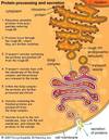
How are proteins processed in the cell?

A
49
Q

How do macromolecules move between different organelles?

A
50
Q

What enzyme removes the pre-sequence from preproinsulin?

A

The pre-sequence is cleaved by signal peptidase

51
Q

What are the steps of insulin production?

A
  1. Preproinsulin is translated on ribosomes and is inserted across the ER membrane.
  2. Inside the ER, the processing begins, the pre-sequence is cleaved by signal peptidase. Disulphide bond A and B chains which stabilises the 3D structure.
  3. Proinsulun then traffics to golgi via vesicular transport.
  4. Proinsulin traffics through the Golgi, before being packaged into secretory granules.
  5. Proinsulin is cleaved into insulin and C-peptide by enzymes called pro hormone convertase PC1/3 and PC2. Carboxypeptidase E removes basic residues at the C-terminus.
  6. High levels of insulin are packaged into the secretory vesicles by making crystalline complex with Zn2+ ions selectively pumped into the secretory vesicles.
  7. The net result = Very high insulin concentrations in specialised packages.
  8. In response to a suitable signal, secretory granules fuse with the plasma membrane and release their contents (insulin and C-peptide).
52
Q
A
53
Q

What is regukated exocytosis?

A

Regulated exocytosis is a process in which the membranes of cytoplasmic organelles fuse with the plasma membrane in response to stimulation.

54
Q

Give a brief overview on how glucose triggers insulin release

A

Increased extracellular glucose is detected in the blood.

Glucose moves from outside of the beta cell to inside using the glucose transporter GLUT2.

Once inside the cell, it undergoes glycolysis. Phosphates get added to produce ATP.

By doing this the ratio of ATP:ADP is increased.

The ATP sensitive postassium channel detects this change and allows the movement of potassium out of the cell. It’s inhibited by ATP causing it to close, depolarising the beta cell.

This is sensed by voltage gated calcium channel causing it to open.

The No. of extracellular Ca2+ is 2-3 orders of magnitude higher than the intracellular Ca2+ and the the ions diffuse into the cell down the diffusion gradient.

Leads to increase in intraxcellular calcium ions causing the fusing of the vesicles to membrane, thus the release of insulin.

55
Q

What is vital to the mechanism of regulated exocytosis of insulin?

A

That extracellular glucose is accurately sensed.

  • Vital to this mexhanism is the ability of the cell to sense the extracellular conc. of glucose and adjust intracellular metabolism in direct proportion.
  • The cell needs to proportionally gauge a response to the increase in blood sugar levels.
  • A key facet of this mechanism is the low-affinity glucose transporter expressed in the beta cell plasma membrane - GLUT2. It is not that effective in its role.
56
Q

Why would getting glucose across the plasma membrane on its own (without a glucose transporter) be difficult?

But how would it be done???

A

it would be difficult because glucose is a polar molecule so it would no tbe able to interact with the plasma membrane enough to be able to pass through its non-polar hydrophobic layer.

However, the way in which the glucose would be transported in this case, the glucose would need to be dehydrated (as it’s usually surrounded by water). The molecule becomes rehydrated within the cell.

This requires a lot more energy than with a transporter.

57
Q

What are the two gradients substances can diffuse down?

A

The two gradients a substance can diffuse down are either the chemical graident (a region of high conc. to low conc.) or the elctrical gradient (a charged particle would move from a high potential difference to a lower potential difference).

58
Q

What are glucose trasnporters AKA

A

Facilitative diffusion transporters

59
Q

What is the alternating conformation model for membrane transport?

A

The principle of which is that the protein switches conformations to present the substrate binding site to alternate sides of the membrane without ever fully opening a channel from one side to the other.

60
Q

Name some differecnes between a Transporter and a Channel NOW!

A

GLUT transporters have a turnover of about 17,000-20,000 glucose molecules per min.

Whereas channels open and allow flow of many ions quickly. This may be as high as 106 ions per second.

Structures are different to reflect this.

There are 12 different GLUTs in the human genome. Even more channels

61
Q

What is a membrane channel?

A

Membrane channels are a family of biological membrane proteins which allow the passive movement of ions (ion channels), water (aquaporins) or other solutes to passively pass through the membrane down their electrochemical gradient.

62
Q

Michaelis-Menton cureve for a catalysed enzyme reaction. Which can double as a velocity/substrate curve for a trasnport protein like a glucose transporter.

Yes, this is a wordy question card

No, I will not ammend it.

A
63
Q

What does the KM in a Michaelis-Menten curve show you?

A

The KM in a Michaelis-Menton curve shows the concentration of extracellular glucose at which we see half of the maximum velocity of glucose entry into a cell.

The KM is a measure of affinity, how well this protein can trasnport glucose.

64
Q

The higher the KM of a trasnporter the…

A

The higher the KM of a transporter the lower the affinity for its substrate. As it takes a longer time for the trasnporter to reach its 1/2Vmax.

65
Q

Using your knowledge of linearity and the Michaelis-menten curve, why would we want GLUT2 to be a low affinity transporter?

A

We would want GLUT2 to be a low affinity transporter because this means that the velocity of glucose entry per concentration of glucose is inherently more proportional than if it were a high affinity transporter with a low KM.

This allows the glucose transporter to respond to increases in extracellular glucose proportionally.

66
Q

How is glucose metabolised in beta cells?

A

In most cells ATP is relatively invariant, not for Beta cells though.

Glucose metabolism in beta cells is inefficient.

As a result, beta cells respond to changes in extracellular glucose concentrations by rapidly changing ATP:ADP ratio.

Glucose phosphorylation in beta cells use the low affinity (high KM) enzyme glucokinase. This enzyme is not as saturated as physiological glucose concentrations. So the phosphorylation rate is directly proportional to intracellular glucose concentrations.

Glycolytic rate therefore depepnds on glucose entry/blood glucose concentrations.

67
Q

What is the michaelis-menten equation?

A
68
Q

Micahelis-menten graph displaying the hexokinase and glucokinase.

A
69
Q

Michaelis-menten graph of GLUT2 and GLUT3 and GLUT4

A
70
Q

What are the 3 main targets of insulin?

A

The 3 main targets of insulin are muscle cells, liver cells and adipose tissue which express an inuslin receptor.

71
Q

What is the receptor that insuin binds to?

And how does it work?

A

The insulin receptor is an alpha2Beta2 polypeptide, held together by disulphide bonds.

Has two alpha subunits and two beta subunits.

Insulin binds to the alpha 2 subunits inducing a conformational change in alpha subunits.

This transmits a signal to the Beta subunits activating an intrinsic tyrosinase kinase within the cytosolic domain.

The signal has crossed the membrane at this point.

The binding is specific - only insulin can bind to the receptor.

72
Q

What are the steps inviolved in insulin signalling

A

Insulin Signalling?

  1. Activation of the receptor occurs only upon insulin binding. Imparts specificity of signal to the tissues that express the receptor.
  2. Amplification - a single molecule of insulin activates a kinase that cxan phosphorylate many target molecules.
  3. Tyrosine kinase receptor phosphorylates specific Tyr resiudes within the receptor - auto-phosphorylation
  4. These p-Tyr residues recruit signalling molecules (IRS-1 shown here) tit he receptor which are then themselves phosphorylated.
  5. This forms a signalling complex.
73
Q

What happens at phosphorylated receptors?

A

Phospho-tyrosine residues are recognised by specific proteins

  • SH2 domains
  • PTB domains

Recruits specific set of target proteins to the activated receptor to form an active signalling complex.

Many of these targets are then phsophorylated by the activated receptors.

74
Q

What haooens in insulin signalling after the insulin receptor auto-phosphorylates/

A

When insulin receptor auto-phosphorylates:

Phosphorylated residues become docking sites for other signalling molecules

Insulin receptor recruits a big adaptor protein:

  • IRS-1
  • (Insulin receptor substrate 1)
  • IRS-1 is phosphorylated

Selectively recruit molecules

Signalling comlex

75
Q

What does the autophosphorylateion of specific tyrosine residues do?

A
  • The autophosphorylation of specific tyrosine residues function to recruit SH2 domain containing proteins to the receptor.
  • These are then activated.
  • Signal propagated.
  • Insulin receptors selectively recruit different SH2-domain containing proteins.
76
Q

Considering SH2 domains and PTB domains are found on many other proteins how is specificity achieved?

A

All SH2 domains bind phsophotyosine.

All SH2 domains have a pocket into which the P-tyr residue fits.

But not all SH2 domains bind all P-Tyr residues; there is specificty dictated by the surrounding sequence in the target (receptor) polypeptide.

77
Q

Detailed diagram on specificty of SH2

A
  • Pocket contains positively charged residues at the bottom to interact with negative phosphate
  • If tyrosine isn’t phosphotylated: affinity for pocket is gone
  • Second binding pocket:
    • E.g. hydrophobic pocket which recognises isoleucine.
    • Confers specificity
78
Q

Diagram of Signalling Complex

A
79
Q

What is an example of a key downstream target of a Ser/Thr kinase known as?

A

A key downstream target ia thw activation of a Ser/Thr kinase known as Akt.

Amplification at all steps

Insulind change the phosphorylation status of 100s of different proteins.

cAkt is an key enzyme which is activsted by insulin.

All signalling pathways amplify.

80
Q

What is the key action of insulin?

A

The key action of insulin is to increase glucose transport into muscle and fat cells.

81
Q

The effect of insulin needs to be independenet of new protein synthesis and requires ATP, how is this achieved?

A

Uses a specialised form of gluvpse transporter, GLUT4, expressed only in fat and muscle.

GLUT4 is capable of moving to the cell surface as a response to insulin binding to its receptor.

This si triggered by cAkt.

Regulated exocytosis.

82
Q

How is GLUT4 stored in the absence of insulin?

A

In the absence of insulin GLUT4 is inside the cells in specialised vesicles - GSVs

GLUT4 is the only GLUT isoform that exhibits this trafficking.

Glucose transport is low in the absence of insulin.

83
Q

Diagram of GLUT4 in GSV

A
84
Q

Insulin has cell specific effects, how is this achieved?

A

In adipose cells cAkt activates GLUT4 trafficking to the cell surface.

In liver/muscle cells cAkt phosphorylates and activates phosphatase - promoting glycogen storage.

Akt us a crucial molecule in insulin signalling. Different tissues express different Akt ‘effectors’.

85
Q

Insulin can regulate gene expression, describe how.

A

Insulin can upregulate and down regulate gene expression.

Upregulated genes are transcribed relatively quickly. However, they have a low sensitivity to insulin and require a higher dose.

Downregulated genes are transcribed slowly but have a high sensitivity to insulin sp a low dose is required.

86
Q

In detail, how does insulin modulate gene expression?

A

Insulin signals through IRS1 to a variety of transcription factors that control gene expression.

Insulin activates the LxR transcription factor (one of many) in liver and fat.

In fat, this drives increased expression of SREBP1 transcription factor.

SREBP1 controls (among others) fatty acid synthase, AcetylcoA carboxylase the lipid metabolism genes turned on.

87
Q

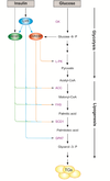
Describe the lipogenesis pathway.

A
  • Acetyl Co-A (under the right conditions) enters the lipogenesis pathway.
  • It then gets converted to triglycerides.

The genes for the enzymes involved in lipogenesis are turned on:

  • Acetyl CoA carboxylase
  • Fatty Acid Synthase
  • SCD1

LxR turns on SREBP1 which turns on the lipid metabolism genes.

This gives co-ordinate regulation of lipogenesis as multiple enzymes upregulated by a single transcription factor.

88
Q

What is a promoter region in a gene?

A

A promoter is a sequence of DNA to which proteins bind that initiate transcription of a single RNA from the DNA downstream of it. This RNA may encode a protein, or can have a function in and of itself, such as tRNA, mRNA, or rRNA.

89
Q

What are the two types of transcription factors?

A

Activators which help general transcription factors and RNA polymerase assemble.

This promotes and increases transcription of target gene.

There are also repressors which blocks the genral transcription factors and RNA polymerase.

90
Q

What can phosphorylation do?

A

It can change the stability of a molecule and its location

91
Q

What can phosphorylated TFs do that non phosphorylated TFs do?

A

Phosphorylated TFs can travel through nuclear pore while non phosphorylated TFs cannot.

92
Q

What is a common feature of signalling cascades?

A

Amplification is a common feature of signalling cascades

93
Q

Glucagon transduction pathway

A
94
Q

What is the role of the hormone leptin?

A

Regulate feeding; it signals to the brain (hypothalamus) to control food intake.

95
Q

What have studies in the early 2000s revealed about adipose tissue?

A
  • Adipose cells aree more than inert energy store, they secrete diffferent polypeptide hormones.
  • These hormones act on other tissues to produce biological effects - adipocytokines; adipokines.
  • Some of these molecukles are constitutively secreted.
  • Most well-characterised is Leptin, but there are many others and the inter-relationships between these molecules and their targets is complex and as yet poorly understood.
  • Likely to be the key to understanding obesity/diabetes
96
Q

What is a paradox of adipose tissue’s production of leptin and obesity?

What does this suggest?

And what are the implications of this paradox?

A
  • Most people who are obese have increased fat depot and increased circulating leptin levels. So why are they obese?
  • This suggests that obesity is asssociated with an inability of leptin to counteract feeding this could be due to leptin resistance?
  • Suggests that key therapies for obesity treatment may need to target complex regulatory networks in the brain, rather than metabolic tissues like liver or fat.
97
Q

What are Endocrine cells?

A

Endocrine cells are responsible for producing and relkeasing hormone molecules into the bloodstream. Endocirne cells are typically grouped together in organs referred to as endocrine glands.

98
Q

What are Neuroendocrine cells?

A

Neuroendocrine cells are cells that recieve neuronal input (neurotransmitters released by nerve cells or neurosecretory cells) and, as a consequence of this input, release message molecules (hormones) to the blood.

99
Q

What are Enteroendocrine cells?

A

Enteroendocrine cells form the basis of the lagest endocrine system in the body. They secrete multiple regulatory molecules which control physiological and hoomeostatic functions, particularly postprandial secretion and motility.

100
Q

Diagram of Lamina propria

A
101
Q

What is GLP-1 secretion dependent on?

A

GLP-1 secretion by ileal L cells (entero-endocrine cells) is dependent on the presence of nutrients in the lumen of the small intestine. It is released in response to major nutrients such as carbs and lipids.

102
Q

Once in circulation, what is the half-life of GLP-1?

A

Once in circulation, it has a half life of only 2 minutes as it is rapidly degraded by an enzyme called dipeptidyl peptidase-4.

103
Q

What is the role of GLP-1?

A

GLP-1 is a major regulator of whole body glucose homeostasis by acting both centrally (brain) and peripherally (pancreas, adipocytes, liver etc.) to regulate metabolic profiles/fucntion of the tissues.

It also slows gastric emptying and thus can indirectly regulate appeptite.

104
Q

What is GLP-1?

What does it act on?

Why is this important?

A

Glucagon-like peptide-1 (GLP-1) is a potentn anti-hypoglycaemic hormone.

GLP-1 acts (via receptors) on the beta cell to enhance the release of insulin in response to a rise in blood glucose levels (and supresses glucagon secretion from alpha cells).

This is important as unregulated release of insulin when plasma glucose is in the normal range can cause hypoglycaemia - a dangerous fall in blood glucose. This does not happen because of GLP-1.

105
Q

How does GLP-1 act within normal blood glucose ranges?

A

When blood glucose is in the normal acting range, there is no GLP-1, and the beta cells no longer secrete insulin; this is important when blood glucose is in thr normal fasting range as it will prevent catastrophic hypoglycaemic episodes.

When you eat a meal, carbohydrates in gut cause a release of GLP-1. ‘Switches the pancreas on’.

106
Q

What is GLP-1 an example of?

A

GLP-1 is an example of homrones called incretins.

107
Q

What are some of GLP-1’s physiological actions?

A

GLP-1 has numerous physiological actions, including: potentation of glucose-stimulated insulin secretion, enhancement of beta cell growth and survival, and inhibition of glucagon release, gastric emptying and food intake.

These anti-diabetic effects of GLP-1 have led to intense interest in the use of this peptide, and other incretins, for the treatment of patients with T2D.

108
Q

What are Incretins?

A

Incretins are a group of metabolic hormones that promote a reduction in blood glucose. They achieve this by several mechanisms:

  • They augment glucose-stimulated insulin release from beta cells.
  • They surpress glucagon release.
  • They regulate the absoprtion of nutrinets from the gut.
109
Q

What are some of the main incretins?

A

Main incretins are Glucagon-like peptide-1 (GLP-1) and gastric inhibitory peptide (GIP). Both are rapidly inactivated by the peptidase dipeptidyl peptidase-4 (DPP-4) and have very short half-life in the curculation.

Because of this, their therapeutic use is limited. Longer-lasting analogues are in clinical use as insulinotropic agents. Another approach is to inhibit the enzyme that activates GLP-1 and GIP, DPP-4.

110
Q

What are some interesting facts about platypus and GLP-1?

A
  • Platypuses lack a functional stomach.
  • They instead have high circulating levels of GLP-1 because its biology is rather unusual.
  • There are conflicting functions of GLP-1 in the platypus: in the gut as a regulator of blood glucose, and in venom to fend off other platypus males during mating season.
111
Q

Why is platypus GLP-1 desired as a type 2 diabetes?

A

While the human version of GLP-1 degrades quickly the platypus version appears to have evolved to be long-lasting which makes it desirable for the tratment of T2D.

112
Q

What is unique about dolphins and insulin response?

A
  • Dolphins and humans have co-evolved metabolic processes. reflecting brain size, - high demands for readily available glucose - and red blood cells that can transport glucose.
  • Studies of populations of dolphins show that animals fasted overnight exhibit blood chemistry akin to T2D.
  • When dolphins have a dextrose meal, they had an insulin deficient response - sustained post-prandial high blood glucose with no insulin. Akin to T1D.
  • Dolphins can switch between T2D and T1D easily.
113
Q

How is hibernation beneficial?

A
  • Hibernating animals store massive amounts of fat each autumn.
  • These serve as the main source of metabolic fuel over winter.
  • Fats are rich and have the advantage of generating metabolic waster as they are catabolised.
  • Fat deposition occurs on an annual basis.
  • This is correlated to the length of daylight.
  • Cycles of natural obesity and insulin resistance.
114
Q

How do animals know when to begin hibernation process?

A
  • Annual cycle of body weight fluctuation is intrinsic rhythm entrained by light.
  • Depends on melatonin secretion from the pineal gland.
  • Independent of temperature or food availability.
  • Liver changes metabolic profile from carbohydrate to fat based metabolism.
115
Q

What is the involvement of the gut in migration?

A
  • Phenotypic flexibility in the digestive system of migrating birds.
    • Digestive organs (gizzard, liver, smalll intestine) increase in size and capacity to convert more food into energy e.g. pre-migration
    • Atrophy when demands fall e.g. during migration.
  • Many other adaptive changes exhibited:
    • Increased muscle size and capacity for oxidative metabolism
    • Liver metabolic profile chnages
  • There is rapid, precise regulation of physiologicaal systems that is often completely reversible.
  • Coordinate regulation of metabolic processes like change in diet from seeds to nectar to accumalate rapid fat deposits.
116
Q

Diagram of sources of ATP during exercise and how long do they last?

A

Stored ATP lasts about a second.

Creatine phosphate lasts for around 4 seconds,

The rest of the exercise will be using the fermentaation of glucose.

117
Q

What is GWAS?

A

Genome Wide Association Studies.

How genes effect the development of a trait/phenotype

118
Q

How to find a gene that affects a trait

A

All you need, e.g. for cystic fibrosis:

  • Trait segregating in (large) families
  • Behaves as a Mendelian, single gene trait
  • Genetic differences across the genome
119
Q

What is a Complex trait/Quantative trait

A

Most traits are not inherited in a simple mendelian fashion.

The risk of developing a disease like autism or diabetes do not display a clear pattern of mendelian inheritnce.

Quantative traits are traits that can be quantified such as height/IQ.

These traits can have multiple genes involved or there could even be no genetic basis at all!

120
Q

What is The mendelian inheritance pattern?

A

The manner by which genes and traits are passed from parents to their children. The modes of Mendelian inheritance are autosomal dominant, autosomal recessive, X-linked dominant, and X-linked recessive. Also known as classical or simple genetics.

121
Q

How can heritability be used to identify of genes are responsible for a trait?

A

If the trait is heritable

It would affect the risk of developing the trait

Trait is not pre-determined

A role for the environment

Could be a small number of genes or many genes which cause the trait.

  • Small effect size (each make a small contribution to risk)
122
Q

How would you deterrmine heritability?

A

By utilising family history?

if a trait runs in a family it may well be tyhe case that the trait is caused by genetics and the heritability of the certain genes, however, it could be due to the shared environment.

123
Q

What sort of study provides insight into heritability of traits?

A

twin studies provide insight into heritabiloity of traits

identical twins

100% shared genes and shared environment

If one twin has multiple sclerosis for example, the odds of the other one having it is 20%

In non identical twins

50% sahared genes and shared environment.

If one twin has MS the odds of the other one having it is 5%

To calculate the heritability from the above

(Monozygiotic twins percentage - Dizygotic twins percentage) x 2 = 30

124
Q

What would 30% heritability of a trait mean? And what does it not mean?

A

30% heritability means:

For population:

30% of risk is attributable to genetics

70% of risk is non-genetic

30% heritability does NOT mean:

Each individual:

30% of trhe trait is genetic

70% of the trait is due to environment.

125
Q

What is the main source of genetic variation between humans?

A
  • 99.9% of the human genome is conserved betyween ppl
  • All variation is from 0.1%
  • SNPs are the main source of common allele differences
126
Q

How would one identify oci contributing to complex traits?

A
  • Cannot use LINKAGE mapping
    • No clear patterns, too few ppl
  • Switch in strategy is needed
    • Study populations rather than families

Pre-disposing alleles should exist on limited number of haplotypes and we shouldbe ble to detect them without genotyping them i.e. if only nearby SNPs are genotyped.

Can find them even if multiple genes contrubute. As each gene contributes a small amount of risk we are esssentially looking for small effects so need huge numbers.

In essence, this is what GWAS (Genome Wide Association Studies) are.

127
Q

How is genetics important in drug discovery?

A
  • New medicines: Drug targets that are backed by genetic info are approximately twice as likely to become a successful medicine
  • Better medicines: Understanding how a medicine is working can help us avoid unwanted side-effects and enhance the probability the medicine will be effective.
128
Q

What is needed in a GWAS?

A

GWAS analyse small effect sizes, therefore one needs huge numbers of people.

The participants need to be separated based on the trait looking to be analysed.

Take care to match the groups for age, sex and ethnicity.

129
Q

What is Genotyping?

A

Genotyping is the technology that detects small genetic differences that can lead to major changes in phenotype, including both physical differences that make us unique and pathological changes underlying disease. It has a vast range of uses across basic scientific research, medicine, and agriculture.

130
Q

Population in GWAS must meet the associations presented in the

Hardy Weinberg equilibrium. What are these 5 assumptions of thew HW equilibrium?

A
  1. Mating is random
  2. No mutations are arising
  3. No gene flow
  4. No natural selection
  5. Population size is infinitely large
131
Q

What plots are used to display GWAS?

A

MAnhattan plots are used to display GWAS.

132
Q

What are the Hallmarks of diabetes?

A
133
Q

Graph detailing the age of diagnosis of diabetes

A
134
Q

What is monogeneiuc diabetes?

A

Maturity Onset Diabetes of the Young (MODY) is an inherited form of diabetes mellitus. It is caused by a change in one of eleven genes. Up to 5% of all diabetes cases may be due to MODY. Just like other people with diabetes, people with MODY have trouble regulating their blood sugar levels.

135
Q

Is MODY diabetes more like T1D or T2D diabetes?

A

This disorder is more like type 1 diabetes than type 2, although it can be confused with either type. In type 1, the pancreas cannot make and release enough insulin. People with type 2 diabetes, on the other hand, usually make enough insulin, but their bodies cannot respond to it effectively (known as insulin resistance). Type 2 diabetes is usually associated with being overweight, but that is not true of type 1 diabetes or MODY. However, obesity does matter. An obese person with a MODY gene mutation may develop symptoms of diabetes sooner than someone of normal weight.

136
Q

What does MODY diabetes stand for?

A

maturity onset diabetes of the young. There are multiple forms (11) which are usually mistaken for T2D or T1D.

137
Q

Name 4 types of monogenic subtypes of diabetes.

A
138
Q

What is the most common subtype of MODY diabetes?

A
  • HNF1A
  • Transcription factor
  • Drives expression of genes in endoderm, including GLUT1 and GLUT2
  • Strong loss of functrion mutations (recessive)
  • Treatment: low dose sulfonylurea
139
Q

What is the 2nd most common subtype of MODY diabetes?

A
  • GCK= Glucokinase
  • catalyses first step in glycolysis
  • “traps” glucose in cells
  • important for rapid uptake and metabolism of glucose
  • STRONG loss of function mutations (recessive)
  • Often asymptomatic requiring no treatment
140
Q

Give a brief overview of Type 1 Diabetes.

A
  • ~10% of all diabetes cases
  • ~0.5% of UK population
  • Autoimmune condition: destruction of pancreatic beta-cells
  • Presentation of insulin as ‘self-antigen’
  • No strong sex bias
  • Onset usually between 5 and 20 yrs old; used to be fatal rapidly
  • Modern therapy – recomb. insulin injection – more or less normal lifespan now; but ….
  • Some familial clustering - 20% familial cases, 80% sporadic – most familial cases are sib-pairs, not large families hints at complex inheritance
141
Q

What is an “Odds ratio”?

A

A measure of INCREASED RISK of having condition conferred by GENOTYPE at each locus/gene

142
Q

How can Genotyping assist with diagnosing diabetes?

A

Clinical signs are well established

•but only evident after development of disease

Genotyping could identify;

  • High-risk subset of children with decent confidence
  • 2-5 years BEFORE development of disease
  • Improved clinical management
  • Stratification: distinguish T1D from other forms of diabetes e.g. MODY
143
Q

Give a brief overview on T2D

A
  • T2D diabetes is a complex disorder
    • Multifactorial
    • Polygenic
  • Biggest known risk factor is obesity/ high adiposity
    • Causal pathway involves ectopic liver fat
  • T2D genes could also affect
    • Behaviour- food intake
    • Metabolism, Glycaemic traits
    • Fat accumulation/ BMI (adiposity genes → T2D genes)
    • Beta-cell responses and survival
144
Q

What is the strongest risk variant for type 2

A

The strongest risk variant is TCF7L2

  • Transcription factor 7-like 2
  • The gene has 4 common spliced variants; when variant SNP rs7903146 is present in two copies (CT/TT risk genotypes)
  • Wnt (wingless/integrated) signalling pathway
    • Pleiotropic effects across tissues and systems
  • Has been identified across multiple ethnic groups
145
Q

Where do the majority of T2D risk variants lie?

A

The majority of T2D risk variants lie in non-coding regions.

146
Q

Can SNPs in non-coding regions affect gene expression?

A

Gene promoter or enhancer regions

Alter gene expression

(Up or Down)

147
Q

How do we calculate an Odds Ratio?

A
  • Let us calculate the increased RISK: the ODDS RATIO: consider TCF7L2
  • 100 people carry the risk allele vs 100 who don’t

T2D Cases Healthy Total

Carriers 7 93 100

No Risk 5 95 100

Odds ratio = ratio of affected/Unaffected

Odds for carrier of risk allele getting T2D = 7/93 = 0.075

Odds for non-carrier getting T2D = 5/95 = 0.053

Therefore the odds ratio = 0.075/0.053 = 1.4

148
Q

How does aerobic exerceise effect diabetes?

A
  • Moderate intensity - unlike healthy individuals, where hepatic glucose production is matched by peripheral glucose uptake; diabetics use more than is produced (reducing blood glucose)
    • Lasts for between 24 - 72 hrs
  • High intensity can cause hyperglycemia due to catecholamine induced glycogen production (lasting 1 - 2 hrs)
    • May be sensitive to duration of intensity?
149
Q

How does Resistance Exercise?

  • Little, if no, data available on single bout of resistance exercise in diabetes.
    • Benefits shown in pre-diabetes (blood glucose elevated during fasting)
    • Lower blood glucose (up to 24 hours)
    • Responds to both volume and intensity
A
150
Q

What changed from the former diet/exercise recommendations to the current one?

A
  • Moderate aerobic activity (2-3 times per week) - metabolic impact for up to 72 hrs
  • Combined with resistance exercise - metabolic impact for up to 24 hrs - increased muscle mass/strength for enhanced insulin independent affect
  • Compliance? - time, access to facilities, boring…. - novel strategies for HIIT
151
Q

At rest, what is the preferred substrate for ATP generation?

And why should a diabetic look to avoid certain foods based on this info?

A

At rest the preferred substrate for ATP generation are FFAs (Free Fatty Acids).

So by default the high carbohydrate content foods increase blood glucose and should be avoided.

The amount of carbohydrates is the primary determinant of blood glucose.

152
Q

The amount of carbohydrates is the primary determinant of blood glucose but what else should be considered?

A
  • The type of carbohydrates (amylose vs amylopectin)
  • Preperation (cooking time or method)
  • Processed foodstuff vs fresh
    • trans fatty acids in energy dense food
153
Q

What are the two components of starch?

And how are these componenets broken down?

A

Starch is made up of two components, amylose and amylopectin.

Amylose is a straight chain of glucose units connected by alpha 1-4 bonds. These bonds are broken by the enzymes maltase and amylase

Amylopectin contains the same chains, but also has “branch points” created by alpha 1-6 bonds, these bonds are broken by the enzyme isomaltase

154
Q

What is steric hinderance?

A

Steric hindrance at a given atom in a molecule is the congestion caused by the physical presence of the surrounding ligands, which may slow down or prevent reactions at the atom.

155
Q

Which of the two starch components is digested more easily?

A
  • Theoretically, amylose should be easier to digest because it does not require isomaltase, and does not have the steric hindrance caused by the branch points
  • However, amylose can form a very compact physical structure, which inhibits digestion
  • Therefore, amylopectin is actually digested better than amylose
156
Q

According to Macciochi what was the reason we evolved as humans?

A

“The whole reason that we evolved as humans is because we started cooking our food. When we only had a raw diet, we had to be eating constantly, because our bodies struggled to get the nutrients out of raw food.”

  • Macciochi

There’s a substantial back catalogue of evidence suggesting that human evolution is directly linked to the use of fire.

157
Q

What was the benefit to our ancestors of cooking their food?

A
  • When our ancestors cooked and processed their food, they made it easier to extract calories and fat (improving the gap between the amount of energy it took to digest their food and that extracted from it)
  • Cooking also kills many of the potentially harmful bacteria that can grow in and on food, protecting from bouts of food poisoning
158
Q

What are the cons of cooking foods at a high temperatures for long periods of time?

A

Some health risks include: obesity, heart disease, high blood pressure and diabetes

159
Q

When is a low carb diet ineffective?

A

A low carbohydrate diet is ineffective without calorie restriction to maintain energy balance.

160
Q
A
161
Q
A