SUGER Histology Flashcards
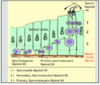
KIDNEY
- pale cortex / dark medulla / 10-15 meduallary pyramids
- cortex contains glomeruli and coils of proximal/distal tubules
- medulla contains pyramids with straight parts proximaal/distal tubule, loop of Henle, collecting duct
Blood
- 5/6 branches renal artery
- arcuate arteries at medullary/cortical border (give off vasa recta - deep to medulla)
- then interlobular arteries penetrate cortex at regular intervals
- affertent to glomerulus
- efferent from glomerulus (filtered blood) - thin walled and between tubules
- acquire fluid and ions -> drain to arcuate veins -> systemic
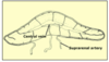
GLOMERULUS
- blood to glomerulus for filtration
- passes primary filtrate to nephron for selected reabsorption
- parallel array of fenestrated capillaries ensheathed by podocytes
- between loops = mesangiel cells (matrix forming)
- afferent arteriole wall produces renin
- next to glomerulus = segment of distal tubule with macula densa
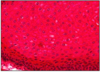
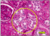
GLOMERULAR TUFT (PAS)
- arise from vascular pole of glomerulus = entrance afferent and exit efferent
- in this angle lies a distal loop of nephron with palisade of macula densa
- capillary loop surrounded by podocyte
- surrounded by urinary space - separates glomerulus from bowman’s capsule
- arrow to distal tubule
- MACULA DENSA - around blood vessels regulate blood flow, provides framework for glomerulus.
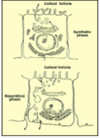
FILTRATION BARRIER
- basement membrane
- synthesised and maintaiend by endothelial capillary cells and epithelial cells (podocytes) that ensheath them
- endothelial = fenestrated
- podocytes stand off membrane with foot processes
- creates physical pores
- pores guarded by filtration membrane
- membrane is charged and resists passage of some molecules
- water and solutes may pass to urinary space = primary filtrate
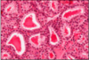
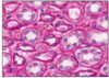
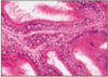
PROXIMAL TUBULES (PAS)
- highly coiled (sometimes with straight projection to medulla)
- prominent brush border and complex invaginations @ basolateral membrane dark pink
- extensive reabsorption here
- Na+ active transport with glucose (cotransporter - GLUT)
- take up protein and polypeptide by endocytosis
- cells contain lyzosomes which break down proteins before returning to circulation
- any small negatively charged protein can enter primary filtrate
PROXIMAL TUBULES 2 (PAS)
- this slide - straight portion of proximal tubule
- leads to loop of Henle
- if glomeruli present = cortex
- some project to medulla
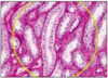
LOOP OF HENLE (PAS)
- mostly @ medulla
- thick/straight descending, thin loop, thick ascending
- thick descend = similar to proximal tubule
- thick ascend = similar to distal tubule
- longest thin loop penetrate deepest to medulla when glomerulus = cortico-medulla junction
- thin descending portion = low permeability to ions and urea, high permeability to water
- thin ascending portion retains water, reabsorption Na+ and Cl-
- this produces dilute/hypotonic filtrate but a hypertonic interstitium
- vasa recta (straight capillaries) run alongside tubules
this slide - thick and thin limbs and vasa recta
DISTAL TUBULE (PAS)
- from medulla to cortex then to vascular pole of glomerulus
- macula densa here monitors sodium levels to influence intitial filtration of glomerulus
- paler than proximal
- deep invaginations of basal plasma membrane with numerous mitochondria
- mitochondria indicative of control acid/base balance and concentration of urine (ATP)
- with aldosterone sodium reabsorbed, potassium excreted
- bicarbonate ions reabsorbed, hydrogen excreted - acidic urine
@ cortex both proximal and distal tubules present
proximal more tightly coiled therefore appear to be more numerous
COLLECTING DUCTS (PAS)
- pale cuboidal cells
- wide lumen
- starts at cortex, filtrate to collecting tubules than larger ducts
- collecting tubules from many nephrons coalesce to larger ducts and form visible streaks - medullary rays
- few organelles
- collecting tubules - dark intercalated cells with high mitochondria
- surrounded by hypertonic medium interstitial from loop of Henle
- ADH increases permeability to water (concentrates urine)
prone to kidney stones and infection
JUXTAGLOMERULAR APPARATUS (PAS)
- afferent/efferent arterioles, macula densa, lacis cells
- afferent - cells produce renin - granules in cytoplasm
- renin catalyses angiotensin 1 (liver) -> angiotensin 2 (conversion at lungs) -> aldosterone release by suprarenal cortex -> reabsorption of sodium and water @ distal tubules and collecting ducts
- lacis cells and macula densa regulate renin secretion by monitoring sodium levels
this slide - obvious macula densa
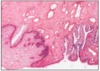
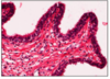
UROTHELIUM
- ureters, bladder and most urethra
- pseudo-stratified epithelium
- surface cells adapted to withstand prolonged urine exposure
- 3-8 layers (distension dependent)
- basal cells = cuboidal
- above = columnar (relaxed)
- surface = large, binucleate umbrella cells
umbrella cells:
- thickened membrane plates joined by thin membrane bands
- lipid composition (unique)
- relaxed plated = perpendicular to membrane
- stretched = drawn to surface of cell
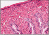
URETER
- epithelial tube with 2 helical layers of smooth muscle
- star shaped lumen
- towards bladder = longitudinal smooth muscle
-
3 constrictions (kidney stones will lodge here)
- origin (pelvis of kidney)
- at sacro-iliac joint (passes to true pelvis)
- as enters postero-inferior bladder surface
- reflux prevented by compression of ureter by muscular wall bladder
- contains blood vessels and adipose
- smooth muscle for peristaltic contraction
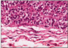
URINARY BLADDER
- wall = thick bundles of smooth muscle with no preferred direction except neck = 3 distinct layers
1. innermost = longitudinal projects inferiorly and turns transversely to form a sphincter around prostatic urethre (male) and external meatus (female)
thrown into folds w/ umbrella cells
walls of bladder contains small nerves (autonomic)
sympathetic NS = mainly blood vessels at bladder
inset = nerves
micturition
when external sphincter relaxes (sympathetic) and muscle wall (detrusor) contracts (para)
PROSTATIC URETHRA
- urethra longer in males
- prostatic
- membranous
- bulbous
- pendulous
- urothelium (pseudo-stratified columnar) except at distal end
- mucus glands along length
- distal urethra = stratified squamous (male and female)
- both sexes = striated (voluntary) muscle sphincter from muscles of pelvic diaphragm around membranous part
- under prostatic urothelium = dense fibrous connective tissue to prevent distension
inset = U-shaped prostatic urethra
TESTES
- within collagenous capsule = tunica albuginea
- within fibrous septa to 250 lobules (each lobule with up to 4 germ cell producing semniferous (tubules)
- semniferous tubule = 50cm loop (open both ends)
- drain to rete testes (channels)
- Leydig cells between semniferous tubules
SEMNIFEROUS TUBULE
- stratified epithelium with support (sertoli) and germ line (developing spermatazoa)
- at periphery = germinal epithelial
- GE produces large cells with speckled chromatin = spermatagonis
- spermatogonia develop to spermatocytes
- spermatocytes pass blood testis barrier created by Sertoli
- Sertoli = blood testis barrier and nurture sperm
- production = 64 days
- maturation wave passes slowly down each tubule
SEMNIFEROUS TUBULES
- thin fibrous capsule
- closest to membrane = germinal epithelium with spermatogonia (speckled)
- between cells are smaller primary spermatocytes and Sertoli cells (pale irregular nuclei)
- towards centre depends on stage in cycle
- sometimes primary/secondary spermatocytes (small dense nuclei)
- otherms more mature (narrow and elongated heads)
- between tubules = leydig clumps
arrow to spermatid
reduction division (2n-1n -> first meiotic) takes place when primary to secondary @ luminal side of blood testis barrier i.e. not in contact with blood stream
EPIDIDYMIS
- rete testis = cuboidal ep
- rete testis to efferent ductules (similar)
- epididymis = single 5m coiled tube
- thick fibrous capsule attached to mediastinum of testes posteriorly - storage and maturation site for sperm aggregated within lumen
- tall pseudo-stratified columnar ep
- small rounded basal cells support tall columnar with microvilli (stereocilia)
- stereocilia (non-motile) reabsorb seminal fluid, phagocytose damaged sperm and cell debris. also nutrients for sperm
- thin layer of smooth muscle thicker as approaches vas
VAS DEFERENS
- spirally arranged smooth muscle
- similar to epididymis but shorter cells and microvilli
- sometimes has longitudinal folds - lamina propria
- sudden and rhythmical contraction expels sperm
thick muscular wall makes vas feel cord like
SEMINAL VESICLE
- highly coiled glands at postero inferior bladder
- double layered capsule of smooth muscle
- long narrow folds
- stimulated by testosterone - enlarge and secrete creamy opalescent fluid with acid pH. rich in globulin, vitamin C, amino acids and sugars
- contraction of smoooth muscle mixes with spermatozoa
- simple columnar epithelium
PROSTATE
- 50 branched tubular glands
- smooth muscle matrix
- smooth muscle capsule around all
- 3 layers (concentric) of glands - mucosal - submucosal - main
- @ ejaculation, contract and mix secretions @ ejaculatory duct
PROSTATIC EPITHELIUM
- glands varied - straight, coiled, branched
- epithelium thrown into broad, branching folds
- with testosterone, cells increase in height and secrete digestive enzymes - acid phosphatase (major component of seminal fluid) - (PSA) prostate specific antigen
- lumen often with sec product / calcified glycoprotein
PROSTATE 2
- this slide L - skeletal muscle at underside of prostate - pelvic diaphragm with levator ani
- this slide R - spiral tubular gland at submucosal level
PENILE URETHRA
- prostatic, membranous, penile
- membranous and penile = non-secreting pseudostratified columnar
- at distal end = stratified squamous epithelium (within glans)
- bulbo-urethral glands within membranous urethra produce watery galactose rich secretion, precedes main ejaculate - can sometimes be a problem with catheterisation
PENIS
- erectile compartments = large endothelium lined blood vessels supported by connective tissue
- each surrounded by compact collagen layer = tunica albuginea
- helicine branches of pudendal artery
- when flaccid arteries transmit very little blood due to artero-venous shunts
- para. stimulus shut shunts (point and shoot)
- most skin = hairy, loosely attached to columns - except prepuce and glans (non-kerat)
VAGINA
- stratified squamous thinly keratinised at vulval end
- thin pre-puberty and post menopause
- beneath epithelium = lamina propria with small blood vessels (moisture by diffusion)
- surrounding epithelium = fibro muschular tube with collagen/elastic and smooth muscle
- sphincter of skeletal muscle at lower end
- bartholin glands - mucus posterolaterally at lower end of vagina
- with age = weaker fibro-muscular tube
EPITHELIAL SQUAMES
- in menstrual cycle surface cells accumulate glycogen and flake off
- desquamated cells rupture and bacteria generate lactic acid
- low pH at vagina
pale green cells - outermost layer, predominate at first half of menstrual cycle
pink cells - deeper layer
CERVIS
- protrudes to anterior wall vagina
- surrounded by gulley - lateral, anterior, posterior ornices
- outer surface - SSNKE (identical to vagina)
- cervical canal (between internal and external os) = tall columnar epithelium thrown into deep gland-like folds
- stroma = fibrous connective and smooth muscle
- this slide - external os
CERVICAL LINING
- secretes mucus - consitency of which varies throughout menstrual cycle
- first half - thin and watery
- second half - viscous after ovulation as plug to prevent microorganism entry
- transitional zone - between sensitive cervical lining and other stratified squamous epithelium prone to infection and lesion
- this slide - mucus glands visible
UTERINE BODY
- 3 poorly defined layers myometrium smooth muscle
- lined by epithelial endometrium
- myometrium hormonally sensitive
hyperplasia and hypertrophy @ pregnancy
large arteries and veins between inner and middle layers, also supply endometrium and give rise to spiral arteries (significant @ menstruation)
ENDOMETRIUM
- uterus = simple columnar with tubular glands
PROLIFERATIVE PHASE - first half, stroma tickens and length glands increase
SECRETORY PHASE - second half of menstrual cycle - glands secrete and stroma differentitates to environment for ovum
MENSTRUATION - most endometrium shef leadving only bases of glands - MENSES = 4 days
PROLIFERATIVE ENDOMETRIUM
- post menses
- 10 days, regrow glands stroma and blood vessels
- at end of proliferation = ovulation, then secretory
- TUBULAR GLANDS APPEAR STRAIGHT and cells do not secrete endometrial mucus
- stroma compact
- uncoiled arteries and veins
- mitotically dividing cells within glands and stroma
- simple glands (often circular)
- loose connective tissue
SECRETORY ENDOMETRIUM
- ovulation @ day 14-16
- cells have large vacuole of glycogen below nuclei
- from these stores syntehsise mucins
- glands = irregular / sacculated with secretions @ lumen
- stroma oedematous
- arteries lengthen and become spiral
- @ end (late phase) spiral arteries close down and blood leaks to stroma = anoxia therfoer decidua detaches and menses begins
UTERINE TUBES
- 10 cm within broad ligament
- 4 regions:
- intramural
- short narrow isthmus
- long ampulla
- wide infundibulum (with fimbria)
- lumen varies in appearance
- UTERINE = star-shaped
- INFUNDIBULUM = complex folds and wide
- ciliated - cilia become longer and beat more strongly soon after ovulation
UTERINE TUBE EPITHELIUM
- two helical layers of smooth muscle (inner circular) and outer (longitudinal)
- simple columnar epithelium - ciliated and mucous secreting
- epithelium is hormonally-sensitive - cels taller and cilia beat more strongly around ovulation
- also secretions are thinner and more copioius at this time
- fertilised at ampulla
- ectopic pregnancy at uterine tube
- surrounded by broad ligament and mesovarium connecting ovary and broad ligament
OVARY
- surface = germinal epithelium (thick BM)
- attached by mesovarium mesentery to upper posterior surface of broad ligament (continuous with ovary)
- 3 parts
- hilum - blood vessels, lymphatics and nerves leave here
- medulla - stromal cells with testosterone secreting cells (similar to Leydig)
- cortex - primordial germ cells and follicles from primary, secondary, atritic
GERM CELL PRODUCTION - to ovary @ w10 of devlopment - immediately start meiosis and reach prophase 1 of meiosis (primary oocutes @ birth)
reawakened with each menstrual cycle - up to 20 cells begin to differentiate but only 1 acheives maturation -remainder resorbed
FOLLICULAR DEVELOPMENT
- @ cortex, many germ cells many = dormant primordial follicles with germ cell in theca from stromal cells
- theca - simple squamous (primordial), later to cuboidal or columnar
- then differentiates into granulosa cells = theca interna that begin to secrete oestrogen and theca externa (flattened) and follicle becomes secondary
- further growth and development -> mature Graafian follicle with fluid-filled antrum
- follices perish as atritic
- once oocyte released from Graafian follicle theca persists and releases oestrogen and progesterone as corpus luteum
this slide - primordial follicles
FOLLICULAR DEVELOPMENT 2
- TL - secondary follicle
- TR - primary follicle
- BL - mature graafian follicle
- BR - primordial follicle
CORPUS LUTEUM
- when Graafian follicle ruptures - antrum collapses = central blood clot
- remaining thecal cells persist as corpus luteum
- with lutinising hormone cells continue to secrete oestrogen and progesterone for 10 days - support secretory endometrium
- if fertilisation occurs, corpus luteum persists for 4 weeks till placenta
- most thecal cells produce progesterone but some at centre synthesise oestrogen - thecal lutein cells
- final involution forms pale fibrous mass - corpus albican
this slide - theca interna and blood vessels
CORPUS LUTEUM 2
- centre contains pale staining amorphous material and blood clot
- surround is wide band of cells from theca interna invaded by blood vessels
- theca externa also contains many blood vessels
theca externa - stellate shape, small, pale
theca interna - large, globular, intense pink
PITUITARY GLAND
- anterior / posterior
POSTERIOR - neuronal origin, down growth of brain, pituitary stalk to median eminence of mid-brain @ floor of V3
supra optic and para-ventricular nuclei
- oxytocin
- vasopressin
ANTERIOR (TROPHIC) - epithelial origin. roof of primitive gut. adheres @ anterior border of posterior pituitary and surrounds stalk (pars tuberalis)
- growth hormone
- thyroid stimulating hormone
- follicle stimulating hormone
PITUITARY PORTAL SYSTEM @ ANTERIOR
- release trophic @ anterior pituitary regulated by factors @ neurons of hypothalamus
- project to medial eminence
- here they release secretions which are taken up by capillaries of pituitary portal and carried to anterior lobe
- then act on hormone secreting cells
“portal system” = one that starts and ends @ capillaries
ANTERIOR PITUITARY
- 3 cell types
- stain heavily with acid dyes - alpha cells / acidophils /eosinophilic (dark red) growth hormone
- stain heavily with basic dyes - beta cells / basophils (dark blue) thyrotrophs/gonadotrophs/cotricotrophs
- stain weakly chromophobes, undifferentiated and no hormone
- cells with no stain may be immature of any type
anterior pituitary is darker staining than posterior
ANTERIOR PITUITARY 2
- peptide based hormones in GRANULES
- acidophils/basophils = small dark hormone containing granules
- chromophobes = few or none
POSTERIOR PITUITARY
- PALE STAIN H&E
- axonal tracts and nerve endings of cell bodies @ hypothalamus (supraoptic or paraventricular nuclei) @ walls of V3
- swollen nerve terminals (Herring bodies) contain dark neuro-secreting granules - brown
- granules contain:
- 1) OXYTOCIN; 2) VASOPRESSIN
- hormones bound to carrier protein that is also secreted
- much tissue (blue staining) is nerve axon/connective tissue
OXYTOCIN - contraction of uterus, dilation of birth canal, contraction of myoepithelial cells around milk ducts + erect nipple for lactation
PINEAL GLAND
- small gland
- backward projection of V3 between corpus callosum
- covered by meninges
- calcium may accumulate making it visible on brain X-Ray
- irregular clumps of neurone-like pinealocytes linked by glial cells
- innervation = autonomic
- secretes melatonin - diurnal rhythms
- UNIFORM
this slide - loose layers of meninges @ bottom, small blood vessels @ pia
THYROID FOLLICLES
- lies between neck of thyroid cartilage and 4th tracheal ring
- midling thyroglossal ligamnet links gland to foramen caecum of tongue
- THYROXINE AND CALCITONIN
- thyroxine producing cells arranged in follicles (hollow spheres)
- simple cuboidal epithelium
- centre of each follicle filled with colloid rich thyroglobulin
- height of cells varies with metabolic activity
- flattened when dormant
- emptying when active/ if full when dormant
- thyroid gland contains lymphocytes
- parathyroid gland is posterior
FOLLICULAR PHASES
- cyclical
- synthesise thyroglobulin and store within follicle
- after dormant period re-sorb and break down colloid and release active hormone (T4) into bloodstream
C CELLS
- calcitonin producing cells
- clumps between follicles
- parafollicular
- sometimes between BM and follicular cells
calcitonin is antagonistic to parathormone
calcitonin @ high blood calcium levels
lowers serum calcium by promoting uptake of calcium in blood to bones (and muscles)
inhibits absorption of calcium from blood
PARATHYROID
- 4 @ dorsal surface of thyroid
- mixture of fat cells and other secretory cells in clusters or short cords
- CHIEF CELL - small an palely stained - very small granules = parathormone (calcium from bone to serum)
- larger pink staining OXYPHIL cells fewer in number
PANCREATIC ISLETS
- 5% of pancreatic mass, 4 types:
- BETA - INSULIN - 70%
- ALPHA - GLUCAGON - 20%
- DELTA - SOMATOTSTATIN - 8%
- PANCREATIC POLYPEPTIDE SECRETING CELLS - 2%
beta cells at periphery of islets form cords to centre. other cell types lie between cords, plexus of capillaries/veins transport hormones to hepatic portal system
ADRENAL GLANDS
- OUTER CORTEX = STEROID HORMONES
- INNER MEDULLA = ADRENALINE AND NORADRENALINE
- centripetal blood supply
- arterioles from capsular arteries to cortical sinusoids and direct links to medulla
- both systems drain to central vein -> to renal vein or direct to IVC
ADRENAL CORTEX
- 3 zones
- OUTER GLOMERULAR (narrow) - ALDOSTERONE AND MINERALOCORTICOIDS
- INTERMEDIATE THICK FASCICULAR long parallel cords responsible for CORTISOL and GLUCOCORTICOIDS
- INNER RETICULAR ZONE anastomosing cords of cells that produce testosterone and other androgenics (DARKER STAINING)
do not store the steroid hormones they synthesise - discharged to sinusoids
accumulate droplets of fat and cholesterol from which hormones are synthesised = pale-staining of many cells
ADRENAL MEDULLA
- irregular clumps interspersed with blood vessels
- 2 types of secretory cells
- ADRENALINE
- NORADRENALINE
stored in small neurosecretory granules in cytoplasm of cell until required
bathed in blood draining from adrenal cortex, thus influenced by steroid hormones
sympathetic stimulation
TESTICLE
- ovaries/testes/placenta are also hormone secreting
this slide - Leydig cells clumped between semniferous tubules
EPIDERMIS
- 4 layers
- GERMINATIVE - single layer
- SPINOUS - thickest
- GRANULAR - narrow with keratohyaline
- DEAD LAYER - dead keratinised squames, thickness varies depending on location and abrasion
DERMIS:
- 2 layers
- rich in collagen (close to epidermis)
- deeper fatty layers
KERATINISED EPITHELIUM
- thickest @ soles of feet and on hands (hairless) - subject to compression
- thin on parts of abdomen/thorax and lips
- epidermis compact and boundary with dermis wavy creating dermal piapillae
- at upper boundary = thin, darkly-staining granular layer, synthesises keratin that surrounds dead squames - prominent keratohyaline
GERMINAL EPITHELIUM AND MELANOCYTES
- majority = keratinocytes (from stem cells at germinal layer)
- also within germinal layer = melanocytes, from neural crest of embryo
- produce melanin - transferred to keratinocytes of basal and prickle cell layers
- amount of melanin proportional to skin colour
SPINOUS (PRICKLE) CELL LAYER
- thickest layer - joined by desmosomes - prevent skin splitting when stretched
- if shrink - desmosomes become obvious and cells appear spiny
- within = LANGERHAN’S CELLS = pale cytoplasm and irregularly shaped nucleus
- @ basal layer of epidermis = MERKEL CELLS - difficult to resolve, synapse with nerve fibres @ dermis and transmit fine touch
HAIRY SKIN
- roots of hair project from dermis, but are of epidermal origin
- arise from geminative epithelium within hair bulb that contains melanocytes - hair colour
- hair lengthens when a root sheath of epidermis with same layers as rest of skin but thicker BM = glassy membrane
- surrounded by collagenous follicle sheath with erector pili (smooth muscle) (& autonomic nerve)
- sympathetic stimulation = stand on end
- to shaft = sebaceous glands (lubrication)
- some parts of body with hairless sebaceous glands - face and groin
FINGERNAILS
- compacted keratin
- arise from nail matrix cell under a fold of skin (eopnychium/cuticle) at proximal end of nail
- nail lengthens across epidermal nail bed, remains firmly attached at distal end where underlain by a skin fold - hyponychium
SWEAT GLANDS
- dermis = 2 layers - outer with collagen and elastic fibres (fibrous layer) deeper = adipose (fatty tissue)
- @ dermis = roots of hairs, glands, plexi of blood vessels, sensory transducers and nerve endings
- most common = ECCRINE SWEAT GLANDS - lined by glycogen rich cells
- tighly coiled knot of fluid secreting cells and straight duct with simple cuboidal epithelium
- duct to surface of skin
- coiled section surrounded by MYOEPITHELIAL CELLS
- @ armpits and anus = apocrine sweat glands - earwax
SENSORY NERVE ENDINGS (OSMIUM TETROXIDE)
- many are too fine to be resolved except:
- PACINIAN CORPUSCLE - pressure @ dermis/pancreas - large structure with nerve ending at core - resembles an onion
- MEISSNER’S CORPUSCLE - numerous with spiral appearance @ dermal papillae
EYELID
- outside = thin hairy skin with row of prominent lashes
- sweat glands discharge to outer surface of eyelid
- inside = conjunctiva = stratified columnar with 2 cell layers
- @ substance = single row of hairs, striated muscle (orbicularis oris) and glands to keep inside surface moist
- largest gland = MEIBOMIAN GLAND modified sweat = lipid rich secretion - reduces evaporation
- smaller sweat and sebaceous
- this slide - meibomian gland
BREAST
- start as mainly adipose tissue, into which grow solid cords from the nipple
- branch out/hollow to form lactiferous ducts but no milk producing alveolar cells
- 15-20 segments formed by fibrous strands from nipple
- suspensory ligaments of Cooper
- ducts open to nipple via lactiferous sinuses
- blind ended before pregnancy
- adult inactive breasts are responsive to fluctuating hormones
- @ 2nd half of each mesntrual cycle PROGESTERON PROMOTED OEDEMA - distension of segments
NIPPLE
- area of skin with raised fibro-muscular core
- 12-20 small openings of lactiferous ducts
- surrounded by areola with raised sebaceous glands = MONTGOMERY’S TUBERCLES
- undergo pigmentation at pregnancy
lacteriferous duct = stratified cuboidal epithelium - if nucleus roughly circular = cuboidal
contains muscle which many change protuberance of nipple
LACTATING BREAST
- @ first trimester, enter proliferative phase stimulated by OESTROGEN, GROWTH HORMONE + CORTISOL
- ducts divide and secretory alveoli develop at ends - columnar surrounded by flattened myoepithelial cells
- alveolar cells mature and secretory material builds up within them and lumena, at the same time the stroma becomes depleted of adipose tissue
- @ latter stages = lymphocytes
lactation suppressed by high levels of oestrogen and progesterone
BREAST TISSUE - LACTATING
- alveoli displace much of fat
- contain secretory cells that produce milk proteins and fats and ion rich watery solution
- milk protein and fat synthesis by same cells within alveoli but discharged differently
- milk fats - apocrine secretion
- milk protein - merocrine process
this slide = fibrous connective tissue - stroma
LACTIFEROUS DUCT
gynecomastia = proliferation of lactiferous ducts
lobar carcinomas = dysplasia in small ducts