Structure and Function of the Eye Flashcards
What is the average anterior-posterior diameter of the orbit?
24 mm
What are the three layers of the eye? Describe their properties and function.
Sclera
Hard and opaque
Maintains the shape of the eye
Choroid
Pigmented and vascular Provides circulation to the eye
Shields out unwanted scattered light
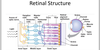
Retina
Neurosensory
Converts light into neurological impulses
What are the two segments of the eye separated by?
Lens separates anterior and posterior segments
Which humours are found in the two segments of the eye?
Anterior = aqueous humour Posterior = vitreous humour
What name is given to the fibrous strands that suspend the lens from the ciliary bodies?
Zonules
Describe the production and drainage of aqueous humour.
Aqueous humour is produced by the ciliary body It is drained via the trabecular meshwork into the canals of Schlemm
What is the role of aqueous humour?
Provides nutrients to the cornea and other tissues in the anterior chamber
Describe vitreous humour.
It is 99% water trapped inside a jelly matrix
What is the function of vitreous humour?
Mechanical support for the eye
Describe how the vitreous humour changes with age.
It loses its jelly consistency, liquefies and can become detached from the retina.
Vitreous detachment in seen as floaters
What are the potentially disastrous consequences of vitreous humour detachment?
Detaching from the retina could cause a small tear in the peripheral retina
If there is a small tear, liquid vitreous could seep into the sub-retinal space and lead to retinal detachment
If untreated, it can lead to blindness
What are the layers of the iris?
Anterior – stromal layer containing muscle fibres Posterior – epithelium
Describe how the retina and choroid contribute to the different parts of the iris and ciliary body.
Retina gives rise anteriorly to the ciliary body epithelium and the posterior (epithelial) layer of the iris Choroid gives rise anteriorly to the ciliary body stroma and the anterior layer of the iris (stromal layer)
What is the Uvea?
Vascular coat of eye ball
Comprised of the choroid, iris and ciliary body
What is the normal range for intraocular pressure?
11-12 mm Hg
What is glaucoma?
Optic neuropathy with characteristic structural damage to the optic nerve, associated with progressive retinal ganglion cell death, loss of nerve fibres and visual field loss
Increased intraocular pressure is a risk factor
What changes can be seen in the retina in glaucoma?
Retinal ganglion cell death Enlarged optic disc cupping
What are the consequences of untreated glaucoma?
Progressive loss of peripheral vision
Blindness
What is the most common type of glaucoma and what is it causedby?
Primary open angle glaucoma It is caused by a functional blockage of the trabecular meshwork
State another relatively common type of glaucoma. What is it caused by?
Closed angle glaucoma This can be acute or chronic It is caused by the forward displacement of the iris-lens complex –narrowing the trabecular meshwork
In what type of patients does closed angle glaucoma tend to occur and what is the treatment?
Small eyes (hypermetropic) Treatment: peripheral laser iridotomy
Describe the structure of the lens.
It has an outer acellular capsule There are regular inner elongated fibres, which give the lens its transparency NOTE: may lose transparency with age

Which two structures provide the majority of the refractive power of the eye?
Cornea = 2/3 Lens = 1/3
What layer of the eye is the cornea continuous with?
Sclera
Other than its role in refracting light, what else is the cornea necessary for?
Physical barrier – protects the eye from opportunistic infection
What are the consequences of prolonged contact lens wear?
Reduce the oxygen supply to the cornea
What are the layers of the cornea?
1 – Epithelium
2 – Bowman’s Membrane
3 – Stroma
4 – Descemet’s Membrane
5 – Endothelium
What is the role of the endothelial layer of the cornea?
It pumps out fluid from the stroma and prevents stromal oedema
What is the role of tear film?
Tear film maintains smooth cornea-air surface
- Oxygen Supply to Cornea – Normal cornea has no blood vessels
- Removal of Debris (Tear film and Blinking)
- Bactericide
What are the three layers of the tear film?
Superficial oily layer
Aqueous tear film
Mucinous layer

What produces the superficial oily layer?
Meibomian glands
‘my-bow-me-an’
What are the roles of each tear film layer?
Superficial Oily Layer:
Reduces tear film evaporation
Aqueous Tear Film:
Oxygen and nutrients
Bactericide
Mucinous Layer:
Ensures that tear film sticks to the eye
Where is the lacrimal gland located?
Anterio-superio-laterally to the orbit
What are the three types of tears?
Basal Tears – produced at a constant level in the absence of irritation Reflex Tears – increased tear production in response to irritation Emotional Tears – crying
Describe the innervation of the cornea.
The cornea is very sensitive and it is innervated by the ophthalmic branch of the trigeminal nerve (CN V)
Describe the drainage of tears.
Tears are drained into two puncta via two tiny holes in the upper and lower medial lid margins It then drains into superior and inferior canaliculi –> common canaliculus –> tear sac Tear sac then drains through the nasolacrimal duct, which opens up inthe inferior meatus
What two things regulate how much light reaches the retina?
Pupil
Pigmented Uvea
What is the term given to perfect focusing ability?
Emmetropia
What is the technical term for long-sightedness?
Hyperopia
How can long-sightedness be corrected and what is it caused by?
Convex lenses
It is usually caused by having a short globe (axial hyperopia)
It is occasionally caused by a flat corneal surface or insufficient refractive power (refractive hyperopia)
What is the technical term for short-sightedness?
Myopia
How can short-sightedness be corrected and what is it caused by?
Concave lenses
It is usually caused by having a long globe (axial myopia)
It is occasionally caused by having a highly curved cornea or excessive refractive power (refractive myopia)
What is astigmatism?
The cornea is oval rather than round
This means that the refractive power varies in different planes (in some planes you will be hypermetropic, and in others you would be myopic)
Parallel rays come to focus in 2 focal lines rather than a single focal point
Cause : refractive media is not spherical–>refract differently along one meridian than along meridian perpendicular to it– >2 focal points ( punctiform object is represent as 2 sharply defined lines)
Which nerve is responsible for accommodation?
Oculomotor Nerve (CN III)
What is the near response triad?
Pupillary Miosis (constriction of sphincter pupillae) – increases the depth of field Convergence – the medial recti of both eyes contract Accommodation – ciliary muscles contract to thicken the lens and increase its refractive power
What is the term given to naturally occurring loss of accommodation with age?
Presbyopia
‘pris-be-opia’
What are the four branches of vessel arcades radiating from the optic disc?
Superior Temporal Inferior Temporal Superior Nasal Inferior Nasal
Describe the difference in perfusion between the outer and inner parts of the retina.
Inner 2/3 of the retina = retinal arteries Outer 1/3 of the retina = choroidal vasculature
What part of the retina is responsible for central vision?
Fovea (it has the highest concentration of cones)
What is peripheral vision responsible for?
Shape, movement, night vision
Describe the structure of the retina.
Just inside the choroid you have the retinal pigment epithelium
Then you have the neuroretina, which consists of:
Outer Layer – Photoreceptors (1st Order Neuron) = Detection of Light
Middle Layer – Bipolar Cells (2nd Order Neurons) = Local Signal Processing to improve contrast sensitivity, regulate sensitivity
Inner Layer – Retinal Ganglion Cells (3rd Order Neurons) = Transmission of Signal from the Eye to the Brain
What is the function of the retinal pigment epithelium?
Transports nutrients from the choroid to the photoreceptor cells and removes metabolic waste from the retina
Phagocytosis of shed discs from photoreceptor cells
Describe how the fovea appears on a cross-section of the macula.
It appears as a foveal pit due to the absence of overlying retinalganglion cells
State the two classes of photo-receptor and their properties.
1) Rods:
Longer outer segment with photo-sensitive pigments
100 times more sensitive to light than cones
Slow response to light
Responsible for night vision (scotopic vision)
120 million rods
2) Cones:
Less sensitive to light
Faster response to light
Responsible for daylight vision and vision and colour vision (photopic vision)
6 million cones
Describe the recycling of photopigments.
Photopigments are synthesised in the inner photo-receptor segment and then are transported to the outer segment discs
The distal discs with deactivated photo-pigments are shredded from the tips and phagocytosed by retinal pigment epithelial cells
The deactivated photo-pigments are regenerated inside the retinal pigment epithelial cells and are then transported back to the photo-receptors
What is the rod photopigment?
Rhodopsin It is a G-protein coupled receptor system
What is the co-factor for rod photopigment?
11 cis retinal (vitamin A derived)
To what wavelength does rhodopsin react maximally in humans?
498 nm
What is another term for night vision?
Scotopic vision
What is another term for day-time vision?
Photopic vision
Describe the distribution of rods and cones across the retina.
Rods have the highest density 20-40o outside the macula
They decrease in density the further you move away from the macula
There are NO rods in the macula
Cones are almost exclusively found in the macula
The highest density of cones is in the fovea

What does a hill of vision represent?
Sensitivity of vision over a visual field
Where is the physiological blind spot located?
20 degrees temporal to central vision
What are the three types of cone photopigment and which colours do they respond maximally to?
S-cone – short wavelength – BLUE
M-cone – medium wavelength – GREEN
L-cone – long wavelength – RED
What is the most common colour vision deficiency and what is it caused by?
Deuteranomaly It is caused by the shifting of the M-cone towards the L-cone
What is the term given to shifted peaks?
Anomalous trichomatism
What test is used to diagnose colour blindness?
Ishihara Test
Describe how light sensitivity changes in dark adaptation.
Light sensitivity increases in dark adaptation
Biphasic Process
- Cone adaptation 7 minutes
- Rod adaptation 30 minutes – regeneration of rhodopsin. Rods become more sensitive than cones after 10 mins
How does retinal light sensitivity change in light adaptation and what is responsible for this effect?
Light sensitivity decrease in dark adaptation
This suppression of light sensitivity is caused by photopigment bleaching and neuro-adaptation inhibiting rod and cone function
What is the reflex pathway of reflex tear production
Cornea irritation V1 afferent PNS efferent
Label this


Label this


Label this


Label this


Label this


What is the conjunctiva?
The conjunctiva is the thin, transparent tissue that covers the outer surface of the eye.
It begins at the outer edge of the cornea, covers the visible part of the eye, and lines the inside of the eyelids.
It is nourished by tiny blood vessels that are nearly invisible to the naked eye.
The conjunctiva has goblet cells that produce mucin