Organisation of the Brainstem and Cranial Nerves Flashcards
Where are the colliculi found and what are their functions?
Roof of the midbrain Superior – important for the coordination of eye and head movements at the same time Inferior – auditory reflexes – turning your head in the direction of a loud noise
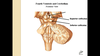
Describe the location of the pons relative to the ventricular system.
The pons is the floor of the 4th ventricle

Name an important unpaired, midline structure on the posterior aspect of the brainstem.
Pineal gland
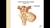
What is the role of the pineal gland?
It produces melatonin, which is involved in regulating the circadian rhythm
Which cranial nerve emerges from the back of the brainstem?
Trochlear nerve

What is the role of this nerve? (CN IV)
It supplies the superior oblique muscle – one of the extrinsic muscles of the eye
What structure defines the medulla in the dorsal aspect and what pathways are found within this structure?
Dorsal Columns – sensory pathways – touch and proprioception
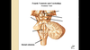
What three significant structures can be seen superior to the pons when viewing the brainstem from an anteroinferior view?
Optic Chiasm Pituitary Stalk (infundibulum) Mammillary Bodies

What are the mammillary bodies?
They are the inferior part of the hypothalamus – it is part of the limbic system
Which cranial nerve emerges in the midline just above the transverse fibres of the pons?
Oculomotor Nerve (CN III)

What are the cerebral peduncles? What is the significance of the word ‘peduncle’?
These are the descending motor tracts coming from the motor cortex. Peduncle is specifically a structure that has a functional AND structural role – it holds the cerebrum onto the brainstem

Name the cranial nerve that emerges from the lateral aspect of the pons.
Trigeminal (CN V)

What is the role of this nerve? (CNV)
Touch and sensation throughout the head and neck - 3 regions of the face from the 3 different branches
It has a small root next to the larger one as it emerges out of the transverse fibres – this is the motor root providing motor innervation of the muscles of mastication (temporalis and massater)
Which three nerves emerge at the pontomedullary junction (from medial to lateral)?
Abducens, Facial, Vestibulocochlear
Briefly state the role of each of these cranial nerves. (CN VI, VII, VIII)
Abducens – innervates the lateral rectus which is involved in abducting the eye Facial – innervates the muscles of facial expression and is involved in taste sensation for the anterior 2/3 of the tongue Vestibulocochlear – involved in balance and hearing
Which three nerves emerge from the lateral medulla?
Glossopharyngeal, Vagus and Accessory

Briefly state the role of each of these cranial nerves. (CN IX, X, XI)
Glossopharyngeal – sensory innervation of posterior 1/3rd of the tongue and pharynx. Innervates the parotid gland
Vagus – main parasympathetic nerve descending down to the viscera
Accessory – supplies the trapezius and sternocleidomastoid
Name and state the role of CN XII
Hypoglossal Nerve – supplies the musculature of the tongue

The motor fibres coming down from the motor cortex come via the cerebral peduncles then disappear behind the transverse fibres of the pons. What structure do they re-emerge as, inferior to the transverse fibres?
Pyramids
What percentage of motor fibres cross to the contralateral side of the body in the brainstem?
90-95%
What are the four functional subtypes of the cranial nerves and what are their actions?
- General Somatic Afferent Sensation from the skin and mucous membranes
- General Visceral Afferent Sensation from the GIT, heart, vessels and lungs
- General Somatic Efferent Muscles for eye and tongue movements
- General Visceral Efferent Preganglionic parasympathetic
What are the special subtypes of cranial nerves and what are their actions?
- Special Somatic Afferent Vision, hearing and equilibrium
- Special Visceral Afferent Smell and Taste (comes from three nerves that converge on the nucleus solitarius)
- Special Visceral Efferent Muscles involved in chewing, facial expression, swallowing, vocal sounds and turning the head
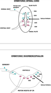
What structure separates the sensory nuclei from the motor nuclei in the spinal cord and brainstem?
Sulcus limitans

What happens in the formation of the rhombencephalon and how does this affect the arrangement of the nuclei?
- The alar plate opens up and a ventricle forms
- This opening of the alar plate results in the motor nuclei being medial in the brainstem and the sensory nuclei are lateral
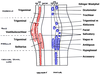
Describe the arrangement of the different groups in columns within the brainstem.
- Motor – Medial in the brainstem
- Sensory – Lateral columns of the brainstem
- Motor – arranged in columns from medial to lateral in this order: GSE, SVE, GVE
- Sensory – arranged in columns from medial to lateral in this order: GVA/SVA, GSA, SSA
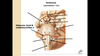
Describe the appearance of a cross-section of the midbrain. What are the key features seen?
- It has a distinctive ‘mickey mouse’ appearance
- Ears of Mickey Mouse are the cerebral peduncles
- At the point where the cerebral peduncles meet the rest of the midbrain you find the substantia nigra
- You will see the cerebral aqueduct in the middle (small diamond shape)
- The two rounded protrusion on the opposite side of the cerebral peduncles are the inferior colliculi

What is the substantia nigra? Explain its appearance Parkinson’s
The substantia nigra is a group of dopaminergic neurons In their normal metabolism they produce neuromelanin, which gives the black colour of the substantia nigra Parkinson’s disease is caused by loss of these dopaminergic neurons so patients with Parkinson’s will have a pale substantia nigra
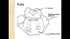
Describe the appearance of a cross-section of the pons. What are the key features seen?
The 4th ventricle will be seen at the dorsal aspect of the pons The most distinctive feature are the transverse fibres On either side you will see the middle cerebellar peduncles
What is the difference between the peduncles seen in the midbrain and the ones seen in the pons?
Midbrain –cerebral peduncles Pons – cerebellar peduncles
Describe the appearance of a cross-section of the medulla. What are the key features seen?
Pyramids will be seen on the ventral aspect The inferior olivary nucleus will be found next to the pyramids The 4th ventricle will still be visible

What is the role of the inferior olivary nucleus?
It is involved in fine tuning motor function
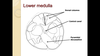
Describe the appearance of a cross-section of the lower medulla. What are the key features seen?
- It will be more round
- The central canal will be seen in the middle
- The dorsal columns will be seen on the dorsal side
- The pyramidal decussation may be seen
Name the two columns that make up the dorsal columns.
Gracilis – more medial – sensory information from the lower limb Cuneatus – more lateral – sensory information from the upper limb
What is lateral medullary syndrome? Describe and explain the symptoms.
It is a constellation of symptoms caused by an occlusion in the vertebral arteries or the posterior inferior cerebellar arteries (PICA) It causes: Horner’s Syndrome– disturbing the sympathetic tract Vertigo– because of disturbing the vestibular nucleus Ipsilateral loss of pain/thermal sensation on the face–disturbing the spinothalamic tract Contralateral loss of pain/thermal sensation on the trunk and limbs– disturbing the spinothalamic tract Ipsilateral cerebellar ataxia– disturbing the inferior cerebellar peduncle
What are the symptoms of Horner’s Syndrome?
Ptosis Loss of sweating around the eye Hoarseness Difficulty Swallowing
Draw the 3 parts of the brainstem

