Spine Flashcards

You may prefer our related Brainscape-certified flashcards:
1
Q

the occiput-c1 (Atlas) joint provides most of what ROM? How much?

A

flexion/extension. 50%

2
Q

c1-c2 joint (Axis) provides most of what ROM? how much?

A

Rotation. 50%

3
Q

surface land marks:

C2-3

C3

C4-5

A

C2-3: mandible

C3: hyoid

C4-5: thyroid cartilage

4
Q

surface landmarks:

C6

C7

T3

A

C6: cricoid cartilage

C7: vertebral prominens

T3: scapular spine

5
Q

surface landmarks:

T4

T7

L2

L4

L4-5

A

T4: nipples (variable)

T7: distal tip of scapula

L2: Renal Arteries

L4: Aortic Bifurcation

L4-5: iliac crest (L4 spinous process is at the level of the iliac crest)

6
Q

what spinal vertebrae have bifid spinous processes?

A

C2-6

7
Q

At what level is the spinal cord largest in the c-spine?

A

C2

8
Q

L-spine vert bodies are what shape?

T Spine?

A

L spine - kidney

T spine - heart

9
Q

mamillary processes occur in what spinal region?

from what structure do they project from?

A

L-spine

from superior articular process

allow for attachment of the multifidis muscles.

10
Q

How many sacral foramina are there?

A

4 pairs dorsal and ventral

The dorsal and ventral primary rami exit respectively

11
Q

how many vertebrae fused embryologically to form the coccyx?

A

usually 4 sometimes 5

12
Q

most common site of disc herniation?

second most common?

A

L5/S1 first, L4/5 second

13
Q

What is the Tectorial membrane?

A

the extension of the PLL from C1 to the skull

14
Q

Where does the transverse ligament of the c-spine live?

A

Posterior to Dens, stabilizes a-a joint and keeps dens up against anterior arch of c1

Part of the cruciate ligament which lies anterior to the tectorial membrane, behind the odontoid process

  • The transverse atlantal ligament is the strongest component, connecting the posterior odontoid to the anterior atlas arch, inserting laterally on bony tubercles
  • Vertical bands extend from the transverse ligament to the foramen magnum and body of the axis.
15
Q

What is the Anterior Atlanto-Occipital membrane?

A

an extension of the ALL from C1 to the skull.

16
Q

alar ligaments joint what to what?

embryologically they are remnants of what?

A

from occiput to tip of dens

remnant of notochord

17
Q

cruciform ligament of atlas is made of what?

A

includes the transverse ligament

plus inferior and superior longitudinal bands

18
Q

What are the two layers of the intervertebral discs and which layer contains nerve endings?

A

annulus fibrosis and nucleus pulposus

The AF contains nerve fibers.

19
Q

What type of fibers compose the annulus fibrosis and how are they oriented?

A

type I collagen fibers oriented obliquely.

20
Q

nucleus pulposis is mostly what collagen type?

A

type II

21
Q

What is the orientation of the facet joints as your progress down the spine?

A

Sagittal Coronal

C 45° 0°

T 60° 20°

L 90° 45°

22
Q

Describe the amount of pedicle screw “intoeing” you need as you go from T1-L5

A

Thoracic spine:

DECREASES at you go down from T1-T12

In males, goes from ~40 deg to ~15 deg

Therefore greatest at T1

Lumbar spine:

INCREASES as you go down from L1-L5

L1 approximately 5-10 deg

increases ~5 deg per level from L1 down to sacrum

23
Q

What are the landmarks for the T-spine pedicle screw start point?

A

Superior ridge of TP

Midpoint of inferior articular facet

24
Q

What is the landmarks for a lumbar pedicle screw start point?

A

midpoint of TP

midpoint of superior articular process

nb: pars lines up with medial aspect of pedicle

25
Q

upper spine largest pedicle?

L-spine largest pedicle?

A

T1

L5

26
Q

What is the smallest pedicle in T spine?

What is the smallest within L-spine?

A

T4

L1

27
Q

What are some what nerve injuries can occur with appication of Halo traction?

A

Supraorbital nerve most common - avoid by

Cranial nerve injuries have also been described. CN 4,6,10,11,12

CN VI - abducens n

can hit it at petrosphenoidal junction. get eyes that look down and in

Glossopharyngeal (4) + vagus (10) + hypoglossal (12)

dysphagia, loss of palatal/pharyngeal reflexes, weakness of tongue

from penetration of jugular foramen

CN eleven accessory n

supraorbital nerve - from anterior pins too medial

28
Q

Describe pin placement for halo

A

anterolateral pins: just below head equator, 1cm above orbit, in lateral 2/3 of orbit (avoid supraorbital nerve)

place anterior pins lateral enough to avoid injury to the frontal sinus, supratrochlear and supraorbital nerves

place pins anterior enough to avoid the temporalis muscle

pinsposterior pins. Usually place just directly above ear pinna

29
Q

list the fixation options for c1-2 fusion

A

transarticular screw

wiring

lateral mass screw (c1) and pars screw (c2)

clamp

30
Q

list the c2 fixation options

A

transarticular screw (with c1)

pars screw

pedicle screw

translaminar screw

clamp

wiring

31
Q

What are 3 contraindications to C1-C2 transarticular screws?

What about contraindications to C1-2 wiring?

A

Screws:

  1. abberant vertebral artery (medial or high riding)
  2. subluxation of C1-2
  3. cervicothoracic kyphosis precludes screw placement

Wiring:

•Need Intact Posterior Elements

  1. Decompression
  2. Trauma
  3. Tumour
32
Q

What is pelvic incidence?

A

fixed parameter describing the tilt of the S1 endplate relative to the centre of the acetabulum

On lateral view, make line from middle of S1 endplate to centre of acetabulum (center of femoral head)

make another line perpendicular to S1 endplate

angle between these lines is pelvic incidence

Geometrically ends up being equal to pelvic tilt + sacral slope

pelvic tilt=angle between vertical and line joining middle of S1 endplate to centre of acetabulum

sacral slope=angle between s1 endplate and horizontal

33
Q

Describe the spinal cord blood supply - only immediately around the cord

A

single anterior spinal artery

two posterior spinal arteries

they have branches that form an anastmosis/plexus around the cord - vaso corona

34
Q

What arteries feed the spinal arteries in c-spine?

A

Vertebral aa

PICA

segmental branches

35
Q

What arteries feed the spinal arteries in the T-L spine?

A

radicular arteries

36
Q

what is the artery of adamkiewicz?

A

principle arterial suply of lower 2/3 of spinal cord - anastomosis with the anterior spinal artery, supplying arterial blood to the spinal cord from T8 to the conus medullaris

usually occurs on left side at T9-12

can be between T7 and L4

AKA arteria radicularis magna

It arises from the radiculomedullary branch of the posterior branch of the intercostal or lumbar artery, which arise from the thoracic or abdominal aorta respectively 1. It has a diameter of ~1 mm (range 0.8-1.3 mm)

37
Q

extension of ALL from C1 to skull is called what?

A

anterior atlanto-occipital membrane

38
Q

extension of PLL from C1 to skull is called what?

A

tectorial membrane

39
Q

what is ligamentum nuchae?

A

c-spine supraspinous ligament

The supraspinous ligament only runs from C7 to sacrum

40
Q

vertebral foramina occur in what vertebrae?

through which does the vertebral artery pass?

A

C1-7

artery exists in c1-6

41
Q

continuation of ligamentum flavum from C1 to skull is called what?

A

posterior atlanto-occipital membrane

42
Q

What are the Basion and Opisthion?

A

basion: anterior point on the foramen magnum
opisthion: posterior point on the foramen magnum

43
Q

What is the presentation and prognosis of anterior cord syndrome?

A

Injury to anterior spinal cord caused by

direct compression (osseous) of the anterior spinal cord

anterior spinal artery injury

anterior 2/3 spinal cord supplied by anterior spinal artery

Mechanism

usually result of flexion/ compression injury

Exam

lower extremity affected more than upper extremity

loss

Corticospinal tract (motor)

Spinothalamic tract (pain, temperature)

preserved

DC (proprioception, vibratory sense)

Prognosis

worst prognosis of incomplete SCI

most likely to mimic complete cord syndrome

10-20% chance of motor recovery

44
Q

What is the presentation of posterior spinal cord syndrome?

A

very rare

Exam:

loss:

proprioception (dorsal columns)

preserved:

motor (corticospinal) pain

light touch (Lateral and ventral spinothalamic)

45
Q

What is the common presentation and prognosis of central cord syndrome?

A

spinal cord compression and central cord edema with selective destruction of lateral corticospinal tract white matter

UE weaker than LE

preserved perianal sensation (sacral sparing)

75% recover (not fully but fairly fuctional)

Epidemiology/incidence

most common incomplete cord injury

demographics often in elderly with minor extension injury mechanisms

due to anterior osteophytes and posterior infolded ligamentum flavum

Pathophysiology

believed to be caused by spinal cord compression and central cord edema with selective destruction of lateral corticospinal tract white matter

anatomy of spinal cord explains why upper extremities and hand preferentially affected

hands and upper extremities are located “centrally” in corticospinal tract

Presentation/symptoms

weakness with hand dexterity most affected

hyperpathia (burning in distal upper extremity)

physical exam

loss:

motor deficit worse in UE than LE (some preserved motor function)

hands have more pronounced motor deficit than arms

preserved –> sacral sparing

late clinical presentation

UE have LMN signs (clumsy)

LE has UMN signs (spastic)

Prognosis/final outcome

good prognosis although full functional recovery rare

usually ambulatory at final follow up

usually regain bladder control

upper extremity and hand recovery is unpredictable and patients often have permanent clumsy hands

recovery occurs in typical pattern

  1. lower extremity recovers first
  2. bowel and bladder function next
  3. proximal upper extremity next
  4. hand function last to recover
46
Q

What is the presentation and prognosis of brown-sequard syndrome?

A

a unilateral cord injury

  • Ipsilateral hypotonic paralysis at the level of injury (loss of LMN at that level) (ANTERIOR HORN CELLS, GREY MATTER)
  • Ipsilateral spastic paralysis below the level of injury (CORTICOSPINAL TRACT)
  • Ipsilateral loss of vibration and proprioception (DORSAL COLUMNS)
  • Loss of pain and temperature contralaterally from the level of 2 below the lesion (SPINOTHALAMIC TRACT)

99% ambulatory at final follow up

best prognosis for function motor activity

47
Q

Arrange the following incomplete spinal cord syndromes from best to worse in terms of prognosis:

Brown-Sequard

Anterior Cord Syndrome

Posterior cord syndrome

Central cord syndrome

A
  1. Brown-Sequard (Best)
  2. Central
  3. Posterior
  4. Anterior (Worst)
48
Q

Explain the ASIA scale ABCDE

A

A = Complete: No motor or sensory function is preserved in the sacral segments S4-S5.

B = Incomplete: Sensory but not motor function is preserved below the neurological level and includes the sacral segments S4-S5.

C = Incomplete: Motor function is preserved below the neurological level, and more than half of key muscles below the neurological level have a muscle grade less than 3.

D = Incomplete: Motor function is preserved below the neurological level, and at least half of key muscles below the neurological level have a muscle grade of 3 or more.

E = Normal: Motor and sensory function are normal.

49
Q

what is the definition of neurological level?

A

The most caudal (lowest) level at which both motor and sensory modalities are intact on both sides of the body.

  1. 􏱑Motor >3 or more w/ levels above being 5/5
  2. 􏱑Sensory intact bilaterally for LT and PP with all sensation above intact
50
Q

where is the watershed region of the spinal cord?

A

T4-9. narrowest spinal canal and poorest blood supply.

51
Q

Smith-robinson approach: from left or right side? Which is better and why?

A

left is better

reasons:

predictable course of recurrent laryngeal n (around aortic arch, runs between trachea and esophagus. Problem: most ppl right handed

c.f. right side: loops around subclavian a and crosses field from lateral to medial to run next to trachea. can be abberant at thyroid level

Incision

Mandible C2-3

Hyoid C3

Thyroid C4-5

Cricoid C6

  • make transverse skin crease incision at appropriate level
  • extend obliquely from the midline to the posterior border of the SCN
  • Superficial Dissection
  • incise fascia over platysma
  • spit platysma with finger
  • identify anterior border of SCM
  • incise fascia and retract SCM lateral
  • identify and retract strap muscles medially (sternohyoid and sternothyroid)
  • identify the carotid pulse and retract carotid sheath lateral
  • cut through pretrachial fascia
  • localize superior and inferior thyroid arteries and tie off if necessary
  • Structures at risk -
  • contents of carotid sheath
  • Recurrent Laryngeal nerve
  • Deep dissection
  • Once thru pretracheal fascia this should bring you down onto the longus colli mm. Place retractors. ?deflate and inflate cuff to decrease risk of RLN palsy, conflicting evidence
  • split longus colli muscles and anterior longitudinal ligament
  • be aware of sympathetic chain that lies on longus colli lateral to vertebral body
  • The prevertebral fascia is divided in the midline using rolled gauze (“peanuts”) in Kelly clamps. The longus colli is elevated subperiosteally on either side of the spine, taking care not to err anterior and lateral to the muscle mass so as to avoid damage to the sympathetic chains flanking the spine
  • subperiostally disect to expose anterior surface of vertebral body
  • retract longus colli muscles and ALL laterally
  • identify level with needle in disc space and lateral xray
  • Structures at risk
  • Sympathetic chain (10.6 mm from medial border of longus coli)s
  • Esophagus
  • recurrent laryngeal nerve tracheoesophageal groove

ACDF

Once we have exposure

check level radiographically

rectangle in annulus

discetomy

bone graft

instrumentation

52
Q

Name the 3 fascial layers you pass through in the smith robinson approach, from superficial to deep

A

deep cervical fascia

pretracheal fascia

prevertebral fascia

53
Q

Describe the smith robinson approach, and identifiy the important intervals.

A

transverse incision

split fibres of platysma (vertical) - CN VII facial n

go through deep cervical fascia

go between SCM (CN XI accessory) and strap muscles (omohyoid, sternothyroid, sternohyoid, thyrohyoid - all ansa cervicalis innervation)

go through pretrachial fascia anterior to carotid sheath (Sheath contains IJV, vagus n, common carotid a)

go between left and right longus colli (segmental n)

go thorugh prevertebral fascia

Reminder of levels

  • Mandible C2-3
  • Hyoid C3
  • Thyroid C4-5
  • Cricoid C6

Full description of approach:

Incision

make transverse skin crease incision at appropriate level

extend obliquely from the midline to the posterior border of the SCN

Superficial Dissection

incise fascia over platysma

spit platysma with finger

identify anterior border of SCM

incise fascia and retract SCM lateral

identify and retract strap muscles medially (sternohyoid and sternothyroid innervation:Ansa cervicalis)

identify the carotid pulse and retract carotid sheath lateral

cut through pretrachial fascia

localize superior and inferior thyroid arteries and tie off if necessary

Structures at risk -

contents of carotid sheath

Recurrent Laryngeal nerve

Deep dissection

Once thru pretracheal fascia this should bring you down onto the longus colli mm. Place retractors. ?deflate and inflate cuff to decrease risk of RLN palsy, conflicting evidence

split longus colli muscles and anterior longitudinal ligament

be aware of sympathetic chain that lies on longus colli lateral to vertebral body

The prevertebral fascia is divided in the midline using rolled gauze (“peanuts”) in Kelly clamps. The longus colli is elevated subperiosteally on either side of the spine, taking care not to err anterior and lateral to the muscle mass so as to avoid damage to the sympathetic chains flanking the spine

subperiostally disect to expose anterior surface of vertebral body

retract longus colli muscles and ALL laterally

identify level with needle in disc space and lateral xray

Structures at risk

Sympathetic chain (10.6 mm from medial border of longus coli)s

Esophagus

recurrent laryngeal nerve tracheoesophageal groove

ACDF

Once we have exposure

check level radiographically

rectangle in annulus

discetomy

bone graft

instrumentation

54
Q

What are the dangers of smith robinson approach?

A

carotid sheath (vagus n, common coarotid a, IJV)

thyroid arteries

trachea

esopahgus

recurent laryngeal n

stellate ganglion/sympathetic chain

vertebral a

55
Q

Explain posterolateral approach to the thoracic spine

AKA costotransversectomy

A

incision adjacent to spinous processes over rib

split trapezius fibres

subperiosteal dissection around rib

watch for intercostal bundle

remove rib up to TP

operate via retroperitoneal space

56
Q

interval of wiltse approach (modern variant)

A

between multifidus and longissimus

57
Q

What is the orientation of the zygopophyseal joints of the cervical, thoracic and lumbar spine?

A

Sagittal Coronal

C 45° 0° - Oriented A to P

T 60° 20° - almost vertical

L 90° 45° - wrapped

58
Q

Is the inferior articular process of the facet joint anterior or posterior in the cervical spine?

A

Posterior

59
Q

In what percent of the population does the vertebral artery run in C7?

A

7.5%

Normally runs from C1-C6

60
Q

What is spinal shock?

A

All phenomena surrounding transaction of the spinal cord that results in temporary loss of all or most spinal reflex activity below the level of injury. Characterized by loss of bulbocavernosus reflex

Spinal shock per Orthobullets

defined as temporary loss of spinal cord function and reflex activity below the level of a spinal cord injury.

characterized by

  • flaccid areflexic paralysis
  • bradycardia & hypotension (due to loss of sympathetic tone)
  • absent bulbocavernosus reflex (reflex characterized by anal sphincter contraction in response to squeezing the glans penis or tugging on an indwelling Foley catheter)

timing

  • variable but usually resolves within 48 hours
  • at its conclusion spasticity, hyperreflexia, and clonus slowly progress over days to weeks

mechanism

  • neurophysiologic in nature
  • neurons become hyperpolarized and unresponsive to stimuli from brain
  • evaluationimportant because one cannot evaluate neurologic deficit until spinal shock phase has resolved
  • end of spinal shock indicated by return of the bulbocavernous reflex
  • conus or cauda equina injuries may lead to permanent loss of the bulbocavernous reflex
61
Q

What is neurogenic shock?

A

characterized by hypotension & relative bradycardia in patient with an acute spinal cord injury ABOVE T6

mechanism:

circulatory collapse from loss of sympathetic tonedisruption of autonomic pathway within the spinal cord leads to

  • lack of sympathetic tone
  • decreased systemic vascular resistance
  • pooling of blood in extremities
  • hypotension
  • Loss of sympathetic tone to the heart due unopposed vagal activity and has been found to be exacerbated by hypoxia and endobronchial suction
62
Q

In what percent of people does the vertebral artery run in C7?

A

7.5% Remember - mostly it’s C1-C6

63
Q

Describe the safe zone for 8mm occipital screws. What are the dangers?

A

The safe zone for screw placement in the occiput for occipitocervical fusion is in a triangular region created by connecting 2 dots 2cm lateral to the EOP and a point 2 cm inferior to the EOP.

6 screws can be placed in a V formation

1st is on Nuchal line 2cm away from center of EOP

2nd is 1cm down and 1cm over

3rd is 2cm down and 5mm over

The dangers are the superior saggital sinus and the transverse sinus

64
Q

Define the motor level

A

Most caudal level with 3/5 power with 5/5 power above

65
Q

What tract is the main descending motor pathway in the cervical spinal cord?

A

Lateral corticospinal tract

66
Q

The occiput is the thickest in what location?

A

The thickest region of the occiput is at a point 5 cm lateral to the External Occipital Protuberance. However, this point is too far lateral to be useful for occipital-cervical fusion, and is instead where you place the posterior inferior pin of a Mayfield holder

67
Q

In the setting of bilateral C5-6 facet dislocation, which of the following structures is usually preserved?

1: Facet joint capsules
2: Ligamentum flavum
3: Posterior annulus
4: Anterior longitudinal ligament
5: Interspinous ligament

A

4: Anterior longitudinal ligament

68
Q

Which of the following pedicles has the smallest transverse diameter in most people?

A: T1

B: T12

C: L1

D: L3

E: S1

A

3: L1

T4 is the smallest overall.

T1 is biggest in thoracic spine

S1 > L3 > L1

T1 > T12

So this questions asks which is bigger T12 or L1. T12 is bigger so L1 is the answer

69
Q

Describe the main blood supply to the spine? What is the name of the collateral blood supply?

A

Main:

1x median longitudinal anterior spinal artery

2x (right & left) longitudinal posterior spinal arteries

These are supplied by various radicular arteries

Collateral: Vaso Corona: anastomosis between the longitudinal vessels forming a fine pial plexus Gives limited blood supply

70
Q

Which area of the spinal cord has the worst collateral blood supply? The best?

A

Worst: T4-T9 (Watershed area)

Best: Cervical and lumbar - they have redundant supply

71
Q

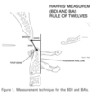
What are Harris’ measurements of the spine? What do they measure?

A

They measure atlanto-occipital dissociation There are 2 measurements:

  1. Basion to the tip of the dens (BDI)
  2. Basion to the posterior axial line (BAI - basion posterior-axial interval)

***Neither one should be >12mm (Rule of 12’s) or this is a sign of AO dissociation

72
Q

What are the signs of sacral dysmorphism? Why is it important and what should you do instead?

A

Signs:

  1. Sacralization of L5
  2. Lumbarization of S1
  3. Presence of mammillary processes
  4. Oval or oblong foramen
  5. Tongue in Groove sign of SI joint

**Important to recognize to avoid iatrogenic injury to L5 during SI screw insertion. If dysmorphic, plan for S2 screw instead (of S1)

73
Q

What is the highest motor level that the patient can have and initially still be ventilator independent? Why?

A

C5

C3,4,5 make up the phrenic nerve, which supplies the motor function of the diaphragm (C3,4,5 keep the diaphragm alive) People with C3-C4 levels can progress to be ventilator independent

74
Q

What is a Jefferson’s fracture? mechanism?

A

fracture thru the anterior and posterior arches of C1, burst type fracture.

Often due to axial loading (diving into shallow water) or forced hyperextension

75
Q

In a Jefferson’s fracture, how much lateral overhang on open mouth x-ray indicates disruption of the transverse ligament?

A

A total overhand (both sides added up) of >8mm indicates disruption of the transverse ligament (>6.9mm on CT, though there is some controversy)

76
Q

What are the risk factors for non-union of odontoid fractures?

A

Displacement >6mm

Angulation >10

77
Q

What is a Hangman’s fracture? mechanism?

A

bilateral C2 pars fracture.

hyperextension and distraction.

traumatic spondylolisthesis of C2 on C3

How is it treated?

Undisplaced - collar

Displaced - Halo or surgery

78
Q

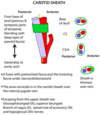
What are the contents of the carotid sheath?

A
  1. Common carotid artery +/- internal carotid artery Internal jugular vein
  2. Vagus nerve
  3. Deep cervical lymph nodes

Artery is medial to vein, with nerve in between

79
Q

What are the ligamentous attachments to the dens?

A
  1. Apical odontoid ligament
  2. Alar ligaments x2

Transverse ligament of the atlas (part of cruciform ligament) - doesnt actually attach to the dens

(Anterior atlantoaxial ligament (membrane) is anterior and not actually attached to the dens)

80
Q

What is autonomic dysreflexia?

A

Develops in individuals with spinal cord injury at or above the level of T6, resulting in acute, uncontrolled hypertension.

Painful stimulus below the lesion is sensed by the spinal cord and transmitted proximally. Interrupted by lesion so pt does not feel sensation.

This causes sympathetic response/discharge

–> vasoconstriction, most notably in the splanchnic circulation.

This is sensed proximally and there is a vagal, parasympathetic discharge to try to counteract this.

This discharge is unable to pass the lesion therefore. You have unopposed hypertension.

Symptoms:

The sympathetic response causes vasoconstriction, resulting in hypertension, pounding headache, visual changes, anxiety, pallor and goosebumps (below the level of injury)

Above the level of injury you get BRADYCARDIA and FLUSHING and NASAL CONGESTION (rhinorhea (cholinergic discharge)

81
Q

A patient comes in post-cervical spine ACDF complaining of a drooping eyelid. On examination, you see that she has constricted pupils, no sweating on one side of the face (ptosis, miosis, anhidrosis). What structure was damaged. Where is this structure located?

A

Sympathetic ganglia in the C-spine was damaged - This caused a Horner’s syndrome The sympathetic ganglia are located on the 10.6 mm from medial border of longus coli muscles

82
Q

Which of the following most accurately describes the medial-lateral (transverse) pedicle diameters in the T/L spine?

  1. Diameters are smaller in the lower T-spine than mid-T spine
  2. Diameters are smaller in the mid-T spine than high T-spine
  3. Diameters are smaller in the high T-spine than the mid-T-spine
  4. Diameters are larger in the upper L-spine than the lower T-spine
  5. Diameters are larger in the high T-spine than the lower L-spine
A
  1. Diameters are smaller in the mid-T spine (T5-7) than the high T-spine (T2-T4)
83
Q

A misplaced screw is recognized after lumbar surgery. The screw has grossly violated the inferior cortex of the left L4 pedicle. What is the most likely neurologic sequelae?

A

L4 weakness (ankle dorsiflexion)

84
Q

What is the highest motor level that the patient can have and still be ventilator INDEPENDENT? Why?

A

C5 C3,4,5 make up the phrenic never, which supplies the motor function of the diaphragm - C3,4,5 keep the diaphragm alive

85
Q

What are Harris’s measurements of the spine? What do they measure?

A

Measure occitio-atlanto dissociation 2 measurements: - Basion to the tip of the dens (BDI) and basion to the posterior axial line (BAI) - Neither one of these should be >12mm - This is the rule of 12’s

86
Q

What is a teardrop fracture?

A

Fracture of the anterior inferior endplate - Must differentiate between flexion and extension types - Flexion is unstable and associated with SCI. Extension is stable

87
Q

What is Swischuk’s Line?

A

Used to differentiate between subluxation and pseudosubluxation in pediatric patients

Connects the anterior aspect of the spinous processes of C1 and C3.

If the anterior aspect of the spinous process of C2 misses this line by ⩾2 mm true subluxation of hangman’s fracture must be considered.

***For true subluxation to be diagnosed then there has to be SUBLUXATION and DEVIATION <2MM from SWISHCUKS LINE

The distance is frequently 2 mm or more if no subluxation is apparent.

Figure:

(A) No subluxation. Therefore, PCL (posterior cervical line) cannot be applied. Anterior aspect of spinous process of C2 commonly misses PCL by 2 mm.

(B) Subluxation is present. The anterior aspect of spinous process of C2 misses the PCL >2 mm. Finding is suggestive of a hangman’s fracture of the neural arches of C2.

(C) Pseudosubluxation is present. The anterior aspect of spinous process of C2 touches or lies within 2 mm of PCL. (Adapted from figure from Fesmire FM and Luten RC.16)

88
Q

Describe the cervicomedullary angle and its significance:

A

The cervicomedullary angle can be measured on MRI by drawing a line along the anterior aspect of the cervical spinal cord and the medulla.

This angle is normally between 135° and 175°

• Cervicomedullary angle <135° linked to myelopathy

89
Q

How do you perform a bulbocavernosus reflex?

A

Normal: - Anal sphincter contraction (anal wink) after squeezing the glans penis or tugging on the foley - In women, squeeze the clitoris or tug on the foley

90
Q

Figures 8a and 8b show the radiograph and MRI scan of a 31-year-old man with severe back pain and intermittent leg pain. Which of the following anatomic measurements has been best correlated with this patient’s condition?

  1. Pelvic tilt
  2. Pelvic Incidence
  3. Lumbar Lordosis
  4. Thoracic Kyphosis
  5. C7 Sagittal plumb line
A

2.Pelvic Incidence

91
Q

Name the intrinsic craniocervical ligaments (3).

A

The intrinsic ligaments, located within the spinal canal, provide most of the ligamentous stability. These ligaments form three layers anterior to the dura. From dorsal to ventral, they include:

  1. ​Tectorial membrane: connects posterior body of axis to anterior foramen magnum. Is the cephalad continuation of the PLL
  2. Cruciate ligament: lies anterior to tectorial membrane, behind the odontoid process
  3. Odontoid ligaments (alar and apical)

(The Atlanto-occipital membrane (continuation of ALL is extrinsic)

92
Q

Name the extrinsic ligaments of the craniocervical spine

A
  1. Ligamentum nuchae: extends from external occipital protuberance to the posterior aspect of the atlas and cervical spinous processes
  2. Anterior Atlantoocciptal Membrane: Continuation of ALL, intervertebral disks, and ligamentum flavum between the occiput and atlas and between the atlas and axis
  3. Atlanto-occipital joint capsule
  4. Atlanto-axial joint capsule
93
Q

Figure 255 shows the axial CT scan of a 33-year-old man with severe neck pain after a motor vehicle collision. Which structure is most important in guiding treatment?

  1. VA
  2. Alar Lig
  3. Apical Lig
  4. Transverse Lig
  5. Post Lig Complex
A
  1. Transverse ligament
94
Q

What osseous structure is seen by the * in Figure 237?

1- Inferior articular facet of C4

2- Inferior articular facet of C5

3- Superior articular facet of C5

4- Pedicle of C5

5- Pars interarticularis of C5

A
  1. Inferior articular process of C4
95
Q

Figures 89a and 89b are the parasagittal and axial CT scans of a 42-year-old patient seen after a motor vehicle collison. What anatomic structure is identified by the asterisk in Figure 89b?

  1. C6 facet
  2. C7 facet
  3. C6 pars
  4. C6 pedicle
  5. C7 pedicle
A
  1. C6 Facet
96
Q

What is the nerve injury shown?

A

Abducens nerve palsy, reported as a complication of Halo. CN VI

No lateral gaze, eye down and in.

97
Q

What is the amount of lateral mass displacement necessary to indicate disruption of the transverse ligament on on imaging?

A

6.9mm vs. 8.1mm

A Jefferson fracture (Type II Atlas Fracture) is characterized by bilateral fractures of both the anterior and posterior arch. They are considered unstable when the transverse ligament is no longer intact. Radiographically, an atlanto-dens-interval (ADI) of >3mm or a sum of lateral mass displacement of >8.2mm both indicate a ruptured transverse ligament. Treatment of Jefferson fractures remain controversial. A stable injury (intact transverse ligament) can be treated with a cervical orthosis.

Spence et al., in a historic anatomic study, showed that lateral mass separation of > 6.9 mm implied rupture of the transverse ligament.

Heller et al. built radiographic magnification into Spence’s findings, and argues that a transverse ligament rupture should not be inferred unless lateral mass separation is > 8.1 mm on open mouth odontoid views.

Orthobullets >7mm QUOTING: “Tears of the transverse ligament of the atlas. A clinical and biomechanical study.” Fielding et al JBJS.

98
Q

What are the synovial joints of the spine?

A

Facet joints

Atlanto-occipital joint

Lateral Atlanto-axial joints

Central atlanto-axial joint

99
Q

Which of the following is most predictive of disability in adult patients with scoliosis?

  1. Coronal plane cobb angle
  2. Retrolisthesis at L3/4
  3. Osteoporosis
  4. Sagittal plane decompensation
  5. Severity and number of levels of disk degeneration
A
  1. Sagittal plane decompensation
100
Q

On a standing lateral radiograph of the spine in which the edge of the film represents a true vertical, a normal C7 plumb line should intersect which of the following anatomic landmarks?

  1. Center of the C7 vertebral body and posterior-superior corner S1
  2. Center of C7 body and anterior-superior corner of S1
  3. Anterior border of C7 vertebral body and anterior-superior corner of S1
  4. Anterior border of C7 body and posterior-superior corner of S1
  5. Posterior border of C7 vertebral body and posterior-superior corner of S1
A

1.Center of the C7 vertebral body and posterior-superior corner S1

101
Q

Figure 169a is the lateral radiograph of a 19-year-old female gymnast with low back and leg pain. Examination reveals exacerbation of back pain with extension. She has a normal motor examination but diminished light touch sensation in an L5 distribution. What measurement shown in Figure 169b has been correlated with this disease?

  1. Slip Angle
  2. Sacral Slope
  3. Pelvic Tilt
  4. Pelvic Incidence
  5. Spinopelvic Angle
A
  1. Pelvic incidence
102
Q

A 27-year-old male is an unrestrained passenger in a motor vehicle accident. He was medically stabilized in the emergency room. His initial injury CT scans are seen in Figures A and B. He is neurologically intact and placed in a halo fixator prior to surgical treatment. What is the most common neurologic complication with halo traction?

  1. Weakness in biting and chewing strength
  2. Deficit in medial and downward eye movement
  3. Deficit in lateral eye movement
  4. Inability to close eyes against resistance
  5. Tongue deviation toward the affected side
A
  1. Deficit in lateral eye movement (CN VI injury). Thought to be due to traction injury

Supraorbital nerve palsy (less common)

Supratrochlear nerve palsy (less common)

103
Q

After application of a halo device, a patient has pain and numbness over the medial one third of the eyebrow. This is most likely the result of injury to which of the following structures?

  1. Second dorsal rami
  2. Facial nerve
  3. Auriculo temporal nerve
  4. Lesser occipital nerve
  5. Supraorbital nerve
A
  1. supraorbital nerve injury
104
Q

What vertebra does the vertebral artery run in?

A

C1-C6

Although all the cervical vertebra have a vertebral foramen

105
Q

What is Chassaignac’s Tubercle ?

A
  • the Carotid tubercle - the anterior tubercle of transverse process of C6
  • This separates the carotid artery from the vertebral artery and the carotid artery can be massaged against this tubercle to relieve the symptoms of supraventricular tachycardia.
  • Important landmark for a plexus block
  • The stellate ganglion lies just inferior, anterior to the transverse process of C7, superior to the neck of the first rib, and just below the subclavian artery.