Spinal Cord Syndromes Flashcards

1
Q

What is a myelopathy?

A

Any disease that affects the spinal cord

2
Q

The spinal cord ends at vertebral body L1-L2.

Spinal taps are done between L4 and S1, don’t worry, you are very far away from the cord!

Most back pain is lumbar. If the patient points to the thoracic area, be concerned!. Bad things happen in the thoracic cord!

A
3
Q
A

Here are the CLINICALLY RELEVANT components of the spinal cord.

YES, you will see deficits in these areas in patients with spinal cord disease.

Note, that spinal thalamic tracts cross within the spinal cord, all other tracts stay ipsilateral within the spinal cord and then cross in the brain.

4
Q

In addition to the spinal cord anatomy, you need to understand the relationship between the spinal cord and surrounding structures, because as these areas are damaged, they are so close to the spinal cord, the cord can easily be damaged. Note the following anatomy from the inside of the cord to the outside, in the following order:

A
  1. The spinal cord itself, also referred to as spinal cord parenchyma
  2. Next is pia mater
  3. Then subarachnoid space
  4. Then the subdural/intradural space
  5. Then the dura
  6. Followed by the epidural/extradural space (fat is in here)
  7. Finally bone (vertebral body)
5
Q

Again, there are lots of synonyms and medical jargon you need to know:

EPIDURAL = EXTRDURAL

INTRADURAL = SUBDURAL

WITHIN THE SUBARACHNOID SPACE = LEPTOMENINGEAL (The pia and subarachnoid space are the ‘leptomeninges’).

WITHIN THE SPINAL CORD PARENCHYMA = INTRAMEDULLARY.

A
6
Q

The cervical spinal cord.

CSF is white.

The spinal cord is gray/black.

Again, note the proximity of the cord to discs, vertebral bodies etc.

A

MRI of the thoracic spinal cord.

7
Q

MRI of the lumbar spine. Note the end of the spinal cord and the roots as they leave the spinal cord

A
8
Q

Patients with spinal cord syndrome commonly develop ‘sensory levels’. That is, they cannot feel sensation at or below this level.

T4 (nipple line) and T10 (umbilicus) are common sensory levels.

A
9
Q

Bladder symptoms are common in spinal cord lesions. (Bladder control is mostly a CNS phenomena). How?

A

Long axons from the frontal lobe of the brain synapse in the thoracic and sacral areas of the spinal cord, thus these tracts are very vulnerable to injury in spinal cord lesions.

10
Q

Two general kinds of bladder symptoms can occur in spinal cord lesions.

A

In acute lesions (left) there is urinary retention with some overflow incontinence.

In chronic lesions, there is a small spastic bladder that does not completely empty with spasms and urge incontinence.

11
Q

Acute myelopathies are true NEUROLOGIC EMERGENCIES AND REQUIRE IMMEDIATE ATTENTION.

Your actions may determine whether or not the patient will ever walk again!

A

Knowing the spinal cord anatomy will help you recognize these syndromes, especially at two o’clock in the morning!

12
Q
A
13
Q
A

Never forget to do these things on the left of the slide.

The neurologic exam is critical and will help you to recognize and localize the lesion.

14
Q

THE PACE OF SYMPTOMS IS CRITICAL AND UNPREDICTABLE.

Some patients can walk into the office, yet if not recognized, can become paralyzed in a matter of minutes as shown in this slide!

A
15
Q

How does an epidural lesion progress?

A

First note the progression from A to B to C.

Let’s review ‘A’ first: Early symptoms include: At the level of the lesion, the dorsal root can be damaged or compressed which can lead to numbness or allodynia = hyper-sensitivity to touch, IPSILATERAL.

If the spinothalamic TRACT is injured at this point, very subtle changes in sensory symptoms can be seen in the CONTRALATERAL lower extremity.

The motor symptoms are extremely important and are due to compression of the corticospinal tract:

Early in the process the patient may NOT be weak, but there WILL BE NEUROLOGIC SIGNS including hyper-reflexia, positive Babinski signs and difficulty walking.

Urinary urgency is COMMON. DON”T FORGET TO ASK THE PATIENT. Many will be embarrassed and will not tell you.

16
Q
A
17
Q

B. Symptoms are worsening:

Motor: In addition to spasticity and hyper-reflexia, the legs are weak.

Sensory symptoms are worse, with numbness as shown.

Definite sphincter dysfunction – bladder and may include bowel dysfunction.

May appear as a Partial Brown Sequard syndrome = hemi-cord syndrome with weakness and numbness to touch and vibration on the ipsilateral side AND loss of pain sensation on the CONTRALATERAL side, due to the crossing of the spinothalamic tract within the spinal cord.

A
18
Q

C. Full blown spinal cord compression – yes you will see this on the wards.

If slow and chronic, there will be spastic paraparesis with a definite sensory level to all modalities.

If acute or hyper-acute, there is so much damage to the spinal cord that ‘spinal cord shock’ occurs, which is FLACCID PARAPLEGIA WITH A COMPLETE SENSORY LEVEL TO ALL MODALITIES.

A
19
Q

What are some extradural causes of myelopathy?

A

disc disease

tumor (mets, lymphoma, sarcoma, plasmacytoma)

scar

abscess

hemangioma

20
Q

What are some intradural causes of myelopathy?

A

neurioma

meningioma

intracranial tumor seeding (ependymoma, medulloblastoma, glioma)

cauda equina lesions

21
Q

What are some intramedullary causes of myelopathy?

A

syringomyelia

tumor (ependymoma, glioma, hemangioblastoma)

myelitis

edema

lipoma

22
Q

Herniated disc and degenerative disc disease can result in cord compression.

The arrows shows multiple discs compressing the spinal cord.

The arrow heads show hyper-intense lesions within the spinal cord parenchyma.

This is edema associated with the compression.

Treatment is steroids and surgery.

A

This is a photo of a vertebral body fracture with acute spinal cord compression.

This patient may present with spinal cord ‘shock’, because of the acuity of the lesion.

This may result in spinal cord infarct or cord transection

23
Q

What is the standard tx for spinal cord injruy?

A

–Methylprednisolone 30 mg/kg IV over 1 hour

–Followed by 5.4 mg/kg/hr over the next 23 hours

24
Q

Cancer can affect the spinal cord at EVERY level we have discussed: intramedullary, leptomeningeal, epidural, subdural.

This slides lists many of the spinal cord problems patients with cancer can develop.

It depends on the primary cancer and the route of metastases.

A
25
Q

Epidural compression is a common complication of metastatic cancer.

The cancer enters the vertebral body, weakens it, expands and then compresses the spinal cord – as shown in this figure.

The most common cancers that do this are: lung, breast, and prostate.

A
26
Q

How should a cancer pt with back pain be handled?

A

neurologic exam: if normal, do a spinal X-ray. If that is negative observe or investigate a retroperitoneal tumor/lymphoma with a spinal MRI. If the spinal X-ray is positive and indicates a lytic or blastic lesion, proceed to spinal MRI

If the neurological exam is abnormal, begin dexamethasone and get a spinal MRI

27
Q

Here is a diagnostic approach to cancer patient with back pain, particularly in the thoracic region.

Most physicians NO LONGER use plain spine x-rays.

If you are clinically suspicious of a spinal cord compression: treat with steroids (dexamathosone) first, arrange for a spinal MRI with gadolinium and follow the patient slowly.A neurosurgical consult is also required in case surgical decompression is needed.

A

CT myelograms are rarely done because of the sensitivity and non-invasiveness of MRI.

If the work-up is negative, cancer cells can still enter the subarachnoid space, cause a meningitis and spinal cord injury.

Therefore, a spinal tap is required if there is NO EVIDENCE of cord compression by imaging techniques.

28
Q

Infections of the spinal column can also result in spinal cord compression.

Remember, most metastatic cancer begins in the VERTEBRAL BODY. In contrast, most spinal abscesses begin in the DISC SPACE. Then develop into an abscess and expand to cause spinal cord compression.

The most common infection is Staph Aureus. Be particularly suspicious in IV drug abusers.

MRI of the spine is the test of choice.

A
29
Q

How might an epidural abscess present?

A

Fever, pain on percussion, elevated white count, elevated ESR

Risk factors (IV drug use, HIV, immunosuppression)

30
Q

Osteomyelitis is an infection of the vertebral bodies. It causes weakness of the bones, collapse of the vertebral body and subsequent cord compression as shown here.

Note the irregular vertebral body end plates (large arrows).

A
31
Q

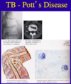
Pott’s disease is a class neurologic syndrome of osteomyelitis resulting from TB infection.

I’ve never seen a board exam without at least one question on this.

A
32
Q

Spinal meningioma is intradural, extramedullary

Common in thoracic spine.

Clinical clue: although the occasional benign herniated disc can occur in the thoracic spine, most diseases of the thoracic spine are bad.

A

Schwannoma and neurofibroma are also extramedullary, intradural

Dumbbell shape is not very common, but it is CLASSIC, in that if you see it, it is usually one of these tumors.

33
Q

Now we are going to shift from lesions and diseases that compress the spinal cord to lesions that begin WITHIN the spinal cord parenchyma itself.

Many of these syndromes are not ‘perfect’. You will see partial syndromes, but there are still clinical and radiological clues that indicate an intra-medullary (=within the spinal parenchyma) lesion or disease process.

As shown in this figure, these lesions begin in the spinal cord.

Many start in the central spinal cord, where the spinothalamic fibers cross, therefore loss of pain and temperature are an EARLY sign.

In the first panel, loss of sensation across the torso as shown is common.

A
34
Q

In the middle panel, the lesion has extended to other areas of the spinal cord:

Dorsal root entry zone – interruption of the afferent arc of the reflexes = loss of reflexes in the arms at the area of the lesion.

Continued loss of pain and temperature.
Involvement of the sympathetic pathways = Horner’ syndrome (ptosis, miosis)

The posterior columns are spared until late. Therefore vibration and joint position sense are preserved compared to pain and temperature.

This is known as a DISSOCIATED SENSORY LEVEL.

Below the lesion, involvement of corticospinal pathways results in spastic paraparesis, hyper-reflexia and Babinski signs.

A
35
Q

Last panel. All worsen. There may be SACRAL SPARING, because these fibers are the most lateral = furthest away from the central cord, thus they are not damaged and sacral dermotomes will maintain normal sensation.

A
36
Q
A
37
Q

The classic lesion that presents this way (central cord syndrome) is a syrinx – a large expanding space in the spinal cord.

Note how each tract results in a patient’s symptoms.

Syrinx can be the result of trauma, or tumor.

A
38
Q

Here are the radiographic and pathologic appearance of spinal cord astrocytoma.

Note the enhancement with gadolinium (D) and the large amount of edema (E).

A
39
Q

Vascular disease can also affect the spinal cord.

A
40
Q

The anterior 2/3 of the spinal cord is supplied by the Artery of Adamkiewicz = the great radicular artery (located in the mid thoracic region). These patients will have symptoms referable to spinothalamic (loss of pain & temperature, sensory level), corticospinal function (weakness) but INTACT POSTERIOR COLUMN FUNCTION (vibration and joint position sense).

A

These patients will have symptoms referable to spinothalamic (loss of pain & temperature, sensory level), corticospinal function (weakness) but INTACT POSTERIOR COLUMN FUNCTION (vibration and joint position sense).

41
Q

Another picture of the anterior spinal artery syndrome

A
42
Q

Our next patient has a spinal cord syndrome not related to epidural compression.

Note that she did NOT have back pain.

Note early urinary retention = suggests a spinal cord lesion!

A
43
Q

This is a review of spinal cord shock.

Although we typically view flaccidity as a peripheral nerve lesion, in the setting of a complete sensory level and urinary retention, this leads to a diagnosis of acute spinal cord injury.

A
44
Q

Describe Brown-Sequard syndrome?

A

We rarely see pure, Brown – Sequard lesions. However, it is common to see them in a partial form (i.e., weakness on one side, loss of pain and temperature on the other side.

45
Q

Chronic myelopathies also occur, which we will describe next.
This is particularly important when spinal imaging is negative.

A
46
Q

There are other causes of myelopathy that are neither infiltrative nor compressive.

These are also important and part of the differential diagnosis of spinal cord disease.

Many are reversible.

A

This is how B12 deficiency can present in the spinal cord = subacute combined (i.e., two tract systems, corticospinal and posterior column) degeneration.

Remember, B12 deficiency can also cause dementia and peripheral neuropathy.

Treatment is intramuscular B12 injections.

47
Q
A
48
Q

Infection with HTLV-1 also causes spastic paraparesis and spinal cord disease.

A

These are the clinical presentation of someone infected with HTLV-1 who has HAM/TSP.

49
Q

HIV patients also get myelopathy.

The pathology, radiology and clinical presentation are displayed in this slide.

A
50
Q
A