Skull Flashcards

1
Q

In anatomical position, what is the plane that is at the level of the bottom of the orbit and the opening of the external auditory meatus?

A

orbitomeatal plane

2
Q

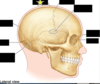
Identify the portion of the cranium that is blacked out and its general functions.

List the (2) parts that make up this cranial segment and the (8) bones it is composed of.

A

Neurocranium

Consists of dome shaped Calvaria (skullcap) and basicranium

Cranial bones:

  1. Frontal
  2. Parietal (2)
  3. Temporal (2)
  4. Occipital
  5. Sphenoid
  6. Ethmoid
3
Q

Identify the portion of the cranium that is blacked out.

What is this structure formed by and what is its function?

Name the (14) bones that make up this structure

A

viscerocranium

Formed from the skeleton of the pharyngeal arches. Functions related to respiration, mastication/jaw apparatus.

Facial bones:

  1. Maxillae (2)
  2. Zygomatic (2)
  3. Nasal (2)
  4. Lacrimal (2)
  5. Inferior nasal conchae (2)
  6. Palantine (2)
  7. Vomer
  8. Mandible
4
Q

Describe the composition of the bone formign the calvaria.

A

outler layer of dense bone, spongy bone sandwitched between the dense cortical bone

spongy bone = diploe (large communicating venous channels)

5
Q

What is the name for fibrous interlocking joints of the skull?

A

sutures (synarthroses)

6
Q

What is the name for cranial bones connected by cartiledge during childhood?

A

synchrondroses

7
Q

Which layer of cranial bone is most susceptible to fracture?

A

The inner layer of compact bone is distinctly thinner and more prone to injury. It can fracture with outer lamina still in tact.

8
Q

What type of calvaria fracture results in a fragment of bone being depressed inward, compressing and/or injurign the brain?

A

depressed fracture

9
Q

What type of calvarial fracture results in the bone breaking into several pieces?

A

comminuted fractures

10
Q

What type of calvarial fracture occurs at the point of impact, but fracture lines often radiate away in two or more directions?

A

linear fractures (most common)

11
Q

What type of calvarial fracture results in a fracture on the opposite side of the trauma?

A

contrecoup fracture

12
Q

What is the name of landmarks that are used radiographically to make cranial meausrments and document abnormal variations?

A

craniometric points

13
Q

What is the name of the craniometric point indicated by the picure. Describe its location with relation to sutures.

A

Nasion: the point on cranium where frontonasal adn internasal sutures meet

14
Q

What is the name of the craniometric point indicated by the picure. Describe its location with relation to other structures.

A

Glabella: smooth prminence most marked in males; on frontal bones superior to root of nose between eyebrows

15
Q

What is the name of the craniometric point indicated by the picure. Describe its location with relation to other structures.

A

Bregma: the point on calvaria at junction of coronal and sagittal sutures

16
Q

What is the name of the craniometric point indicated by the picure. Describe its location with relation to other structures.

A

Vertex: the superior point of the neurocranium, in middle with cranium oriented in an anatomical plane

17
Q

What is the name of the craniometric point indicated by the picure. Describe its location with relation to other structures.

A

Lambda: the pont of calvaria at junction of lambdoid adn sagittal sutures

18
Q

What is the name of the craniometric point indicated by the picure. Describe its location with relation to other structures.

A

Pterion: a line/suture junction of greater wing of spenoid, squamous temporal, frontal and parietal bones (H)

19
Q

What is the name of the craniometric point indicated by the picure. Describe its location with relation to other structures.

A

Asterion: star-shaped; located at junction of three sutures:

  1. parietomastoid
  2. occipitomastoid
  3. lambdoid
20
Q

What is the name of the craniometric point indicated by the picure. Describe its qualifying characteristic.

A

Inion: most prominent point of external occipital protuberance

21
Q

Why can a fracture at the pterion be life-threatening?

A

It overlies the anterior branches of the middle meningeal vessels, which runs in the grooves on the interal aspect of the lateral wall of the cranium. A rupture of these bones could rupture the meningeal artery, resultign in an epidural hemtoma.

22
Q

What is the name for “soft spots” on the head present in neonates?

A

frontanelles

23
Q

Name this fontanelle and describe its location.

A

Anterior: junctin of parietal adn frontal bones

24
Q

Name this fontanelle and describe its location.

A

Posterior: junction of parietal and occipital bones

25
Name these fontanelles and describe their location. What muscle are they overlain by?
(left) _Mastoid_ (right) S**_phenoidal_** They are formed at the junction of sphenoid, temporal and frontal bones. They fuse during infancy. They are overlain by the temporalis muscle.
26
What is the name for premature closure of the cranial sutures?
craniosynostosis
27
What is the name for premature closure of the sagittal suture? What are signs/symptoms for this?
Scaphocephaly First sign is an abnormal head shape, increased intracranial pressure, developmental delays leading to mental retardation, seizures, & blindness if not recognized & surgically corrected
28
What is the facial aspect of the cranium (anterior view) formed from?
1. frontal bones 2. zygomatic bones 3. orbits 4. nasal region 5. maxillae 6. mandible
29
Name the bone and indicated feature.
Squamous part of frontal bone Forms skeleton of the forehead
30
Name this bone and associated feature.
Nasion of frontal bone It is the point on the cranium where frontonasal and internasal sutures meet
31
Name this bone and its indicated feature.
orbital part of the frontal bone Forms the roof of the orbit
32
Name this bone and its indicated feature. What is the function of this feature?
Supraorbital foramen/notch for the passage of supraorbital neurovasculature to penetrate the supraorbital margin
33
Name this bone and its indicated feature.
Supraorbatal margin of the frontal bone
34
What is the name of the pneumatic space found in the frontal bone with apetures in the nasal cavity?
frontal sinus
35
Name this bone. Name its articulation points.
Zygomatic bone. Form prominence of the cheeks, articulate superiorly with frontal bone, inferiourly with maxillae, and posteriorly with zygomatic process of temporal bone
36
What feature of the frontal bone form elevated ridges above the orbital rim? Are they more expressed in males or females?
supraciliary arches (more expressed in males)
37
What feature of the frontal bone articulates with the zygomatic bone?
zygomatic process
38
What is the indicated bone and name the suture that connects its partner.
Maxilla -- maxillae are connected with the intermaxillary suture
39
Name this bone and its indicated feature. What are its contents?
Infraorbital foreamen of the maxilla. Contains the infraorbital neurovasculature
40
What is the name of the processes that project from the mandible and maxilla to the teeth?
alveolar process
41
What is the large pyramid-shaped space in the maxillary bone, and where does it drain to?
maxillary sinus--\> drains to the middle meatus of the nose
42
What is the name of the opening for the nasal cavity? What divides this cavity and what 3 structures is this division formed from?
Piriform apeture it s divided into two halves by the nasal septum, which is formed by 1. nasal crests maxillae 2. perpendicular plate of the ethmoid 3. vomer
43
What is the name of the curved bony plates on the lateral sides of the nasal cavity?
nasal conchae
44
Name all 6 identified features on the skull provided.
1. nasal bone 2. Ethmoid bone, perpendicular plate 3. middle nasal concha 4. vomer 5. Inferior nasal concha 6. anterior nasal spine
45
What is the name of the joint that connects the mandible to the remainder of the cranium? What type of joint is this?
Tempromandibular joint (TMJ), synovial joint
46
What is the name of horizonal portion fo the mandible? What are the three features of this bone?
The body is the horizontal part of the mandible 1. _alveolar process_ --(contains teeth) 2. _Mental protuberance_ -- 1-2 tubercles form the prominent part of the chin 3. _Mental foramina_ for mental nerves/vessels inf. to second premolar teeth
47
What is the name of vertical portion fo the mandible? What are the indicated features of this bone?
Ramus = vertical portion of mandible From left to right 1. Mandibular foreamen (leads into the mandibular canal that contains the inferior alveolar nerve) 2. Coronoid process (attachment of temporalis muscle) 3. Mandibular notch 4. Condylar process (forms condyle for articularion with temporal bone TMJ) 5. Angle of mandible (where ramus meets body) 6. Ramus of mandible
48
Why would pressure from a dental prosthesis produce pain during eating?
Extraction mandibular teeth = alveolar part resorb. Eventually, mental foreamen lies near superior border of body of mandible, and sometimes the mental foramina disapears, esposing mental nerve to injury.
49
Name the four components of the orbit and what structures form them. Be able to identify these bones on the image provided.
1. Floor (orbital process maxilla) 2. roof (orbital part frontal bone) 3. medial wall (orbital plate ethmoid bone, frontal process maxilla, lacrimal and sphenoid bones) 4. lateral wall (anteriorly by zygomatic bone and posteriorly by sphenoid bone)
50
What are the three types of facial fractures?
1. Le Fort I = horizontal fracture of maxillae, superior to maxillary alveolar process, crosing bony nasal septum 2. Le Fort II = passes obliquely from posteolateral parts maxillary sinuses superomedially through infra orbital foramina, lacrimals, or ethmoids to the bridge of teh nsoe. The entire central part of the face is separated. 3. Le Fort III = horizontal fracture that passes through the orbit and the nasal bones and extends laterally through teh greater wings of the sphenoid adn teh frontozygomatic sutures. Concurrent fractureing of zygomatic arches causes maxillae and zygomatic bones to separate form rest of cranium
51
Name the two lateral fossa separated by the zygomatic arch
temporal and infratemporal fossa (deep to zygomatic arch)
52
Identify this bone and indicated features.
Temporal bone 1. Squamous part (lateral wall cranium) 2. Zygomatic process-- articulate wiht temporal proces zygomatic bone to form zygomatic arch2 3. Mastoid process, attachment of important musculature ie. SCM 4. Styloid processneedle-like ventral projection, attachem point muscles/ligaments 5. Petrous part (inner ear apparatus)
53
What is the name for the back posterior portion of the head? Identify the indicated structures.
Occiput
54
What is the name of the line that runes inferior from the external occiptal protuberance? Wht are teh parallel lines that run transversely across this structures?
The external occipital crest The superior nuchal line runs through the external occipital protuberance
55
Identify the features of the superior view of the skull
56
What are the two components of the hard palate? What is its posterior border?
The hard palate is formed by the *palantine process of the maxillae* anteriorly and the *horizontal plates of the palantine* bones posteriorly. Its posterior boundary is formed by the **chonae** (posterior nasal apetures)
57
Where is the loction of the incisuve foramen? What is it's function?
The incisive foramen is located in the incisive fossa and allows communication between the hard palate adn teh nasal cavity. (typically one foramen)
58
What is the function of the greater and lesser palantine foramina?
passage of the **descending palatine artery and vein** and **greater and lesser palatine nerves** into the **oral cavity.**
59
Identify the comonent from the inferior view of the skull
60
Name the three processes of the sphenoid bone and describe their features. Name and describe the other two unique structures of the sphenoid bone. What is contained within the sphenoid bone?
**Greater wings** **Lesser wings** **Pterygid processes:** inf. portion sphenoid-- **medial plate** (post. continuation of the lateral walls of the nasal cavities-- ends inferiorly w/ hook-like projections called haulus) and the l**ateral plate** (broader w/ scalloped edges) **pterygoid fossa** is the depression between the medial and lateral plates **Foramen ovale** found posteriolateral to pterygoid process. Passage of CN V3 (mandibular nerve), lesser petrosal n. and accessory meningeal artey **Foramen spinosum** posterolateral to foramen ovale, serves as passage for the middle mengeal artery It is pneumatic (air-filled) and contains the **spenoidal sinus**
61
Identify the indicated features of the inferior aspect of the skull
62
What aspect of the temporal bone is wedged between the splenoid and occipital bones? What opening exists at the junction of these three bones?
**The petrous portion** of the temporal bone **foramen lacerum**, which in life is filled with cartilage
63
What feature lies within the groove between the sphenoid and petrous part of the tempral bone?
The cartilaginous part of the **pharyngotympanic** (auditory or Eustachian) **tube**
64
Which canal opens lateral to the foramen lacerum? What contens are found within this canal?
Carotid canal, allows for passage of the internal carotid artery
65
Which foramen is a notch between the petrous portion and basilar part of the occiptal bone? What are the contents of this foramen?
Jugular foramen, cranial nerves CN IX, CN X, CN XI, and internal jugular veing pass through it
66
Which foramen is located posterior to the based of the styloid process? What contents pass through this foramen?
stylomastoid foramen, passage of the facial nerve (CN VII) and stylomastoid artery
67
Identify the indicated features of the inferior view of the skull.
68
What is the largest foramen in the skull? What bone is it located within? What contents pass through this foramen?
occipital bone, foramen magnum, spinal cord with accompanyign structurs
69
What are the four parts of the occipital bone? Describe the characteristics of each.
1. **basilar part**- directly posterior to sphenoid (pharyngeal tubercle = bony protuberance and is the attachment of pharynx) 2. **lateral parts (2**) - prominent occipital condyles for articulation with atlas. 3. **squamous part**- posterior to foramen magnum
70
Which canal opening is anterior and superior to the occipital condyles? What contents pass through this canal? Whic canal opening is posterior to the occipital condyles? What contents pass through this canal?
**hypoglossal canals** (passage hypoglossal nerve (XII) **condylar canal** (connects emissary veins to the main venous system)
71
Identify the indicated features of the inferior view of the skull.
72
What is the name for air-filled extensions of the respiratory part of the nasal cavity? Name the four examples of these structurs found withitn the skull.
**Paranasal sinuses** 1. **Frontal sinuses**- between inner adn outer tables of frontal bone posterior to superciliary arches. Drain to the middle nasal meatus. 2. **Ethmoidal sinuses**- small invaginations ethmoid bone between nasal cavity and orbit. Ant/middle open into middle nasal neatus; post open into superior meatus 3. **Sphenoidal sinuses**- in the body of the sphenoid. unevenly divided and separated by a bony septum 4. **Maxillary sinuses**- larges of paranasal sinuses. Occupy bodies of maxillae adn communicae with middle nasal meatus
73
What is the name for inflammation adn swellign of the mucosa of the sinuses? What is the etiology of this reaction? Symptoms?
Sinusitis Infection, allergies, or something else local pain, fever, congestion, and a feeling of pressure
74
Identify the indicated, air-filled extension of the respiratory part of the nasal cavity.