session 2: cells/tissues + connective/ adipose tissue Flashcards

1
Q

what are cell junctions?

types?

A

structures found between adjacent cells, making sure they are tightly adhered together

2
Q

What are the 3 cell junctions?

A

Tight junction
Desmosomes
Gap junctions

3
Q

What are tight junctions ?

A

impermeable junction that prevents moleculs from passing

4
Q

What are desmosomes ?

A

anchoring junctions that bind adjacent cells together

5
Q

What are gap junctions?

A

communicating junctions that allow ions and small molecules to pass

6
Q

All cells sit on..?

A

basement membrane

7
Q

How are cells attached to the basement membrane?
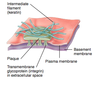
A

Hemi-desmosomes

Focal adhesion

8
Q

Hemi-desmosome?

A

While desmosomes link two cells together, hemidesmosomes attach one cell to the extracellular matrix

9
Q

What are the functions of focal adhesions?

A

Anchor intraellular actin filaments to the basemment membrane

play a role in call movement “migration of epithelial cells of skin”

10
Q

What are the functions of integrins?

A
  1. attach cell to ECM
  2. activate signal transductionpathways from ECM to cell upon ligand binding
  3. immune patrolling
  4. cell migration
  5. binding to cells by certain viruses
11
Q

what are integrins?

A

transmembrane receptors that facilitate cell-extracellular matrix (ECM) adhesion.

12
Q

Which of the following is involved in cell migration?

(Gap junctions, desmosomes, focal adhesions, hemi-desmosomes, integrins)

A

focal adhesions

integrins

13
Q

which of the following in NOT involved in lateral domain cell contact?

(desomosomes, gap junctions, hemi-desmosomes, tight junctions)

A

hemi-desmosomes

14
Q

What is this communication called?

A

autocrine

15
Q

Why are cultured cells different than normal cells?

A

when they come into contact with other cells they stop growing “Contct inhibition

they have a limitd life span “senescence

16
Q

Explain Senescence?

A

loss of a cell’s power of division and growth

17
Q

What are gap junctions made of?

A

channels of Connexon

18
Q

Explain the paracrine communication?

A

cell-cell communication in which a cell produces signals/ chemicals to induce changes in nearby cells

19
Q

Explain the endocrine communication?

A

Communication between endocrine organs, as they are ductless and secrete directly into the blood stream

20
Q

Explain the synaptic communication?

A

Communication between neurons is achieved at synapses by the process of neurotransmission and the release of neurotransmitters

NA - sympathetic

Ach - parasympathetic

21
Q

Explain the neurocrine communication?

What 3 areas can we find them in our body?

A

electrical signaling traveling along the nerve and releases chemicals into the blood stream instead of at a synapse

Examples: hypothalamus, posterior pituitary, adrenal medulla

22
Q

What process involved in cell replacement?

A

mitosis

23
Q

Necrosis vs. apoptosis?

A
24
Q

explain necrosis?

A

Physical distrusting through cell injury,
bacterial toxins, or nutritional deprivation
-> cell swells then bursts -> cytotoxic
cellular components spill out -> damage
and inflammation

25
explain apoptosis?
Bcl-2 protein on outer mitochondrial membrane inhibits apoptosis, deactivating Bcl-2 initiates apoptosis -\> catabolic reactions take place + enzymes digest cytotoxic compounds and DNA fragments-\> **Caspases**: target specific proteins in nuclear lamina and cytoskeleton -\> cell repackaged for safe removal -\> cell shrink and fragments into small apoptotic bodies to be phagocytized
26
What type of cell death causes **1. inflammation**? **2. Caspases activation**?
1. necrosis 2. apoptosis
27
What is meant by **Stable cells**? Where can we find them?
**Stable cells** are cells that multiply only when needed. They spend most of the time in G0 phase fibroblasts, endothelium, SMCs liver, the proximal tubules of the kidney, and endocrine glands
28
What is meant by **Static/ permenant** cells? Where can we find them?
cells that are incapable of regeneration This includes **brain** cells, **neurons**, **heart** cells, **skeletal** muscle cells and **(RBCs?)**
29
What is inherited exclusively through the maternal lineage?
mitochondia
30
What is meant by **Labile** cells? Where can we find them?
cells that multiply constantly throughout life including **hepatocytes**, **skin** cells, **GI** tract, and **blood** cells in the bone marrow
31
which cell type is associted with cancer and why?
**labile/ renewing cells**, bcuz constantly dividing cells have a higher risk of becoming malignant and develop cancer, dividing uncontrollably
32
which cells that are rarely cancerous? why?
**muscle cells**, since they are not constantly dividing cells "static/ permenant"
33
which of the following methods of ell communication involve scretion of hormone? ## Footnote paracrine, endocrine, autocrine, meurocrine, direct contact
endocrine neurocrine
34
where can we find anaerobic prokaryotes in our body?
colon
35
prokaryotes vs. eukaryotes?
36
Explain **Endosymbiosis**?
**_aer_**obic bacteria wa taken into an **_ana_**erobic eukaryote and fomed the **mitochondria**
37
what are the 4 basic tissue types?
epithelial muscle nerve connective tissue
38
what are the 6 specialized connective tissues?
adipose lymphatic blood hemopoietic cartilage bone
39
What is an Epithelium? Function?
A tissue composed of cells that covers the exterior body surface and lines internal closed cavities and body tubes that communicate with the exterior. Epithelium also forms the secretory portion of glands and lines their ducts. specialised epithelium functions as receptors for the special senses (smell, taste, hearing and vision)
40
What are **epithelioid**? Where can we find them?
activated macrophages resembling epithelial cells, but lack an apical surface found in: 1. leyding cells (testis) 2. lutein cells (ovary) 3. islets of langerhans (pancreas) 4. parenchyma (adrenal gland)
41
what structures can we find on th **apical domain** of epithelial cells?
microvilli stereovilli cilia
42
what are **Microvilli**? location?
membrane protrusions that increase the surface area for diffusion (brush borders), found in: intesitines kidney tubules eggs (sperm attachemnt) WBCs (migration)
43
what are **Stereovilli**?
microvilli limited to epididymis and sensory hair cells of ears
44
what are **cilia**?
motile ctoplasmic processes that can bat synchrony with rapid forward movement "effective stroke", and slow return "recovery stroke" found in: tracheobrachial tree oviducts
45
What is a connective tissue?
connective tissue is sparsely populated by cells and contains an extensive extracellular matrix consisting of protein fibers, glycoproteins, and proteoglycans. The function of this type of tissue is to provide structural and mechanical support for other tissues, and to mediate the exchange of nutrients and waste between the circulation and other tissues
46
Give examples of wondering and fixed cells?
Wondering: plasma cells, macrophages, WBCs, mesenchymal cells Fixed: melanocytes, macrophages, mast cells, fibroblasts, adipocytes
47
What are the 3 types of **loose** connective tissue?
Areolar, adipose, reticular
48
What are the **functions** of connective tissue?
1. **Connects** cells to form tissues, connects tissues to form organs and connects organs to form the body. Some tissues provide support as well as connecting (cartilage and bone) 2. **Transportation** - provide a medium for diffusion of nutrients and wastes 3. **Protection** - provide a cushion between tissues and organs and provides insulation (adipose tissue) 4. **Storage** (adipose tissue) 5. **Defense** against infection (blood, lymph, fixed and wandering cells) 6. **Wound healing** (macrophages, fibroblasts, myofibroblasts)
49
What are the 3 main components of connective tissue?
Cells, fibers, ground substance
50
What are the 3 main **fibers** found in connectives tissues?
**Collagen** – Flexible with high tensile strength **Reticular** – Provide a supporting framework/sponge (type III collagen) **Elastin** – Allows tissues to recoil after stretch or distension
51
Describe the **ground substance** in connective tissues? Components?
Ground substance is a viscous, clear substance with a slippery feel. It has a high water content. Composed of **proteoglycans** * A **proteoglycan** is a large macromolecule consisting of a core protein to which glycosaminoglycans are covalently bound * **Glycosaminoglycans** (GAGs) are long-chained polysaccharides. GAGs attract water to form a hydrated gel that permits rapid diffusion but also resists compression.
52
What type of **Glycosaminoglycans** is found in cartilage? Function?
**hyaluronic acid** that is bound to proteoglycans by a link protein to form giant hydrophilic macromolecules. The swelling pressure or turgor that occurs in cartilage ground substance allows it to resist compression without inhibiting flexibility
53
What is the extracellular matrix?
Complex meshwork of proteins and carbohydrates that consists of ground substance and fibres
54
Loose vs Dense connective tissue?
**Loose** – Many cells – Sparse collagen fibres – Abundant ground substance – Viscous, gel-like consistency – Important role in transport (by diffusion) **Dense** – Few cells, nearly all fibroblasts – Many collagen fibres – Little ground substance
55
components of connective tissue
56
Where can we find **loose** connective tissues?
beneath epithelia (to facilitate diffusion) Associated with epithelium of glands around small blood vessels
57
what type of connective tissue is found in the **superficial** layer of the skin?
loose
58
what type of connective tissue is found in submucosa of the colon?
Loose
59
What are the types of **dense** CT?
**Regular** – Collagen fibres are arranged in parallel bundles and are densely packed. Between the bundles are fibroblasts – Designed to withstand stress in a single direction – Seen in tendons, ligaments and aponeuroses **Irregular** – Collagen fibres are arranged in bundles orientated in various directions. Between the bundles are fibroblasts – Designed to withstand stress in multiple directions – Examples are submucosa of intestine and deep layers of dermis
60
Name what the arrows are pointing at?
Rows of elongated flattened fibroblasts lie between the collagen bundles
61
What is the **myotendinous junction** MTJ?
complex specialized region located at the muscle-tendon interface that represents the primary site of force transmission
62
What type of connective tissue is found in the anterior cruciate ligament?
Dense regular
63
label
64
What type of CT is found in the **Dermis**?
Dense irregular
65
gylcosaminoglycans repel water **X**
66
it contain many collagen fibers
67
all exept the last
68
What are **fibroblasts**?
Fibroblasts synthesise and secrete both ground substance and the fibres that lie within the ground substance They are very important in wound healing and are the cells primarily responsible for the formation of scar tissue
69
What are **myofibroblasts**?
modified fibroblasts that contain actin. They are responsible for wound contraction when tissue loss has occurred
70
What do fibroblasts secrete? Region of synthesis?
Procollagen synthesized from the RER, which assembles into collagen fibrils
71
What are **macrophages**? Functions ?
Macrophages are derived from blood monocytes which move into loose connective tissue, especially when there is local inflammation Macrophages are phagocytic and can degrade foreign organisms and cell debris Macrophages are ‘professional antigen presenting cells’ (i.e. they can present foreign material to the T lymphocytes of the immune system)
72
What are **mast** cells? What do they secrete?
Found in connective tissue near blood vessels, but absent from the central nervous system to avoid damaging effects of oedema there Mast cell cytoplasm contains abundant granules These granules contain: • **Histamine** (increases blood vessel wall permeability) • **Heparin** (an anticoagulant) • **Substances** that attract eosinophils and neutrophils
73
Mast cell degranulation induces which type of hypersensitivity?
Type I “ immediate”
74
Explain the process of mast cell degranulation?
Mast cells become coated with Immunoglobulin E (IgE), molecules which specifically bind allergens. When an allergen cross-links these surface-bound IgE molecules, the contents of the granules are all rapidly released from the cell. The secretions of the granules can result in immediate hypersensitivity reactions, allergy and anaphylaxis.
75
What is **collagen**? How many type?
main structural protein in the extracellular space in the various connective tissues 28 types identified
76
What type of collagen is **reticulin**?
Type III collagen
77
What type of collagen is the most abundant in our body?
Type I collagen
78
Which type of collagen that does not form fibers? Where can we find it?
Type II collagen Fibrils form fibres around muscle and nerve cells and within lymphatic tissues and organs. It is called reticulin
79
Which type of collagen found in hyaline and elastic cartilage ?
Type II collagen
80
What type of collagen found fin skin?
Type I collagen
81
Which type of collagen is found in the basement membrane?
Type IV collagen in the basal lamina
82
Explain the collagen fibril production process?
Fibroblasts secrete procollagen that is converted to collagen molecules outside the cell. The collagen molecules are then aggregated to form the final collagen fibrils In some tissues fibrils group together to form collagen fibers Note that Vitamin C is required for the intracellular production of procollagen. Vitamin C deficiency leads to **scurvy** which include poor wound healing and impaired bone formation
83
What is **scurvy**? Main cause?
Vit. C deficiency = not enough collagen = bruises easily * muscle and joint pain * tiredness * the appearance of red dots on the skin * bleeding and swelling of the gums
84
What is **osteogenesis imperfecta**? Main cause?
genetic bone disorder (autosomal dominant) characterized by fragile bones that break easily, and caused by abnormal type I collagen
85
the picture shows dense iregular tissue.. label
86
TEM of macrophage.. label
87
What type of collagen is found in lymph nodes?
Type III
88
Where can we fin elastic fibers?
Dermis Artery walls Lungs
89
What is Marfan’s syndrome? Main cause? Features?
Marfan’s syndrome is an autosomal dominant disorder in which expression of the fibrillin gene is abnormal such that elastic tissue is abnormal. Sufferers are abnormally tall, exhibit arachnodactyly, have frequent joint dislocation and can be at risk of catastrophic aortic rupture.
90
What layer of blood vessels can we find elastic fibers, and collagen?
Tunica media - elastic fibers Tunica adventitia - collagen
91
What cells are responsible of producing elastin, collagen and matrix in the tunica media of the aorta?
Smooth muscle cells
92
What is the main cause of pneumothorax?
Loss of lung elastic recoil
93
What are the 2 types of fat cells?
White and brown
94
Why do adipose tissue show as empty balloons on histology?
because the toluene and xylene, used in tissue preparation, have dissolved away the lipid.
95
Main functions of adipose tissue?
Adipose tissue contains fat, a fuel reserve, but also has a role in thermal insulation and in shock- absorbtion.
96
White vs brown adipose tissue?
* *White** - unicellular * *Brown** - multicellular: each contain many lipid droplets and a central nucleus
97
What is the main function of brown adipose tissue?
These cells are found close to the scapula, sternum and axillae, especially in the newborn. They Are also present in the upper chest and neck of adult The brown colour is due to the rich vascular supply and abundant mitochondria. There is thus a high respiratory capacity for the generation of heat ‘Non-shivering thermogenesis’ is important for babies and for hibernating animals