S4) The Knee Flashcards
What is the knee joint?
The knee joint is a bicondylar type synovial join formed by articulations between the patella, femur and tibia

Describe the two articulations of the knee joint
- Tibiofemoral – medial and lateral condyles of the femur articulate with the tibia
- Patellofemoral – anterior and distal part of the femur articulate with the patella

Describe the arterial supply of the knee joint
Arterial supply via genicular anastomoses around the knee, which arise from the genicular branches of the femoral and popliteal arteries

Describe the innervation of the knee joint
Nerves which cross the knee joint – femoral, tibial and common fibular nerves

Identify and describe the three types of ligaments found in the knee joint
- Patellar ligament – a continuation of the quadriceps femoris tendon distal to the patella
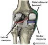
- Collateral ligaments – tibial (medial) collateral ligaments and fibular (lateral) collateral ligaments
- Cruciate ligaments – anterior and posterior cruciate ligaments

Compare and contrast the structure of the medial and lateral collateral ligaments
- Medial collateral ligament is wide and flat
- Lateral collateral ligament is thinner and rounder

State the function of the collateral ligaments

The medial and lateral collateral ligaments act to stabilise the hinge motion of the knee, preventing any medial or lateral movement
State the origin and attachment of the medial collateral ligament
- Origin: medial epicondyle of the femur
- Attachment: medial surface of the tibia

State the origin and attachment of the lateral collateral ligament
- Origin: lateral epicondyle of the femur
- Attachment: depression on the lateral surface of the fibular head

State the respective functions of the ACL and PCL

- ACL: prevents anterior dislocation of the tibia onto the femur
- PCL: prevents posterior dislocation of the tibia onto the femur
State the origin and attachment of the anterior cruciate ligament
- Origin: anterior intercondylar region of the tibia
- Attachment: intercondylar fossa of femur

State the origin and attachment of the posterior cruciate ligament
- Origin: posterior intercondylar region of the tibia
- Attachment: femur in the intercondylar fossa

What are the menisci and what do they do?
The medial and lateral menisci are fibrocartilage structures in the knee that deepen the articular surface of the tibia, stabilising the joint and acting as shock absorbers

Describe the attachments of the menisci

- Both menisci are attached at both ends to the intercondylar area of the tibia
- The medial meniscus is also fixed to the tibial collateral ligament and the joint capsule (less mobile)
Identify the 4 main movements possible at the knee joint and the muscles involved
- Extension: quadriceps femoris muscles
- Flexion: hamstrings, gracilis, sartorius and popliteus
- Lateral rotation: biceps femoris
- Medial rotation: semimembranosus, semitendinosus, gracilis, sartorius and popliteus
What is the patella?
The patella (knee-cap) is a sesamoid type bone located at the front of the knee joint, within the patellofemoral groove of the femur

State the superior and inferior attachments of the patella
- Superior: quadriceps tendon attaches to patellar base
- Inferior: patellar ligament attaches to tibial tuberosity

The patella has a triangular shape, with anterior and posterior surfaces.
Describe its bony landmarks
- The apex is situated inferiorly on the bone
- The base forms the superior aspect of the bone

What does the posterior surface of the patella articulate with?
The posterior surface of the patella articulates with the femur

Describe the features of the posterior surface of the patella
The posterior surface of the patella is marked by two facets:
- Medial facet – articulates with the medial condyle of the femur
- Lateral facet – articulates with the lateral condyle of the femur
Identify and describe the 2 functions of the patella
- Leg extension – enhances the leverage that the quadriceps tendon can exert on the femur
- Protection – protects the anterior aspect of the knee joint from physical trauma
Identify and describe the following bursae related to the knee:

- Suprapatellar bursa: between femur & quadriceps tendon
- Prepatellar bursa: between patella & skin
- Deep infrapatellar bursa: between tibia & patella ligament
- Subcutaneous infrapatellar bursa: between tibial tuberosity & skin
- Popliteal bursa: between popliteus tendon & capsule

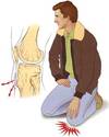
What is the abnormality observed in the image below?

Patellar dislocation

What is a patellar dislocation?
- A patellar dislocation occurs when the patella bone is displaced out of the patellofemoral groove
- Most occur laterally and are caused by high force impact on the patella or forceful sudden twisting of the knee