S3) The Hip Flashcards
What is the hip joint?
The hip joint is a ball and socket synovial joint which connects the lower limb to the pelvic girdle
Describe the articulating surfaces of the hip joint
- Head of femur articulates with the acetabulum of the pelvis
- Acetabular labrum is a fibrocartilaginous collar which deepens the concavity

Describe the arterial supply of the hip joint
Arterial supply via the medial and lateral circumflex femoral arteries which are branches of the profunda femoris artery

What is the clinical significance of the medial circumflex artery?
- Medial circumflex femoral artery is responsible for the majority of the arterial supply (lateral artery has to penetrate through the thick iliofemoral ligament)
- Damage to the medial circumflex femoral artery can result in avascular necrosis of the femoral head
Describe the innervation of the hip joint
The hip joint is innervated by the femoral nerve, obturator nerve, superior gluteal nerve, and nerve to quadratus femoris
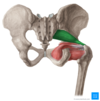
The only intracapsular ligament of the hip is the ligament of the head of the femur.
Describe its structure, function and anatomical location
- Structure: relatively small ligament
- Function: increases stability, encloses a branch of the obturator artery (artery to head of femur)
- Location: runs from the acetabular fossa to the fovea of the femur

Identify the 3 extracapsular ligaments of the hip joint
- Iliofemoral ligament
- Pubofemoral ligament
- Ischiofemoral ligament
Describe the structure, function and location of the iliofemoral ligament
- Structure: ‘Y’ shaped appearance
- Function: prevents hyperextension of the hip joint
- Location: spans between the anterior inferior iliac spine and the intertrochanteric line of the femur

Describe the structure, function and location of the pubofemoral ligament
- Structure: triangular shape
- Function: prevents excessive abduction and extension
- Location: spans between the superior pubic rami and the intertrochanteric line of the femur

Describe the structure, function and anatomical location of the ischiofemoral ligament
- Structure: spiral orientation
- Function: prevents excessive extension
- Location: spans between the body of the ischium and the greater trochanter of the femur

Identify and describe the factors that stabilise the hip joint
- Acetabulum – encompasses nearly all of the femoral head (decreases chance of dislocation)
- Acetabular labrum – increases depth provides a larger articular surface
- Intracapsular & extracapsular ligaments
- Joint capsule
Explain how the muscles and ligaments work in a reciprocal fashion at the hip joint
- Anteriorly, where the ligaments are strongest, the medial flexors are fewer and weaker
- Posteriorly, where the ligaments are weakest, the medial rotators are greater in number and stronger
Identify the possible movements at the hip joint as well as the muscles involved
- Flexion: iliopsoas, rectus femoris, sartorius
- Extension: gluteus maximus, semimembranosus, semitendinosus, biceps femoris
- Abduction: gluteus medius, gluteus minimus and deep gluteals
- Adduction: adductors longus, brevis & magnus, pectineus, gracillis
- Lateral rotation: biceps femoris, gluteus maximus, deep gluteals
- Medial rotation: gluteus medius & minimus, semitendinosus, semimembranosus
Identify and describe the three articulations of the hip bone
- Sacroiliac joint – articulation with sacrum
- Pubic symphysis – articulation with the corresponding hip bone
- Hip joint – articulation with the head of femur

Which three parts compose the hip bone?

How do the bones composing the hip joint vary before and after puberty?
- Prior to puberty, the triradiate cartilage separates these constituents
- After age 15-17 the three parts begin to fuse (forms acetabulum)

The superior part of the hip bone is formed by the ilium, the widest and largest of the three parts.
Describe its structure

- Body: forms the superior part of the acetabulum
- Wing: expansion above the acetabulum
- Iliac fossa: concave inner surface
- Gluteal surface: convex external surface
- Iliac crest: thickened superior margin (extends from ASIS to PSIS)

Which muscles attach to the ilium?
- Gluteal muscles attach to the gluteal surface of the Ilium (at anterior, posterior and inferior gluteal lines)
- Iliacus muscle attaches medially at the iliac fossa
Why is the anterior superior iliac spine clinically relevant?
The ASIS serves as the attachment site of the inguinal ligament, which runs from the ASIS to the pubic tubercle

The most anterior portion of the hip bone is formed by the pubis
Describe its structure

- Body – located medially & articulates with opposite pubic body at the pubic symphysis
- Superior ramus – extends laterally from the body, forming part of the acetabulum (encloses the obturator foramen)
- Inferior ramus – projects towards and joins the ischium (encloses the obturator foramen)

The posterioinferior part of the hip bone is formed by the ischium.
Describe its structure

- Inferior ischial ramus – combines with inferior pubic ramus to form the ischiopubic ramus (encloses part of obturator foramen)
- Ischial tuberosities – found on posterorinferior aspect of the ischium (carries body weight when sitting)
- Greater sciatic notch – found on the posterior aspect of the ischium

Which ligaments attach to the ischium and what do they do?
- Sacrospinous ligament which runs from the ischial spine to the sacrum, forming the greater sciatic foramen
- Sacrotuberous ligament which runs from the sacrum to the ischial tuberosity, forming the lesser sciatic foramen

What is fascia?
Fascia is defined as a sheet or band of fibrous tissue lying deep to the skin that lines, invests and separates structures within the body

The fascia lata is a deep fascial investment of the whole thigh musculature.
Describe its structure and location
- Structure: strong, extensible and elasticated
- Location: begins around the iliac crest and inguinal ligament, ends at the bony prominences of the tibia (becomes crural fascia)

Identify and describe the 3 types of fascia
- Superficial fascia: blends with the reticular layer beneath the dermis
- Deep fascia: envelopes muscles, bones and neurovascular structures
- Visceral fascia: provides membranous investments that suspend organs within their cavities

The width of the fascia lata varies considerably at different regions of the thigh.
Where is it thinnest and where is it thickest?
- Thickest: superolateral aspect of the thigh
- Thinnest: adductor muscles of the medial thigh
Describe the divisions of the fascia lata in the leg
- The deepest aspect of fascia lata gives rise to three intermuscular septa that attach centrally to the femur
- This divides the thigh musculature into three compartments: anterior, medial lateral

Which of the intermuscular septa is strongest?
The lateral intermuscular septum is the strongest of the three due to reinforcement from the iliotibial tract

What is the saphenous opening and what does it do?
- The saphenous opening is an ovoid hiatus which is found in the fascia lata inferior to the inguinal ligament
- It is an entry point for efferent lymphatic vessels and the great saphenous vein, draining into superficial inguinal lymph nodes and the femoral vein respectively

Describe the structure and location of the Iliotibial Tract (ITT)
- Structure: a longitudinal thickening of fascia lata, strengthened posteriorly by fibres from the gluteus maximus
- Location: lies laterally in the thigh, extending from the iliac tubercle to the lateral tibial condyle

Describe the three functions of the Iliotibial Tract (ITT)
- Acts as an extensor, abductor and lateral rotator of the hip
- ITT extends centrally to form the lateral intermuscular septum
- Forms a muscular sheath for the tensor fascia lata
What is the tensor fascia lata?
The tensor fascia lata is a superficial gluteal muscle involved in hip flexion, abduction, internal rotation

State the origin and attachment of the tensor fascia lata?
- Origin: iliac crest (descends down superolateral thigh)
- Attachment: anterior aspect of ITT
When stimulated, the tensor fasciae lata tightens the iliotibial band and braces the knee.
Why is this clinically important?
- When the fascia lata is pulled taut, it forces muscle groups closer together within their intermuscular septa towards the femur
- This centralises muscle weight and limits outward expansion, which reduces the overall force required for movement at the hip joint

What effect does tensing the fascia lata have on veins?
- Makes muscle contraction more efficient in compressing deep veins
- Ensures adequate venous return to the heart from the lower limbs
What are the superior attachments of the fascia lata?
- Posterior: sacrum, coccyx
- Lateral: iliac crest
- Anterior: inguinal ligament, superior pubic rami
- Medial: inferior ischiopubic rami, ischial tuberosity, sacrotuberous ligament
Describe the inferior attachments of the fascia lata
The fascia lata atttaches at bony prominences around the knee joint (femoral & tibial condyles, patella, head of fibula, tibial tuberosity) then becomes the deep fascia of the leg (crural fascia)
Describe the lateral attachments of the fascia lata
The lateral thickening of fascia lata forms the iliotibial tract which descends the lateral thigh and attaches to the lateral tibial condyle
Describe the central attachments of the fascia lata
The deep aspect of fascia lata produces three intermuscular septa which attach centrally to the femur
The fascia lata is also continuous with regions of deep and superficial fascia at its superior aspect.
Explain this.
- The deep iliac fascia from the thoracic region covers the iliopsoas and blends with the fascia lata superiorly
- Superficial fascia from the inferior abdominal wall and perineal region both blend with the fascia lata just below the inguinal ligament
Identify the superficial gluteal muscles
- Gluteus maximus
- Gluteus medius
- Gluteus minimus
- Tensor fascia lata
Describe the structure, function and innervation of the gluteus maximus muscle

- Structure: largest & most superficial of the gluteal muscles
- Function: hip extension, lateral rotation
- Innervation: inferior gluteal nerve

State the origin and attachment of the gluteus maximus muscle

- Origin: gluteal surface of the ilium, sacrum and coccyx
- Attachment: iliotibial tract and the gluteal tuberosity of femur

Describe the structure, function and innervation of the gluteus medius muscle

- Structure: fan-shaped, lies between to gluteus maximus and minimus
- Function: abduction, medial rotation
- Innervation: superior gluteal nerve

State the origin and attachment of the gluteus minimus muscle

- Origin: gluteal surface of the ilium
- Attachment: lateral surface of the greater trochanter

Describe the structure, function and innervation of the gluteus minimus muscle

- Structure: deepest and smallest of the superficial gluteal muscles
- Function: abduction, medial rotation
- Innervation: superior gluteal nerve

State the origin and attachment of the gluteus minimus muscle

- Origin: ilium
- Attachment: anterior side of the greater trochanter

Describe the structure, function and innervation of the tensor fascia lata muscle

- Structure: small superficial muscle, lies towards anterior edge of the iliac crest
- Function: abduction, medial rotation (tightens fascia lata)
- Innervation: superior gluteal nerve

State the origin and attachment of the tensor fascia lata muscle

- Origin: anterior iliac crest & ASIS
- Attachment: ITT (lateral condyle of tibia indirectly)

Identify the deep gluteal muscles
- Piriformis
- Obturator internus
- Superior & inferior gemelli
- Quadrator femoris
Describe the structure, function and innervation of the piriformis muscle

- Structure: most superior of the deep muscles
- Function: lateral rotation, abduction
- Innervation: nerve to piriformis

State the origin and attachment of the piriformis muscle

- Origin: anterior surface of the sacrum (travels through greater sciatic foramen)
- Attachment: greater trochanter of the femur

Describe the structure, function and innervation of the obturator internus muscle

- Structure: forms the lateral walls of the pelvic cavity
- Function: lateral rotation, abduction
- Innervation: nerve to obturator internus

State the origin and attachment of the obturator internus muscle

- Origin: pubis and ischium at the obturator foramen (travels through lesser sciatic foramen)
- Attachment: greater trochanter of the femur

Describe the structure, function and innervation of the gemelli muscles

- Structure: superior and inferior gemelli are two narrow triangular muscles, separated by the obturator internus tendon
- Function: lateral rotation, abduction
- Innervation: superior – nerve to obturator internus, inferior – nerve to quadratus femoris

State the origin and attachment of the gemelli muscles

- Origin: superior – ischial spine, inferior – ischial tuberosity
- Attachment: greater trochanter of the femur

Describe the structure, function and innervation of the quadrator femoris muscle

- Structure: flat, square-shaped muscle, most inferior of the deep gluteal muscles
- Function: lateral rotation
- Innervation: nerve to quadratus femoris

Describe the arterial supply of the gluteal region
Arterial supply via superior and inferior gluteal arteries (arise from internal iliac artery) which enter the gluteal region via the greater sciatic foramen

Describe the venous drainage of the gluteal region
Venous drainage via inferior and superior gluteal veins (empty into the internal iliac vein)
State the origin and attachment of the quadrator femoris muscle

- Origin: lateral side of the ischial tuberosity
- Attachment: quadrate tuberosity on the intertrochanteric crest

What is osteoarthritis?
- Osteoarthritis is a non-inflammatory chronic disease of the synovial joints in the musculoskeletal system (no systemic involvement)
- No joint ankylosis is observed in the course of the disease

Compare and contrast the aetiology of primary and secondary osteoarthritis
- Primary OA: aetiology is unknown
- Secondary OA: trauma, infection (septic arthritis), inflammation (RA), metabolic (gout), endocrine (DM)
Describe how articular cartilage changes in osteoarthritis
- Increased tissue swelling
- Cartilage fibrillation
- Cartilage erosion down to subchondral bone

What are the risk factors for arthritis?
- Obesity
- Past injury in a joint
- Occupational factors
- Genetics
- Age
What are the signs and symptoms of arthritis?
- Joint pain
- Crepitis (grinding)
- Joint deformity
- Osteophytes
- Joint stiffness
Outline the investigation of osteoarthritis in terms of bloods, imaging and radiological findings.
- Bloods – FBC, U&Es , LFTs, CRP
- Imaging – look for subchondrial sclerosis, osteophytes, narrowing of joint space, subchondrial cysts
What is a pelvic bone fracture?
- Pelvic bone fractures are fractures which result from direct trauma to the pelvic bones or indirectly through forces transmitted from the lower limb
- Fractures often occur at the weaker points of the bones (pubic rami, acetabulum, region of sacroiliac joint)

A common complication of pelvic fractures is soft tissue injury.
Which structures are at high risk?
- Bladder
- Urethra