S3) Somatosensory Pathways/System Flashcards
What are the two types of sensation?
- General sensation, referring to the body wall and viscera (including parietal layer of serous membranes and mucosa of pharynx, nasal cavity and anus)
- Special sensation, referring to the special senses of vision, hearing, balance, taste and smell
What are the two types of general sensation?
- Somatic sensation (conscious)
- Visceral sensation (unconscious)
What are sensory modalities?
Sensory modalities are different forms of sensory experience/ somatic sensation e.g. pain, temperature which exists due to various types of receptors
A modality can be thought of as a ‘unit’ of sensation, relying on a distinct receptor type
Besides pain and temperature, identify five other sensory modalities
- Pressure (crude touch)
- Vibration
- Distension
- Proprioception (kinesthetic sense)
- Fine touch
- 2 point discrimination
Different modalities travel along different trajectories via the nervous system on their way up to the brain.
What are the two different systems?
- Spinothalamic system
- Dorsal column-medial lemniscus system

What modalities are carried by the spinothalamic system?
- Temperature (thermoreceptors)
- Pain (nociceptors)
- Pressure/ crude touch (mechanoreceptors)

What modalities are carried by the dorsal column-medial lemniscus system?
- Vibration (mechanoreceptors)
- Proprioception, or joint position sense, or kinaesthetic sense (detected by a variety of receptors such as muscle spindles and Golgi tendon organs)
- Fine touch (mechanoreceptors)
- Two point discrimination (mechanoreceptors)

What is two point discrimination?
It is the ability to resolve/ discriminate between two simultaneous stimuli
What are primary sensory neurones aka?
aka dorsal root ganglion neurones or primary afferents or first order sensory neurones or psueudunipolar (one process emanating from the body) neurones

Where are primary sensory neurones found?
The cell body of primary sensory neurones lies in the dorsal root ganglion.
They receive information from receptors from the skin/ elsewhere and are responsible for the inital encoding of sensory information.
And the axon transmits information to the dorsal horn of the spinal cord where the neurone synapses

Describe the relationship between the cell body and axon for primary sensory neurones
For primary sensory neurones, the axon runs ipsilaterally to the cell body

Identify 3 main features of the primary sensory neurone.
- Each individual primary neurone receives input from a single receptor type
- Primary sensory neurones have their cell body in the dorsal root ganglion, and collect information from a single dermatome along their peripheral axon
- Primary sensory neurones project into the spinal cord along their central axon
The strength of receptor activation is converted from one type of signal to another. What are they?
converted from an analogue signal (related to ion flux during the generator potential) to a digital signal (which is the frequency of action potentials in the primary sensory neurone)
What is the relationship between the strength of the receptor activation and the frequency of the action potentials?
Strong receptor activation causes high frequency of action potentials in the primary sensory neurone
Weak receptor activation causes a low frequency of action potentials in the primary sensory neurone

We have two types of primary sensory neurones.
What are they?
- Rapidly adapting receptors
- Slowly adapting receptors
What are rapidly adapting receptors?
(e.g. mechanoreceptors)
respond best to changes in strength of stimulation.
However, their frequency of firing diminishes rapidly after the initial stimulus (i.e. they rapidly adapt).
Adaptation of these receptors explains why you are not aware of your clothes on your skin
Identify an example of rapidly adapting receptors.
e.g. mechanoreceptors on your bottom - initially aware that you are sitting due to pressure when sat down
But eventually, no longer aware when sat down for long
What are slowly adapting receptors?
e.g. nociceptors
change their frequency of firing very little after the initial stimulus.
This explains why pain can be so persistent, and you never really get ‘used to’ having pain
What are receptive fields?
A single primary sensory neurone supplies a given area of skin (it’s receptive field)
What is the relationship between the size of the receptive fields and the sensory acuity?
- If an area of skin is supplied by sensory neurones with relatively large receptive fields, this area will have low sensory acuity (it would have poor two-point discrimination where two points would need to be far apart to be distinguished). e.g. The skin of the back has relatively low acuity
- If an area of skin is supplied by sensory neurones with relatively small receptive fields, this area will have high sensory acuity (it would have great two-point discrimination where two points could be very close together to be distinguished). e.g. The skin of the fingertip has relatively high acuity
- Acuity ∝ 1/size of receptive field

Why are dermatomes said to have ‘fuzzy’ boundaries?
The overlap of receptive fields of primary sensory neurones from adjacent dermatomes is one of the reasons why dermatomes can have ‘fuzzy’ boundaries
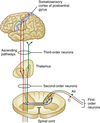
The somatosensory system conveys a system of 3 neurones.
It carries conscious sensation from the body wall e.g. skin, lining of pharynx, parietal pleura/ peritoneum
What are the 3 neurones?
- First order sensory neurones
- Secondary order sensory neurones
- Third order neurones (thalamocortical neurones)
Describe the three types of neurones in the somatosensory system in terms of location of their cell bodies, where they project into and their path/ communicate with.
– First order sensory neurones
- Have their cell bodies in the DRG
- Communicate with a receptor
- Their central axon projects ipsilateral to the cell body
- Project onto second order neurones
– Second order sensory neurones
- Have their cell bodies in the spinal cord dorsal horn or medulla
- Decussate
- Project onto third order neurones
– Third order neurones
- Have their cell bodies in the thalamus
- Project to the primary sensory cortex (postcentral gyrus)

Describe the topographical representation in the sensory system
- Principle that every point on the surface of the body is equivalent to an area along the sensory pathway w some exceptions: adjacent body regions map to adjacent regions of the sensory system e.g. sensory cortex, the hand is represented adjacent to the wrist.
- This way of organising the pathways → minimises the amount of ‘wiring’ needed to transmit sensory information
- The motor system has similar organisation but running in reverse
- Info becomes re-organised as we move upwards through the neuraxis such that as the level of spinal nerves and spinal cord, we have a dermatomal organisation but at the levels of the thalamus and above, we have a homuncular pattern
- At the levels of the sensory homunculus, all modalities converge (i.e. the head area of the sensory cortex deals with pain, temperature, vibration etc. all at the same time)

What is the purpose of the dorsal column-medial lemniscus system (DCML)?
- Responsible for carrying impulses concerning modalities such as light touch, vibration, two point discrimination and proprioception
Observe this image and understand. - The dorsal column-medial lemniscus system

We have three neurones involved in the dorsal column-medial leminscus pathway.
What are they and briefly describe them.
- Concerning first order neurones of the DCML system:
→ Axons of first order neurones ascend ipsilaterally through the dorsal columns of the spinal cord
→ Those from the lower body (T7 and below) ascend through the gracile fasciculus to the gracile nucleus in the medulla)
→ Those from the upper half of the body (T6 and above) ascend through the cuneate fasciculus to the cuneate nucleus in the medulla
- Concerning second order neurones of the DCML system:
→ Neurones in the gracile nucleus project to the contralateral thalamus in the medial lemniscus
→ Neurones in the cuneate nucleus project to the contralateral thalamus in the medial lemniscus
- Concerning third order neurones of the DCML system:
→ Thalamic neurones receiving information ultimately from the lower half of the body (via gracile nucleus) project to the medial part of the primary sensory cortex
→ Thalamic neurones receiving information ultimately from the upper half of the body (via cuneate nucleus) project to the lateral part of the primary sensory cortex

Just for further clarification, what is the dorsal column pathway for the lower half of the body i.e. T7 and below?
First order neurone → ascends through the gracile fasciculus to the gracile nucleus in the medulla.
Then second order neurones in the gracile nucleus project to the contralateral thalamus in the medial lemniscus
Then the third order neurone/ thalamic neurones receive info ultimately from the lower ½ of the body via (gracile nucleus) project to the medial part of the primary sensory cortex.
Just for further clarification, what is the dorsal column pathway for the upper half of the body i.e. T6 and above?
First order neurone → ascends through the cuneate fasciculus to the cuneate nucleus in the medulla.
Then second order neurones in the cuneate nucleus project to the contralateral thalamus in the medial lemniscus
Then the third order neurone/ thalamic neurones receive info ultimately from the upper ½ of the body via (cuneate nucleus) project to the lateral part of the primary sensory cortex.
What is the topographical organisation of the dorsal columns?
- Axons from the lower parts of the body run most medially
- Axons from progressively superior body segments are added laterally to the dorsal columns
- i.e.CTL (from lateral to medial)

What is the purpose of the spinothalamic pathway or antero-lateral system or spinothalamic tract (STT).
Responsible for carrying impulses concerning modalities e.g pain, temperature and crude touch
Observe this image and understand. - The spinothalamic system

We have three neurones involved in the spinothalamic pathway.
What are they and briefly describe them.
Overall: Axons of first order neurones project to the ipsilateral dorsal cord, but the spinothalamic tract supplies the contralateral half of the body
– Concerning first order neurones of the STT:
- They project onto second order neurones in the ipsilateral spinal cord dorsal horn in the segment at which they enter the cord through the dorsal root (generally)
– Concerning second order neurones of the STT:
- Their cell bodies are in the dorsal horn
- Their axons decussate in the ventral white commissure of the cord and then go on to form the spinothalamic tract
- The spinothalamic tract projects to the thalamus
– Concerning third order neurones of the STT:
- Thalamic neurones receiving information ultimately from more inferior parts of the body project to the medial part of the primary sensory cortex
- Thalamic neurones receiving information ultimately from more superior parts of the body project to the lateral part of the primary sensory cortex

What is the topographical organisation of the spinothalamic tract?
- Axons from the lower parts of the body run most laterally/superficially
- Axons from progressively superior body segments are added medially/deeper onto the spinothalamic tract
- This is the opposite of the situation for the dorsal columns, and is due to the decussation of the STT second order neurones at the level of entry of the first order neurones
- i.e. LTC (from lateral to medial)

In terms of lesions, if there was damage to DCLM vs STT, which side of body affected?
Damage to DCLM → Ipsilateral sensory loss below level of lesion .: IPSILATERAL FEATURES
Damage to STT → Contralateral sensory loss below level of lesion due to decussation of the second order neurones .: CONTRALATERAL FEATURES
What is the overall somatotopic organisation of spinal cord tracts?

What is Lissauer’s tract? (aka Fasciculus proprius)
It enables axons of 1st order neurones to ascend a couple of segments before synapsing onto a second order neurone within the spinal cord/ grey matter.

What is the concept of the Lissauer’s tract? What is its clinical relevance?
- 1st order sensory neurones of the STT have a no. of options - some synapse immediately at the level of the dorsal root + some of them ascend in Lissauer’s tract to synapse higher up
- Clinical relevance: It allows us to bypass certain types of spinal cord lesions because we can ascend up a couple of segments in Lissaeur’s tract to synapse with a second order neurone.: we typically see that the sensory level for STT modalities is a couple of segments lower down than what we see for the sensory level for dorsal column modalities. (allows the ability to bypass hemisections of the spinal cord) – AKA Brown-Sequard syndrome.

In Brown-Sequard syndrome (limited to sensory features), if we considered a complete cord hemisection causing destruction of one lateral half of the spinal cord segment resulting from trauma/ ischaemia, what structures will be completely destroyed unilaterally?
- The dorsal horn
- The ventral horn
- All other cord grey matter
- All white matter pathways
- Dorsal and ventral roots

In Brown-Sequard syndrome (limited to sensory features), if we do have a complete cord hemisection (destruction of one lateral half of a spinal cord segment), what signs will you see?
Destruction of the dorsal root and dorsal horn → Ipsilateral complete segmental anaesthesia affecting a single dermatome
Contralateral loss of spinothalamic modalities at and below the destroyed segment (although level can be up to a couple of segments lower due to ascent of some primary afferents in Lissauer’s tract –advanced, so only pursue if you are keen!)

Which fibres are involved in the descending modulation of pain?

- C fibres project into the dorsal horn of the spinal cord - they carry the pain impulses from the nociceptors
- A fibres (A-beta fibres) carry impulses from the mechanoreceptors in the skin (e.g. rubbing the site of pain) and they stimulate mainly the encephalingeric inhibitory interneurones which inhibits the 2nd order neurone in the pain pathway .: switching off pain transmission.
Second order neurones of the spinothalamic system dealing with pain receive nociceptive primary afferents as well as inhibitory interneurones which contain the endorphin encephalin
These encephalinergic interneurones can be activated by incoming impulses from mechanoreceptors (hence explaining why rubbing a sore area relieves the pain)

How else can the encephalingeric interneurones be activated?

Activated by descending inputs from higher centres such as the periaqueductal grey matter or the nucleus raphe magnus
e.g. Hypnosis - They activate cortical neurones known to project down into the midbrain. These are excitatory neurones .: they stimulate a 2nd order neurone which sits in the Peri-aqueductal grey matter of the midbrain. Then these neurones will project down into the medulla where they stimulate neurones in the Reticular formation of the medulla called Nucleus Raphe magnus.
These neurones descends down the cord and inhibits the 2nd order sensory neurone in the STT by stimulating the inhibitory interneurone.

The modality of the stimulus is dependent on the type of receptor activated.
What are the different types of receptors?
- Nociceptor
- Mechanoreceptors
- Thermoreceptors
Different types of receptors adapt in different ways.
What is a tonic receptor?
Tonic receptors are slow adapting receptors, respond to the stimulus as long as it persists, and produce a continuous high frequency of action potentials e.g. all nociceptors

Different types of receptors adapt in different ways.
What is a phasic receptor?
- Phasic receptors are rapidly adapting receptors and respond quickly to stimuli but stop responding upon continual stimulation
- Action potential frequency decreases during prolonged stimulation and the receptor remains sensitive to a change/removal of stimulus

What is a nociceptor?
Nociceptors are tonic receptors, which respond to noxious stimuli (stimuli that would cause tissue injury if they were to persist) and result in the sensation of pain
What is a mechanoreceptor?
A mechanoreceptor is a sensory receptor that responds to mechanical pressure or distortion by the means of pressure, touch, vibration or stretch
What is a thermoreceptor?
Thermoreceptors are tonic receptors that respond to warmth and cold
What is a dermatome?
A dermatome is an area of skin supplied by a single spinal nerve

Where are second order sensory neurones found?
The cell body of second order sensory neurones lies in the dorsal horn of the spinal cord

Where are third order sensory neurones found?
The cell body of third order sensory neurones is located in the thalamus and the axon extends into the somatosensory cortex
Where are the majority of the ascending tracts found?
The destination of the majority of the ascending tracts is the somatosensory cortex in the postcentral gyrus of the parietal lobe
What is the sensory homunculus?
The somatosensory cortex corresponds to the sensory homunculus, which is a map of brain areas dedicated to sensory processing for different anatomical divisions of the body

Explain how CNS lesions can show varied patterns
- Dermatomal pattern of sensory loss suggests a lesion at the level of the spinal nerve
- Homuncular pattern of sensory loss suggests a lesion at the level of the cortex
Describe the somatotropic organisation of the spinal cord tracts
- In the dorsal column pathway, the lower body maps to the medial portion of the tract
- In the spinothalamic tract, the lower body maps to the lateral portion of the tract

What is the significance of the varying somatotropic organisation of the spinal cord tracts?
A central cord lesion can affect dorsal column and spinothalamic tracts differentially
Different ascending tracts are found in specific locations in the spinal cord, and each of the ascending pathways carry information about specific sensory modalities.
Identify two ascending tracts of interest and their location
- Dorsal column-medial lemniscus (DCML)
- Spinothalamic tract

Information for which sensory modalities is relayed by the DCML?

The dorsal column-medial lemniscus relays information relating to conscious proprioception of the limbs, fine touch and vibration

How do isolated lesions of the DC pathway present?
Isolated lesions of DC pathway in the cord lead to ipsilateral signs below the lesion
Describe the significance of the following in the dorsal column pathway:
- Gracile/cuneate nucleus
- Medial leminiscus
- The cell bodies lie in the gracile/cuneate nucleus
- The fibres decussate to the contralateral side at the great sensory decussation in the medulla oblongata, and then ascend via the medial leminiscus and terminate at the thalamus

Describe and illustrate the organisation of the homunculus in the dorsal column pathway
In the sensory and motor homunculus, the lower limb is represented medially and the upper limb and face are represented laterally

Information for which sensory modalities is relayed by the spinothalamic tracts?

The spinothalamic tract relays information relating to pain, temperature, and pressure

How do isolated lesions of spinothalamic tract present?
Isolated lesions of spinothalamic tract in cord lead to contralateral signs below the lesion
What is the ventral white commissure and what is its significance in the spinothalamic pathway?
The axons of the tract cells decussate to the other side of the spinal cord via the anterior/ventral white commissure and to the anterolateral corner of the spinal cord

Illustrate the ascension of axons in the spinothalamic pathway

Describe and illustrate the organisation of the homunculus in the spinothalamic pathway
In the sensory and motor homunculus, the lower limb is represented laterally and the upper limb and face are represented medially

What is Brown-Sequard syndrome?
- Brown-Sequard syndrome is the clinical syndrome that emerges when one destroys one half of the spinal cord
- It demonstrates the rare occurrence of a hemisection (only right or left is severed) of the spinal cord
In Brown-Sequard syndrome, consider a complete cord hemisection causing destruction of a single cord segment resulting from trauma or ischaemia and identify the five structures that will be completely destroyed unilaterally
- The dorsal horn
- The ventral horn
- All other cord grey matter
- All white matter pathways
- Dorsal and ventral roots

How does Brown-Sequard syndrome present?
- Ipsilateral complete segmental anaesthesia affecting a single dermatome (due to destruction of dorsal root and dorsal horn)
- Ipsilateral loss of dorsal column modalities below the lesion
- Contralateral loss of spinothalamic modalities below the lesion

Describe the respective functions of A fibres and C fibres in analgesia
- A fibres carry impulses from mechanoreceptors in the skin
- C fibres carry pain

Explain how the activation of mechanoreceptors alleviates pain
Activation of mechanoreceptors alleviates pain (i.e. rubbing a painful area helps) by exciting inhibitory enkephalinergic interneurones in the cord

