S1) Topography of the Nervous System Flashcards
What are the four basic components of the central nervous system?
- Cerebral hemispheres
- Brainstem
- Cerebellum
- Spinal cord
What is the function of cerebral hemispheres?
Higher functions
motor and sensory (conscious)
emotion
memory
What is the function of brainstem and cerebellum?
Communication via cranial nerves including functions such as eye movement, swallowing and cardiorespiratory homeostasis
Cerebellum involved with motor sequencing and co-ordination
What is the function of spinal cord?
Ascending (sensory) and descending (motor) pathways
Spinal reflex arcs
Control of upper and lower limbs at level of cervical and lumbosacral enlargements
What are the four basic components of the peripheral nervous system?
- Dorsal and ventral roots
- Spinal nerves
- Peripheral nerves
- Ganglia
Distinguish between the composition of grey matter and white matter

- Grey matter is composed of cell bodies and dendrites (it is highly vascular due to high metabolic activity) There is axons too but volume is predominantly composed of cell bodies + dendrites.
- White matter is composed of (unmyelinated + myelinated) axons (+ supporting cells) with no cell bodies.

Why does grey matter contain axon terminals?
Grey matter contains axons to allow communication with white matter
Why is white matter white?
White matter is white due to the presence of fatty myelin (a lipid emulsion like mayonnaise)
In the peripheral nervous system, identify the equivalent structures of the following:
- Grey matter
- White matter
- The PNS equivalent of grey matter is a ganglion (collection of cell bodies)
- The PNS equivalent of white matter is a peripheral nerve (or root)
What is a function of the white matter pathways?
- connects areas of grey matter like cables between components of a computer
How many segments does the spinal cord consist of?
The spinal cord is composed of 31 segments , each supplying a given dermatome and myotome on each side

Describe the structure of a spinal cord segment

- Central core of grey matter
- Outer shell of white matter
Each segment connects with a mixed spinal nerve through dorsal sensory roots and ventral motor roots
(.: knowledge of dermatomal and myotomal supply allows localisation of lesions to a given cord segment/s)

A sensory deficit across multiple segments may suggest …
Spinal cord lesion
A sensory deficit in a homuncular pattern may suggest. ….
A lesion above the thalamus
Identify three components of white matter
- Funiculus
- Tract
- Fasciculus
What is a funiculus?
- A funiculus is a segment of white matter containing multiple distinct tracts
- Impulses travel in multiple directions - ascending and descending pathways
Some examples: the dorsal funiculus contains the dorsal column tract (ascending), the lateral funiculus contains the lateral corticospinal tract (descending) and spinothalamic tract (ascending) and the ventral funiculus contains the ventral corticospinal tract (descending)

What is a tract?
- A tract is an anatomically and functionally defined white matter pathway connecting two distinct regions of grey matter
- Impulses travel in one direction
Examples include: spinothalamic tract (connecting spinal cord dorsal horn to thalamus), corticospinal tract (connecting cerebral cortex to spinal cord ventral horn)

What is a fasciculus?
A fasciculus is a subdivision of a tract supplying a distinct region of the body
Examples include: gracile fasciculus (subdivision of dorsal column tract supplying lower half of body) and cuneate fasciculus (subdivision of dorsal column tract supplying upper half of body, excluding the head)

Give an example of funiculus and the corresponding constituents.
Doral funiculus (ascending) → contain dorsal column tracts → splits into 2 fasiculi: 1. Fasciulus cuneatus (supplies upper ½ of body) 2. Fasciulus gracilis (supplies lower ½ of body)

Summary slide: look at white matter.

Identify the three different regions of grey matter

The spinal cord 3- Grey matter slide.
The cell bodies of the grey matter in the cord are organised into cell columns (N.B these are not pathways, but columnar shaped collections of neuronal cell bodies e.g. ventral horn is not a 2D slice of a sausage, but it is a 3D sausage)
These regions of grey matter were organised by Rexed into laminae

What is a nucleus?
A nucleus is a collection of functionally related cell bodies (grey matter)
e.g. thalamus is a nucleus, containing the cell bodies of third order sensory neurones.

What is a cortex?
A cortex is a folded sheet of cell bodies found on the surface of a brain structure (grey matter)
(refers to outer shell of grey mater found on cerebral hemispheres and cerebellum. (Brain: the same basic pattern as the cord i.e. central grey with peripheral white but with an extra layer of grey on the outside)
What is a fibre?
A fibre is an axon in association with its supporting cells e.g. oligodendrocytes (synonymous with axon) (white matter)
What are the three types of fibres found in the nervous system?
- Association fibres
- Commissural fibres
- Projection fibres
What do association fibres do?
Association fibres connect cortical regions within the same hemisphere e.g. arcuate fasciculus or U fibres connecting adjacent gyri

What do commisural fibres do?
Commissural fibres connect left and right hemispheres or cord halves e.g corpus callosum (connects both hemispheres) or the ventral white commissure of the cord.

What do projection fibres do?
Projection fibres connect the cerebral hemispheres with the cord/brainstem and vice versa
ie. they are longitudinally arranged e.g. corticospinal fibres connecting motor cortex to spinal cord ventral horn or spinothalamic fibres.

What are the three components of the brainstem?
- Midbrain
- Pons
- Medulla

What is the function of the midbrain (mesencephalon)?
The midbrain regulates eye movements and reflex responses to sound and vision (visual/ auditory stimuli)

Label these structures in the midbrain:
Substantia niagra
Cerebral peduncle/ Crus Cerebri
Red nucleus
Fibres of CNIII
Edinger-Westphal nucleus
Oculomotor nucleus
Peri-aqueductal grey matter
Cerebral aqueduct
Superior colliculus
Medial leminiscus
Spinothalamic fibres

Note: to make it easier, rotate the picture to see a Mickey mouse view! Then label structures

What are the cerebral peduncles?
Part of the midbrain
white matter, contains descending corticospinal fibres from the ipsilateral hemisphere.
What is substantia niagra?
Part of the midbrain
grey matter, contains dopaminergic neurones that project to the striatum (nigrostriatal fibres)
What is red nucleus?
Part of the midbrain
Grey matter, well distinct region that gives rise to axons that travel to the cord in the vestigial rubrospinal tract
Also has some other less important motor functions.
What is the occulomotor nucleus?
Part of the midbrain
Grey matter, contains lower motor neurone cell bodies that project through the oculomotor nerve to all bar two of the extra-ocular muscles.
What is the Edinger-Westphal nucleus?
Part of the midbrain
Grey matter, contains parasympathethic preganglionic neurones that project to the ciliary ganglion in the orbit to cause pupillary constriction.
What is peri-aqueductal grey matter?
Part of the midbrain
an area surrounding the cerebral aqueduct that has roles in pain transmission and micturition.
What is cerebral aqueduct?
Part of the midbrain
Connects the 3rd ventricle (found between the halves of the thalamus) and the further ventricle (found beneath the cerebellum) inv. in passageway of drainage of CSF.
What is the medial lemniscus?
Part of the midbrain
Connects gracile/ cuneate nucleus to the thalamus
What is spinothalamic tract?
Part of the midbrain
Connects spinal dorsal horn to thalamus
What is the superior/ inferior colliculus?
Part of the midbrain
Grey matter, which regulate reflex responses to visual and auditory stimuli respectively
i.e. superior colliculus → visual stimuli inferior colliculus → auditory stimuli
Which processes are regulated by the pons?
- Feeding (circuits involving the trigeminal nerve)
- Sleep

What are the key features of the Pons?
- Trigeminal nerve exits from its lateral aspect
- Corticospinal fibres travel ventrally (hence susceptible to damage by basilar artery occlusion causing locked in syndrome)
- Sits beneath the fourth ventricle so can get compressed if this ventricle expands
- Contains reticular formation (grey matter) regions important for sleep.
Which key centres are found in the medulla?
- Cardiovascular (BP) and respiratory centres (pH, O2)
- Major motor pathway (medullary pyramids)

What are medullary pyramids?

white matter, ventral swellings on each side containing corticospinal fibres from ipsilateral hemisphere.
These decussate in the caudal medulla at the decussation of the pyramids

What are gracile and cuneate nuclei?
Part of medulla of brainstem
grey matter, relays onto second order neurones in the dorsal column-medial lemniscus pathway

What are the solitary nucleus and dorsal motor nucleus of the vagus?
Part of the medulla of the brainstem
Important nuclei (grey matter) for cardiorespiratory homeostasis
Label the structures


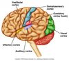
What is the gyrus pl. gyri?

A raised fold of cerebral cortex
What is a sulcus pl. sulci?
valley between the adjacent gyri
Where is the central sulcus found?

Central sulcus is a key landmark separating the frontal and parietal lobes (coronal plane). It seperates the precentral (motor, anterior) and postcentral (sensory, posterior) gyri.
It is a continuous sulcus running from the temporal lobe up to the midline.

Which cortices are found in the following locations:
- Precentral gyrus
- Postcentral gyrus
- Precentral gyrus: contains primary motor cortex
- Postcentral gyrus: contains primary sensory cortex

Where is the lateral/ Sylvian fissure found?

The lateral fissure is a key landmark separating the temporal lobe from the frontal and parietal lobes
Opening the lateral fissure allows us to see deep insular cortex - which sits superficial to the putamen

Where is the parieto-occipital sulcus found?

Parieto-occipital sulcus is a key landmark separating the parietal lobe from the occipital lobe
Only visible on the medial aspect of the hemisphere

Where is the calcarine sulcus found?

The calcarine sulcus is a key landmark surrounded by the primary visual cortex
Visual cortex above the calcarine sulcus supplies the (contralateral) inferior field
Visual cortex below the calcarine sulcus supplies the (contralateral) superior visual field

Overall summary slide: Key features of the brain- gyri and sulci

What is the optic chiasm?
The optic chiasm is a site where fibres in the visual system cross over
It is the site at which fibres from the nasal retinae i.e lateral fields decussate

What is the uncus?
- The uncus is the most medial part of the temporal lobe that can herniate below the tentorium cerebeli, compressing the adjacent midbrain (uncal herniation)
- It has an important olfactory role- due to it being the site of primary olfactory cortex.

What are the medullary pyramids?
The medullary pyramids are a location of descending motor fibres

What is the parahippocampal gyrus?
The parahippocampal gyrus is a part of the medial temporal lobe that provides input to the underlying hippocampus.
It is a key cortical region for memory encoding

Overall summary slide: Key features of the brain - inferior aspect

What is the corpus callosum?
The corpus callosum consists of fibres connecting the two cerebral hemispheres

What is the thalamus?
The thalamus is a sensory relay station projecting to the sensory cortex.
The thalamus is an important gateway for conscious sensation. It contains the cell bodies of third order neurones and projects to the primary sensory cortex in a homuncular pattern

What is the cingulate gyrus?
The cingulate gyrus is a cortical area important for emotion and memory
(participates in an important circuit involving the hippocampus and thalamus - Papez circuit)

What is the hypothalamus?
The hypothalamus is an essential centre for homeostasis

What is the fornix?
The fornix is a major key output pathway from the hippocampus and is part of the Papez circuit.
(damage here → amnesia)

What is the tectum?
The tectum is the dorsal/ posterior part of the midbrain involved in involuntary responses to auditory and visual stimuli
Contains 2 swellings: Superior and inferior colliculi (4 in total, forming corpora quadrigemina)
Superior colliculi → Visual reflex
Inferior colliculi → Auditory reflex

What is the cerebellar tonsil?
The cerebellar tonsil is a the inferior part of the cerebellum that can herniate down the foramen magnum in cases of raised intracranial pressure and compress the medulla (often leading to death due to cardiorespiratory compromise)
(tonsillar herniation → can herniate into foramen magnum and compress the medulla .: compromise CVS and Resp centres)
Overall summary slide: Key features of the brain - midline

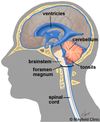
Having developed from a hollow tube, the brain is itself hollow.
What are the cavities in the brain called?
The cavities in the brain are called ventricles
Describe the layout of the ventricular system

What is found inside the brain ventricles?
The ventricles each contain choroid plexus, which makes a total of 600-700ml of cerebrospinal fluid per day

What types of functions does CSF have?
CSF has both metabolic (contains many messenger molecules) and mechanical functions (supporting brain + buoyant within CSF)
What produces CSF?
Choroid plexus in ventricles
What is the function of CSF?
CSF has both metabolic and mechanical functions
– Contains glucose and maybe even hormones
– Shock absorbs the brain and renders it effectively weightless
- Supports weight of brain and spinal cord
- Has nutrition - high levels of glucose → nourishes cells on surface of brain and spinal cord
- Constant turn over of immune cells in CSF
Out of all the ventricles, which one is the biggest and what is the significance of this?
(Large) Lateral ventricles - therefore produces largest amount of CSF
How are the 2 lateral ventricles connected (also where CSF will then accumulate to be drained)?
Interventricular foramen
Why does the 3rd ventricle have a flattened, squashed like appearance?
Due to thalamus on each side squashing it flat in the midline
Describe the drainage system of CSF.
- Lateral ventricles drain CSF at common point - interventricular foramen (where both lateral ventricles meet)
- Then CSF drained into 3rd ventricle
- Then directly drained into aqueduct of Silvius/ midbrain
- Then drained into 4th ventricle
- CSF escapes ventricular system in 2 ways – majority: via 3 apertures in the 4th ventricles: 2 lateral recesses/ apertures and 1 midline aperture. – minority: via the central canal of spinal cord (negligible drainage via the spinal cord central canal)
- These apertures are direct holes in the brain .: permit CSF to drain from the ventricular system into the subarachnoid space.
- Once in the subarachnoid space, CSF percolates around the superficial surfaces of the brain and spinal cord before being reabsorbed in the arachnoid granulations
- From arachnoid granulations , then drain into the dural venous sinuses .: in venous circulation

How does CSF get drained from 3rd ventricle to 4th ventricle?
- CSF gets drained from 3rd ventricle into the the aqueduct of midbrain and then into 4th ventricle
- therefore CSF doesn’t go directly from 3rd to 4th ventricle!!!
There are 2 ways in which CSF leaves the ventricular system from 4th ventricle. What are they?
Majority: via 3 apertures in the walls of the 4th ventricle: 2 lateral recesses/ apetures (Foramen of Luschka) and 1 midline aperture (Foramen of Magendie)
Minority: Central canal of spinal cord (as high resistance .: less flow)
Summary slide: observes the ventricles and labels.

What are arachnoid granulations?
- evaginations of arachnoid into the dural venous sinuses
- Function: CSF enters here and passively enters the venous blood. H20 crosses wall of arachoid granulation → venous blood → right atrium → kidneys → excreted
Arachnoid granulations resemble little cauliflowers, projecting into the superior sagittal sinus. Here CSF crosses the wall of the granulation and enters venous blood

Describe the circulation of cerebrospinal fluid
CSF circulates through the ventricular system and subarachnoid space before being reabsorbed at the arachnoid granulations in the superior sagittal sinus (and some other sites)

What will occur if there is a blockage of the ventricular system?
- Blockage of a part of the ventricular system will lead to upstream dilatation and potential damage to structures surrounding the dilated ventricles
Where is the common site for blockage to occur in the ventricular system? What is the significance of this?
The cerebral aqueduct is a common site for such occlusions, maybe due to congenital stenosis or tumour
Blockage of the aqueduct would cause dilatation of the lateral and third ventricles but with a normal fourth ventricle (downstream)
