S10) Anatomy and Physiology of Stroke Flashcards
What is a stroke?
A stroke is the damaging/killing of brain cells starved of O2 as a result of blood supply to part of the brain being cut off
What is a TIA?
A transient ischaemic attack is a stroke that recovers within 24 hours from the onset of symptoms
What is stroke syndrome?
Stroke syndrome is the constellation of signs and symptoms produced due to occlusion or damage of an artery supplying part of the brain
What are the two main types of stroke?
- Ischaemic (85%) (thromboembolic)
- Haemmorhagic (10%) (intracerebral)
- Other – sissection, venous sinus thrombosis (5%)
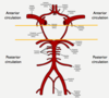
In a stroke, what are the possible locations where the clot could have come from?
- Brain
- Carotid arteries
- The vertebral / basilar arteries
- Aorta
- Heart
Provide four possible conditions which could lead to blood clots arising from the heart and causing a stroke
- Atrial Fibrillation
- Valvular disease / prosthetic valves
- Septic emboli (endocarditis)
- Intra-cardiac thrombus
Identify three unusual conditions which could lead to a stroke
- Vasculitis
- Sickle cell anaemia
- Cocaine (coke stroke)
Which part of the brain does the anterior cerebral artery supply?
The anterior cerebral artery supplies the medial aspects of the frontal and parietal lobe and the anterior part of corpus callosum
What does the part of the brain supplied by the ACA do?
infarct:
→ contralateral weakness of lower limb (much worse than face and upper limb)
→ contralateral sensory weakness similar to motor
How could an anterior cerebral artery stroke affect the parietal lobe and corpus callosum?
- Corpus callosum – split brain syndrome, alien hand syndrome
- Parietal lobes – loss of voluntary control of micturition
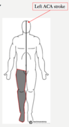
How would a patient present with a left ACA stroke?
- Sensory – contralateral loss of all sensory modalities in the lower limb
- Motor –contralateral paralysis in lower limb more so than upper limb (initially flaccid paralysis then spasticity, UMN signs)
what are some ACA stroke presentations
Urinary incontinence due to paracentral lobules being affected
• Paracentral lobules are essentially the most medial part of the motor/sensory cortices and supply the perineal area
Apraxia
Inability to complete motor planning. Often caused by damage to left frontal lobe
Dysarthria / aphasia
A very unusual sign in ACA infarcts compared with MCA infarct. frontal lobe damage
Split brain syndrome / alien hand syndrome (both rare)
Caused by involvement of corpus callosum which is normally supplied by the ACA
Which part of the brain does the middle cerebral artery supply?

Majority of the hemisphere:
- Basal ganglia
- Internal capsule
- Macular cortex
What would be the result of a main trunk occlusion in the middle cerebral artery?
Considerable cerebral oedema:
- May lead to coma/death
- Malignant MCA
What does the part of the brain supplied by the middle cerebral artery do?
lecticulo striate vessels involved

How does a patient present with a left MCA stroke?
- Sensory – contralateral loss of all sensory modalities in the upper limb and face
- Motor – contralateral upper limb and face affected more than lower limb (initially flaccid paralysis then spasticity UMN signs)

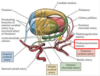
What will be affected by an occlusion at arrow 1?

Occlusion at this point will affect:
→ all Branches effected,
- Lateral motor cortex – responsible for the conralateral face and arm
- Internal capsule – carries descending motor fibres from the entirety of the motor cortex (face, arm and leg may be affected), → contralateral sensory loss as the fibres pass down internal capsule
→ Contralateral homonymous hemianopia
→ can get left sided neglect of its on the right side
→ global aphasia ( combination of Brocas and wernickers) if on the left

What will be affected by an occlusion at arrow 2?
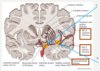
Occlusion at this point spares the internal capsule but still damages the lateral motor cortex, thus only the face and arm are affected

what happens if lenticulostriate arteries are blocked

-
lacunar strokes
1. Cause destruction of small areas of internal capsule and basal ganglia - Essential distinguishing feature from proximal MCA infarct; they do not cause cortical features (e.g. neglect or aphasia)
Types
- Pure motor (face, arm and leg affected
- Pure sensory (face, arm and leg affected equally, caused by damage to sensory fibres travelling through internal capsule probably due to occlusion of thalamoperforator arteries and maybe also lenticulostriate)
- Sensorimotor (mixed, caused by infarct occurring somewhere at boundary between motor and sensory fibres)

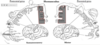
What are the visual effects of proximal and distal occlusions of the middle cerebral artery respectively?
- Proximal occlusion of MCA leads to contralateral homonymous hemianopia
- Distal occlusion of the MCA may lead to contralateral homonymous superior or inferior quadrantanopia (rare)
How does a middle cerebral artery occlusion affect speech?
Symptoms depend on:
- Dominant hemisphere
- Which branch of MCA is occluded
difference between occulusion in MCA inferior and superior division
Inferior: supplies lateral parietal and superior temporal
→ Including primary sensory cortex,Wernicke’s area and both optic radiations, Occlusion will cause contralateral sensory change in face and arm, receptive aphasia if left hemisphere and contralateral visual field defect without macular sparing (often homonymous hemianopia as both radiations damaged)
Superior: supplies lateral frontal lobe
→ Including primary motor cortex and Broca’s area Occlusion will cause contralateral face and arm weakness and expressive aphasia if left hemisphere affected
What happens if the dominant hemisphere (most likely left) is affected by a middle cerebral artery occlusion?
- Global aphasia caused by main trunk occlusion
- Broca’s (expressive) aphasia
- Wernicke’s (receptive) aphasia
What happens if the non-dominant hemisphere (most likely right) is affected by a middle cerebral artery occlusion?
- Hemispatial neglect
- Tactile extinction: touch with sides of the body at the same time they can’t feel left
- Visual extinction: can’t see left
- Anosognosia; failure to acknowledge anything is actually wrong with them
Which part of the brain is supplied by the posterior cerebral artery?

- Occipital lobe
- Inferior temporal lobe
How does a patient present with a PCA stroke?
Contralateral homonymous hemianopia with macular sparing
contralateral sensory loss due to involvement of thalamoperforator/thalamogeniculate branches
Which part of the brain do the cerebellar arteries supply?

- Cerebellum
- Brain stem
What does the part of the brain supplied by the cerebellar arteries do?
Cerebellum – plays a role in the coordination, precision and timing of purposeful movement
How will a patient present with a cerebellar artery stroke (cerebellar signs)?
- Dysdiadochokinesia
- Ataxia
- Nystagmus
- Intention tremor
- Slurred speech
- Hypotonia
Nausea
• Vomiting
• Headache
• Vertigo / dizziness
→ Ipsilateral cerebellar signs (remember DANISH)
→ Possible ipsilateral brainstem signs since cerebellar arteries supply brainstem as they loop round to the cerebellum
→ Possible contralateral sensory deficit / ipsilateral Horner’s (once again due to brainstem involvement)
Account for the variation in presentation of a patient with a cerebellar artery stroke
Cerebellar arteries also supply the brainstem:
- Proximal occlusion may cause brainstem and cerebellar signs
- Distal occlusion may cause cerebellar signs alone
Cranial nerve nuclei reside in brainstem.
In light of this, explain the possible brainstem signs that a patient could present with a cerebellar artery stroke
- Damage to ascending / descending tracts affects contralateral side of body
- Damage to cranial nerves or their nuclei gives ipsilateral signs
A typical feature is that contralateral limb weakness is seen with ipsilateral cranial nerve signs due to damage of corticospinal tracts
The severity of a basilar artery stroke depends on location of occlusion.
What is the presentation for a distal and proximal occlusion respectively?

- Distal occlusion
I. Bilateral occipital lobe infarction (visual and occulomotor deficits)
II. Bilateral thalamic infarction, midbrain infarction
III. Behavioural abnormalities Somnolence, hallucinations and dreamlike behaviour
IIII. Motor dysfunction usually absent as cerebral peduncles ‘rescued’ by PComm
- Proximal occlusion – ’locked-in-syndrome’ (body + facial muscles paralysed), eye muscles work and fully conscious
What part of the brain does the lenticulostriate artery (small branch of the MCA) supply?

Internal capsule (amongst other things)
What does the internal capsule do?
Posterior limb – carries descending motor fibres
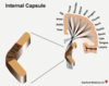
How does a left lenticulostriate artery stroke present?
Pure motor hemiparesis:
- Isolated contralateral paralysis (initially flaccid followed by spasticity)
- Involving face, upper limb and lower limb

What part of the brain does the thalamoperforator artery (small branch of PCA) supply?
Part of the thalamus
What does the part of the brain supplied by the thalamoperforator artery do?
Thalamus – relays sensory information to the left post-central gyrus (somatosensory cortex)
How does a left thalamoperforator artery stroke present?
Pure sensory stroke:
- Isolated contralateral sensory loss of all modalities
- Involving face, upper limb and lower limb

Not every case of dysphasia and/or weakness is a stroke.
Identify four other conditions that mimic the presentation of a stroke
- Hypoglycaemia
- Epilepsy
- Migraine (hemiplegic)
- Intracranial tumours/infections
Describe the findings in the following CT scan of the brain
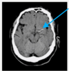
Direct visualisation of the clot (hyperdense artery)

Describe the findings in the following CT scan of the brain:

- Early parenchymal changes
- Grey matter becomes hypodense (dark)
- Loss of grey/white matter differentiation

what is the emergency management of stroke
- are they within the window for thrombolysis
- do a CT head scan to determine if it is a bleed
- CT: ischameic area becomes more established as time moves on → bleed will show up as white
- MRI: ischameia shows up as a high signal area
Bamford stroke classification

