S10) Acute Kidney Injury Flashcards
What is acute kidney injury?
- AKI is a clinical syndrome wherein there is an abrupt decline in actual GFR (days to weeks)
- urea and creatine rise rapidly
- associated with oliguria (no urine) or anuria (no urine)
- usually reversible but not always
- This disrupts homeostasis (ECF volume, electrolyte and acid-base) and leads to an accumulation of nitrogenous waste products
In terms of laboratory findings, provide three definitions of AKI
- Increase in serum creatinine by ≥ 26.5 μmol/L within 48 hours
- Increase in serum creatinine by ≥1.5 times baseline within 7 days
- Urine volume <0.5 ml/kg/h for 6 hours
Define Stage 1 AKI in terms of serum creatinine and urine output
- Serum Cr criteria: ↑ Cr ≥ 1.5- 2x from baseline
- Urine output criteria: <0.5 mL/kg/hr for >6 h
Define Stage 2 AKI in terms of serum creatinine and urine output
- Serum Cr criteria: ↑ Cr > 2-3x from baseline
- Urine output criteria: <0.5 mL/kg/h for >12 h
Define Stage 3 AKI in terms of serum creatinine and urine output
- Serum Cr criteria: ↑ Cr >3x from baseline or initiated on RRT
- Urine output criteria: <0.3 mL/kg/h for 24 h or anuria for 12 h
Identify the three causes of AKI
- Pre-renal failure (volume responsive)
- Intrinsic renal failure
- Post-renal failure
Describe the epidemiology of AKI in high income countries in terms of:
- Typical patients
- Location
- Causes
- Treatment
- Patients: older people
- Location: hospital-acquired AKI
- Causes: dehydration and hypotension
- Treatment: dialysis withheld most commonly due to futility
Describe the epidemiology of AKI in low income countries in terms of:
- Typical patients
- Location
- Causes
- Treatment
- Patients: younger people (often children)
- Location: community-acquired AKI
- Causes: dehydration, hypotension, obstetric causes, snake and insect bites
- Treatment: dialysis withheld due to lack of resources
In 6 steps, outline the pathophysiology of pre-renal AKI
⇒ Decreased renal blood flow
⇒ Reduced actual GFR
⇒ Kidneys work hard to restore blood flow (no cell damage)
⇒ Reabsorb Na+ & H2O (aldosterone, ADH release)
⇒ If mild hypoperfusion, autoregulation preserves renal blood flow
⇒ If overwhelmed compensatory responses, AKI occurs

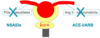
Explain how prescription drugs can affect renal perfusion
- NSAIDS and ACEi can override intrinsic autoregulatory mechanisms
- Disease of the afferent arteriole (BP, DM) can result in too great or too little a response to these stimuli
Describe the causes of pre-renal AKI in terms of:
- Reduced effect arterial blood volume
- Impaired renal autoregulation
- Reduced effective arterial blood volume:
I. Hypovolaemia
II. Systemic vasodilation – sepsis, cirrhosis, anaphylaxis - too low a blood pressure
III. Cardiac failure
- Impaired renal autoregulation:
I. Pre-glomerular vasoconstriction – sepsis, NSAIDs
II. Post-glomerular vasodilatation – ACEi, ARBs
Acute tubular necrosis is volume unresponsive AKI.
Identify three of its causes
- Ischaemia (depletion of cellular ATP)
- Nephrotoxins
- Sepsis
Describe the pathophysiology of ATN (acute tubular necrosis)
⇒ Death of tubular epithelial cells
⇒ Damaged cells cannot reabsorb / expel excessive H2O efficiently
⇒ Aggressive fluid resuscitation risks fluid overload
Describe the course of renal blood flow

Identify the sites of tubular injury in ATN

Compare and contrast the urinary biochemistry of Pre-renal AKI and ATN in terms of:
- Specific gravity
- Osmolality
- Urinary Na+

ATN is much more likely if there is reduced perfusion and a nephrotoxin.
What do nephrotoxins do?
Nephrotoxins damage the epithelial cells lining the tubules and cause cell death
Nephrotoxins can be endogenous or exogenous.
Identify some examples of each

What is rhabdomyolysis?
- Rhabdomyolysis is a serious syndrome due to a direct or indirect muscle injury e.g. crush injury
- Muscle necrosis cases releases of myoglobin into the bloodstream
When is rhabdomyolysis likely to occur?
- AKI in wars / natural disasters (earthquakes)
- Drug users (unconscious so don’t move)
- Elderly (fall & can’t get up)
Explain the association of rhabdomyolysis with AKI
- Released myoglobin is filtered at the glomerulus and toxic to tubule cells
- Released myoglobin can also cause renal obstruction
What is myoglobinuria and how does it present?
Myoglobinuria is the presence of myoglobin in the urine usually associated with rhabdomyolysis or muscle destruction

Besides ATN, identify two other intrinsic renal causes of AKI
- Thrombotic microangiopathy
- Acute (tubule)-interstitial nephritis
Identify 4 clinical conditions which present with thrombotic microangiopathy
- Haemolytic uraemic syndrome
- Malignant hypertension
- Scleroderma
- Pre-eclampsia
In 5 steps, explain how thrombotic microangiopathy leads to microangiopathic haemolytic anaemia
⇒ Endothelial damage
⇒ Platelet thrombi
⇒ Partial obstruction of small arteries
⇒ Destruction of RBC’s
⇒ Microangiopathic haemolytic anaemia occurs

Identify and describe the two causative mechanisms of acute interstitial nephritis
- Toxin induced: antibiotics, NSAID’s, PPI’s
- Infections: due to inflammatory response (not direct effect of infection)
Post-renal failure accounts for 5 - 10% of AKI cases.
How does it occur?
- Occurs commonly in elderly
- Obstruction must block both kidneys or a single functioning kidney
Describe the pathophysiology of post-renal AKI
⇒ Obstruction with continuous urine production
⇒ Rise in intraluminal pressure
⇒ Dilatation of renal pelvis (hydronephrosis)
⇒ Decrease in renal function

Identify and describe the three different causes of post-renal AKI
- Within the lumen (kidney, ureter, bladder) – stones, blood clot, tumours
- Within the wall (usually cause CKD rather than AKI) – congenital megaureter, post-TB stricture
- Pressure from outside – enlarged prostate, tumour, aortic aneurysm
Identify 5 clinical signs in serum biochemistry which are indicative of AKI
- Metabolic acidosis
- Hyperkalaemia
- Hyponatraemia
- Hypocalcaemia
- Hyperphosphataemia
In 3 steps, describe how metabolic acidosis occurs in AKI
⇒ Reduction in GFR
⇒ Impaired reabsorption & regeneration of HCO3-
⇒ Impaired acid excretion
Identify and describe 4 causes of hyperkalaemia
- Excessive intake
- Movement out of cells (acidosis / tissue damage)
- Reduced urine loss (reduced GFR, reduce distal deliver of Na+, reduced secretion)
- Drugs (ACE-Inhibitors, spironolactone, NSAIDs, trimethoprim, amiloride)
Can cause life-threatening cardiac arrhythmias.
Identify 5 clinical signs on an ECG that indicate hyperkalaemia
- Tall T waves
- Small/absent P waves
- Increased P-R interval
- Wide QRS complex
- Asystole
Identify and describe the 5 investigations used for AKI
- Urinalysis (blood, protein, leucocytes)
- Urine microscopy
- CXR (fluid overload ± infection)
- Ultrasound scan (only for obstruction – within 24 hours)
- Kidney biopsy (systemic inflammatory symptoms/ igns)
In terms of volume overload and hyperkalaemia, outline the management of AKI
- Volume overload – restrict dietary Na+ / water < 1L/day
- Hyperkalaemia – calcium gluconate, restrict dietary K+, stop K+-sparing diuretics, ACEi, ARB
What are the 5 indications for dialysis?
- Hyperkalaemia (refractory to treatment)
- Fluid overload (refractory to diuretics)
- Metabolic acidosis (NaHCO3- not appropriate)
- Signs of uraemia (pericarditis, reduced consciousness)
- Dialysable nephrotoxin e.g. aspirin overdose, ethylene glycol
How can one prevent AKI?
- Identify risk factors
- Monitor ‘at risk’ patients closely
- Ensure adequate hydration
- Avoid nephrotoxins
- Detect early and identify cause
Identify 5 risk factors for AKI in terms of susceptibility
- Advanced age
- CKD
- Heart disease
- Liver disease
- Diabetes Mellitus
Identify 5 risk factors for AKI in terms of exposure
- Dehydration
- Sepsis
- Burns / trauma
- Cardiac surgery
- Nephrotoxins (NSAIDS, ACE-I, ARB)
what are the 4 main complications of an AKI
- Metabolic Acidosis
- hyperkalemia
- volume overload
- uraemia
what investigation can be taken at the beside
- bladder scan
- urinalysis
- microscopy culture and specimen
what bloods can be taken from a patient
- VBG - to show if the patient is becoming acidotic
- CK - breakdown of muscle
- immunology screening - see if its autoimmune
what imaging could be done
- USS KUB
- CT
both to show is there is any post renal cause
- CXR - to show is there is any pulmonary oedema
what procedures can be carried out
- nephrostogram
- cystoscopy (procedure to look into the bladder)
what is the management for pre renal hypovolemia
- IV fluid replacement to correct hypovolameia and optimise renal blood flow
- hold nephrotoxic medication
- diuretics
management of renal hypovolemia
correct electrolytes
renal replacement therapy
management of post renal issues
- urinary or supra pubic catheter
- ureteric stents to open the ureter
- nephrostomy - tube in renal pelvis
what is a nephrostomy
- urine is backed up in the ureter
- so urine is drained out via a bag that is attached to the kidney
post renal causes
- calculus → renal stone
- ureteric stricture → scarring in the ureter due to a stone
- Benign prostatic hyperplasia → prostate gland swells so urethra gets squeezed
- tumour - blocks
- retro peritoneal blockage - fibrotic tissue
left hydronephrosis
pre renal causes
sepsis - systemic vasodilation
hypovolaemia - low blood volume so reduced perfusion
shock - low BP
renal artery stenosis - narrowing
CCF - reduced CO, so reduced blood flow to kidney
NSAIDS - inhibit prostaglandin
ACEi -
how does ACEi cause pre renal Aki
- causes vasodilation of the efferent arteriole
- this causes low perfusion rates
left hydronephrosis on X-ray
the kidney normally has some black In the middle, but here the kidney is enlarged

what does a baseline xray of the kidneys look like
the grey diagonal lines coming from the pine are the two posts major muscles
this is also taken on a coronal plane

